This website uses cookies to ensure you get the best experience on our website.


- Table of Contents
Plan TNS3 IHC in paraffin sections using glandular cells with high reported staining as positive references (HPA tissue IHC). Compare membranous and cytoplasmic staining while accounting for the reported low consistency between antibody staining and RNA expression (HPA tissue IHC).

Expected localisation, validated protocols, controls and antibodies — the at-a-glance facts below, then the full design guide.
| Expected localisation | Membranous and cytoplasmic tissue staining (HPA tissue IHC) | |
| Staining pattern | Membranous/cytoplasmic staining in glandular cells (HPA tissue IHC) | |
| Antigen retrieval | EDTA pH 8.0 HIER, heat-mediated (datasheet A07345-1) | |
| Positive control | Appendix+4 more · see all | |
| Negative control | Adipose tissue+4 more · see all |
| Fixation | Keep fixation consistent across sections. (standard IHC practice; not target-specific) | |
| Caveat | Antibody staining has low consistency with RNA data (HPA tissue IHC) | |
| Regulation | EGF down-regulates TNS3 (UniProt) | |
| Isoform / epitope | 4 isoforms; check epitope coverage across variants (UniProt) |
Compare the catalog antibody’s IHC-P protocol with 4 published TNS3 IHC protocols (PMC9392296; PMC8146480; PMC8221360; PMC13048582).
| Sample | Paraffin-embedded human colon adenocarcinoma tissue; fixative not specified (datasheet A07345-1) |
| Fixation | Image fixative and duration unreported (datasheet A07345-1); verify before use. |
| Sectioning | 4–5 µm sections on charged slides (standard) |
| Deparaffinisation | Xylene, graded ethanol series to water (standard) |
| Antigen retrieval | Heat retrieval: EDTA pH 8.0 (datasheet A07345-1); 20 min, 95–100 °C (standard) |
| Peroxidase block | 3% H2O2, 10 min, room temperature (standard) |
| Blocking | 10% goat serum (datasheet A07345-1) |
| Primary antibody | Rabbit anti-TNS3, 2-5 μg/ml (datasheet A07345-1) |
| Primary incubation | Overnight at 4 °C (datasheet A07345-1) |
| Detection | HRP-conjugated secondary, DAB chromogen (datasheet A07345-1) |
| Counterstain | Hematoxylin, blue, dehydrate and mount (standard) |
| Expected result | TNS3-positive staining in glandular cells of appendix (HPA tissue IHC: High). HPA tissue profile: Membranous and cytoplasmic expression in several tissues. No signal in the no-primary control. |
TNS3 localizes to focal and fibrillar adhesions and podosomes (UniProt Q68CZ2); it has no transmembrane segment (UniProt Q68CZ2 topology). In paraffin sections, expect membranous and cytoplasmic staining in selected glandular and other positive cell populations (HPA tissue IHC). Treat tissue predictions cautiously: HPA rates its IHC profile Approved but reports low consistency between antibody staining and RNA expression, pending external verification (HPA tissue IHC).
| Membranous and cytoplasmic signal in colon, appendix, duodenum or gallbladder glandular cells. | This matches the reported tissue pattern; HPA scores these cell populations High (HPA tissue IHC). Assess the named cells within the section, rather than treating a uniformly stained field as confirmation. HPA's Approved profile still has low staining–RNA consistency, so this pattern supports interpretation without independently proving antibody specificity (HPA tissue IHC). |
| Predominantly nuclear staining, with little signal at cell edges or in cytoplasm. | A nuclear-only pattern conflicts with the reported membranous and cytoplasmic tissue profile (HPA tissue IHC) and adhesion-associated localization (UniProt Q68CZ2). Suspect nonspecific staining or a detection artefact; compare positive and negative control sections before assigning biological meaning. Adhesion-scale puncta may be difficult to resolve by chromogenic tissue IHC (general IHC practice). |
| Strong signal in adipocytes, cardiomyocytes or liver cholangiocytes. | Those specific populations are listed as Not detected (HPA tissue IHC). Check the cell identification and staining controls; cross-reactivity or endogenous detection activity could explain unexpected signal (general IHC practice). HPA's low staining–RNA consistency means an unexpected result warrants verification, rather than automatic rejection as impossible (HPA tissue IHC). |
| Diffuse stain spans cells and extracellular space without a discernible cellular pattern. | This does not resemble the reported membranous and cytoplasmic expression (HPA tissue IHC). In chromogenic IHC, incomplete blocking, excess primary antibody or residual detection background can produce such appearances (general IHC practice). Review a no-primary control and the section's morphology before scoring diffuse colour as TNS3. |
| No signal in glandular cells of a well-preserved colon or appendix section. | HPA reports High staining in those populations (HPA tissue IHC), so first investigate a technical false negative using the assay's controls (general IHC practice). Recheck that the relevant glandular cells are present, then review the antibody dilution, retrieval conditions and detection run. HPA's Approved rating is pending external verification (HPA tissue IHC). |
| Subcellular location | TNS3 is associated with focal and fibrillar adhesions, predominantly the latter, and podosomes (UniProt Q68CZ2). HPA describes tissue staining as membranous and cytoplasmic (HPA tissue IHC). A chromogenic section need not resolve individual adhesion sites to agree with those observations (general IHC practice). |
| Cell population and tissue | HPA reports High staining in kidney glomerular cells, pancreatic exocrine glandular cells and fallopian tube ciliated-cell rootlets, alongside several glandular populations (HPA tissue IHC). Select and score the stated cell population; a tissue name alone does not define where signal should appear (general IHC practice). |
| Antibody evidence | Both listed antibodies, HPA055338 and HPA056015, have Approved IHC status (HPA antibodies). The tissue profile nevertheless has low consistency with RNA expression and awaits external verification (HPA tissue IHC). Interpret staining with morphology and controls; Approved status alone does not make every unexpected pattern specific. |
| Isoforms and epitope | UniProt lists 4 TNS3 isoforms and domains spanning different parts of the protein (UniProt Q68CZ2). Without an epitope location in these payloads, the isoforms recognized by a particular antibody cannot be predicted. Do not attribute a staining difference to isoform selection from these data alone. |
| IF/ICC interpretation | If an IF/ICC image is being compared with the IHC slide, HPA places TNS3 mainly at focal adhesion sites and lists Supported ICC status for both antibodies (HPA subcellular; HPA antibodies). The IF localization offers a compartment check, but its spatial detail should not be required of chromogenic IHC (general IHC practice). |
| Situation | Likely cause | Next action |
|---|---|---|
| Positive-control glandular cells are blank. | The assay may have lost detectable signal; colon and appendix glandular cells are reported High (HPA tissue IHC). | Confirm the expected cells are present, then review the IHC-validated antibody's specified dilution, retrieval and detection controls (general IHC practice). These sources do not establish a TNS3-specific fixation sensitivity. |
| Signal appears chiefly in nuclei. | That compartment conflicts with HPA's membranous and cytoplasmic tissue profile (HPA tissue IHC). | Check the no-primary control, section morphology and detection background; repeat with an independently validated antibody when available (general IHC practice). Avoid calling nuclear-only colour a confirmed TNS3 pattern. |
| Negative-reference cells stain strongly. | Adipocytes and cardiomyocytes are reported Not detected, so background, endogenous activity or cross-reactivity is plausible (HPA tissue IHC; general IHC practice). | Verify cell identity and compare no-primary and appropriate detection controls before changing interpretation (general IHC practice). HPA's staining–RNA discordance calls for verification of unexpected biology (HPA tissue IHC). |
| Brown colour is diffuse across the section. | A field-wide haze lacks the reported cell-associated membranous and cytoplasmic pattern (HPA tissue IHC); excess reagent or inadequate background control can contribute (general IHC practice). | Inspect the no-primary control, blocking and washes; adjust assay conditions within the antibody's documented IHC procedure (general IHC practice). Score only interpretable cellular staining. |
| Signal varies between neighboring cell types. | HPA assigns levels to specific populations, such as High glomerular cells and Not detected liver cholangiocytes, rather than to every cell in an organ (HPA tissue IHC). | Annotate morphology and score each relevant population separately (general IHC practice). Check the HPA cell-level expectation before treating heterogeneity as assay failure (HPA tissue IHC). |
| Does IF/ICC have to show the same appearance as chromogenic IHC? | HPA reports focal adhesion localization by ICC-IF, while its tissue IHC summary is membranous and cytoplasmic (HPA subcellular; HPA tissue IHC). | Use the IF observation as a localization cross-check, then judge the paraffin IHC slide against its own cell-level tissue pattern and controls (HPA tissue IHC; general IHC practice). |




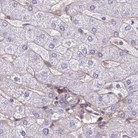
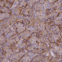


Comprehensive Human Protein Atlas IHC scoring per tissue (reliability: Approved — Low consistency between antibody staining and RNA expression data. Pending external verification.). Rows are taken directly from the HPA tissue chart — click any row's HPA link to view the source.
| Tissue | Cell type | Level | Evidence | Source |
|---|---|---|---|---|
| Appendix | Glandular cells | High | Protein (IHC) | HPA → |
| Colon | Glandular cells | High | Protein (IHC) | HPA → |
| Duodenum | Glandular cells | High | Protein (IHC) | HPA → |
| Endometrium | Glandular cells | Medium | Protein (IHC) | HPA → |
| Fallopian tube | Ciliated cells (ciliary rootlets) | High | Protein (IHC) | HPA → |
| Tissue | Cell type | Level | Evidence | Source |
|---|---|---|---|---|
| Adipose tissue | Adipocytes | Not detected | Protein (IHC) | HPA → |
| Adrenal gland | Glandular cells | Not detected | Protein (IHC) | HPA → |
| Caudate | Glial cells | Not detected | Protein (IHC) | HPA → |
| Cerebral cortex | Glial cells | Not detected | Protein (IHC) | HPA → |
| Esophagus | Squamous epithelial cells | Not detected | Protein (IHC) | HPA → |
Troubleshoot TNS3 staining in paraffin-section chromogenic IHC by checking retrieval, controls, compartment-specific signal and scoring before interpreting tissue differences.
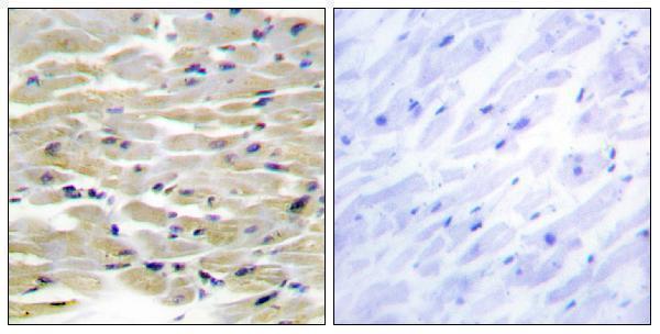
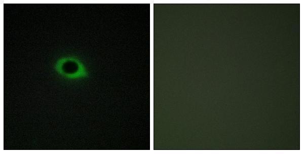
Both antibodies have human paraffin-section IHC images and cell IF images (catalog image captions). Listed reactivity is human for A07345-1 and human, mouse, and rat for A30511 (catalog reactivity).
A07345-1 shows IHC in paraffin-embedded human colon adenocarcinoma and IF in U2OS cells (A07345-1 image captions). A30511 shows IHC in paraffin-embedded human heart and IF in COS7 cells, each with a peptide-blocked comparison (A30511 image captions).
Which to pick: For tissue IHC, choose A07345-1 when its documented EDTA pH 8.0 retrieval and 2 μg/ml overnight incubation fit your workflow; A30511 has a peptide-blocked human heart IHC image but fewer processing details (respective IHC captions). For IF/ICC, both have cell images; choose A30511 if listed mouse or rat reactivity matters, while its illustrated IHC sample is human (catalog reactivity; respective image captions). The fixative is unreported for both paraffin-section IHC images (respective IHC captions).