This website uses cookies to ensure you get the best experience on our website.


- Table of Contents
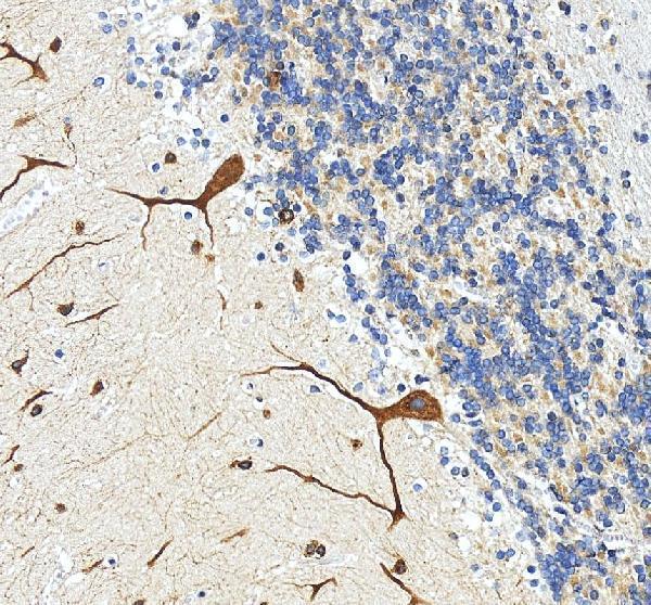
Plan TOE1 staining in paraffin sections with the IHC-validated antibody at 2–5 μg/ml (datasheet A09852-2). Compare the nuclear pattern with tissue IHC evidence, using cerebellar Purkinje cells as a high-staining reference (HPA tissue IHC).

Expected localisation, validated protocols, controls and antibodies — the at-a-glance facts below, then the full design guide.
| Expected localisation | General nuclear staining (HPA tissue IHC) | |
| Staining pattern | Nuclear staining in many cell types, including Purkinje cells (HPA tissue IHC) | |
| Antigen retrieval | EDTA pH 8.0 HIER, heat-mediated (datasheet A09852-2) | |
| Positive control | Adipose tissue+4 more · see all | |
| Negative control | None in HPA (detected in all 45 tissues); use no-primary + isotype controls |
| Fixation | Matched tissue-IHC evidence does not establish the fixation claim. Validate the specimen-specific method before use. (selected-SKU IHC image A09852-2) | |
| Caveat | Cholangiocytes may stain weakly (HPA tissue IHC) | |
| Regulation | Widely expressed; no regulator annotated (UniProt) | |
| Isoform / epitope | 2 isoforms; epitope coverage is unspecified (UniProt) |
The catalog antibody’s IHC-P protocol uses EDTA pH 8.0 retrieval (datasheet A09852-2). Two published TOE1 IHC protocols offer additional conditions (PMC8092974; PMC12764802).
| Sample | Paraffin-embedded human cerebellum tissue; fixative not specified (datasheet A09852-2) |
| Fixation | Image fixative and duration unreported (datasheet A09852-2); verify before use. |
| Sectioning | 4–5 µm sections on charged slides (standard) |
| Deparaffinisation | Xylene, graded ethanol series to water (standard) |
| Antigen retrieval | Heat retrieval: EDTA pH 8.0 (datasheet A09852-2); 20 min, 95–100 °C (standard) |
| Peroxidase block | 3% H2O2, 10 min, room temperature (standard) |
| Blocking | 10% goat serum (datasheet A09852-2) |
| Primary antibody | Rabbit anti-TOE1, 2-5μg/ml (datasheet A09852-2) |
| Primary incubation | Overnight at 4 °C (datasheet A09852-2) |
| Detection | HRP-conjugated secondary, DAB chromogen (datasheet A09852-2) |
| Counterstain | Hematoxylin, blue, dehydrate and mount (standard) |
| Expected result | TOE1-positive staining in adipocytes of adipose tissue (HPA tissue IHC: High). HPA tissue profile: General nuclear expression. No signal in the no-primary control. |
TOE1 should appear chiefly in nuclei across many cell types (UniProt Q96GM8: nucleus, nucleolus and nuclear speckles; widely expressed; no transmembrane segment). HPA describes general nuclear tissue staining, including High staining in adipocytes, hematopoietic cells and several epithelial or neural cell populations (HPA tissue IHC: Supported). That assessment reflects agreement with RNA data and awaits external verification (HPA tissue IHC: reliability description).
| Nuclear chromogen in adipocytes, bone marrow hematopoietic cells or bronchial respiratory epithelial cells. | This matches the reported High cell-specific staining and general nuclear profile (HPA tissue IHC). Score staining in the named cells and their nuclei, rather than treating every cell in the section as an equally strong positive (HPA tissue IHC: cell-level observations). |
| Predominantly cytoplasmic, membranous or extracellular staining, with little nuclear signal. | That compartment pattern conflicts with TOE1's nuclear localization and lack of a transmembrane segment (UniProt Q96GM8). Check localization against the counterstain and a reported positive cell population before interpreting the deposit as TOE1 (HPA tissue IHC: general nuclear expression; standard IHC practice). |
| Strong signal in an unexpected cell population, especially where HPA reports Low staining. | HPA reports Low staining in parathyroid glandular cells and liver cholangiocytes; it supplies no negative tissue here (HPA tissue IHC). Strong staining there warrants checks for antibody cross-reactivity and endogenous detection activity (standard IHC practice). Low does not mean absent (HPA tissue IHC: Low). |
| Diffuse color across nuclei, cytoplasm and tissue spaces. | A widespread deposit that obscures cell boundaries prevents a reliable nuclear call (standard IHC practice). Assess background with an appropriate primary-omission control and inspect detection and blocking steps before assigning it to TOE1 (standard IHC practice; UniProt Q96GM8: nuclear localization). |
| No nuclear signal in a reported High cell population. | First check that the expected cells are present and that the detection run worked (standard IHC practice). An absent result disagrees with HPA's High observation for that population, but one failed section does not establish loss of TOE1 expression (HPA tissue IHC: reported High cells; Supported, pending external verification). |
| Compartment and tissue distribution | TOE1 is nuclear, including nucleolar and nuclear-speckle annotations, and is widely expressed (UniProt Q96GM8). HPA reports general nuclear tissue staining with different cell-level intensities (HPA tissue IHC). Use the named cells when judging a section; wide expression alone does not predict uniform intensity in every cell. |
| Strength of the IHC evidence | The tissue profile is Supported because staining and RNA expression are highly consistent, with external verification pending (HPA tissue IHC: reliability description). Of the listed antibodies, HPA069119 has Supported IHC validation; HPA053775 has no IHC status in this payload (HPA antibodies). Do not transfer the former's IHC status to the latter. |
| Isoforms and processing | UniProt lists 2 isoforms and a chain spanning residues 2–510, with no signal peptide or propeptide reported (UniProt Q96GM8). These annotations do not identify the antibody epitope or establish isoform-specific staining. Interpret compartment and cell distribution without claiming that a particular isoform explains a staining difference. |
| Retrieval and detection controls | For paraffin IHC, optimize retrieval and check detection with a reported positive population and suitable controls (standard IHC practice). No TOE1-specific retrieval condition, fixation effect or dilution is supplied. Endogenous detection activity can produce misleading color, so a control lacking primary antibody helps investigate diffuse or unexpected signal (standard IHC practice). |
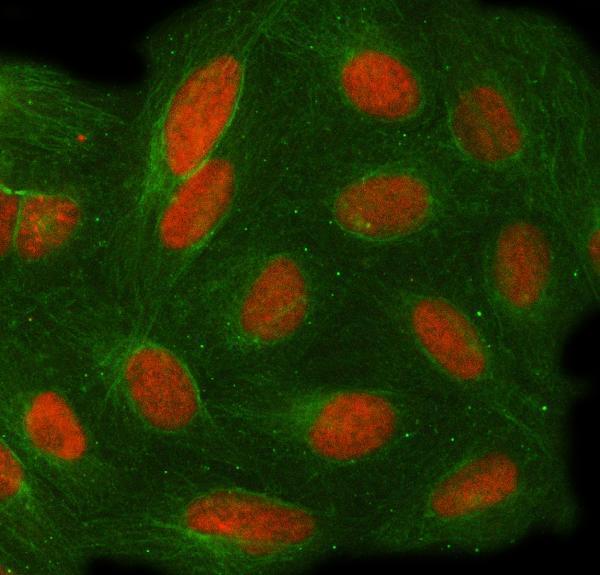
| IF/ICC Q&A: where should fluorescence appear? | Mainly in nucleoplasm, with possible nuclear-body signal; both HPA subcellular assignments are marked uncertain (HPA ICC-IF). UniProt also annotates nucleolus and nuclear speckles (UniProt Q96GM8). HPA lists Enhanced ICC validation for both antibodies (HPA antibodies). These IF/ICC observations do not supply an IHC protocol or override the tissue IHC pattern. |
| Situation | Likely cause | Next action |
|---|---|---|
| Reported High cells are present, but nuclei remain blank. | The section or detection run may have failed; absence in one section alone is inconclusive (standard IHC practice; HPA tissue IHC: High cell observations). | Confirm cell identity on the counterstain, inspect a processed positive section and detection controls, then reassess the nuclear signal (standard IHC practice). |
| Color concentrates in cytoplasm or at cell borders. | The distribution conflicts with nuclear TOE1 and its lack of a transmembrane segment (UniProt Q96GM8); nonspecific staining is possible (standard IHC practice). | Compare with a reported High cell population and a primary-omission control; score TOE1 only where the compartment assignment is credible (HPA tissue IHC; standard IHC practice). |
| Strong staining appears in parathyroid glandular cells or liver cholangiocytes. | HPA calls these populations Low, so a strong result needs scrutiny; the payload does not classify them as negative (HPA tissue IHC). | Check the named cells, nuclear localization and control staining before attributing the signal to TOE1 (HPA tissue IHC: general nuclear expression; standard IHC practice). |
| Diffuse color obscures nuclear boundaries. | Background from blocking or detection steps can hinder localization calls, including through endogenous detection activity (standard IHC practice). | Inspect a primary-omission control and review blocking, detection and counterstain conditions; repeat scoring only when nuclei can be distinguished (standard IHC practice). |
| Two antibodies give different IHC staining patterns. | The listed antibodies have different recorded IHC statuses: HPA069119 is Supported, while HPA053775 has no IHC status here (HPA antibodies). | Record which antibody produced each pattern and compare nuclear staining in the same reported High cells; do not cite ICC validation as IHC validation (HPA antibodies; HPA tissue IHC). |
| An IF image shows nuclear bodies, while IHC looks broadly nuclear. | HPA's ICC-IF nuclear-body and nucleoplasm assignments are uncertain, whereas its tissue IHC profile is general nuclear expression (HPA subcellular; HPA tissue IHC). | Interpret the paraffin section against its cell-level IHC profile and counterstain; reserve detailed subnuclear comparisons for the separate IF/ICC assessment (HPA tissue IHC; standard IHC practice). |


Comprehensive Human Protein Atlas IHC scoring per tissue (reliability: Supported — High consistency between antibody staining and RNA expression data. Pending external verification.). Rows are taken directly from the HPA tissue chart — click any row's HPA link to view the source.
| Tissue | Cell type | Level | Evidence | Source |
|---|---|---|---|---|
| Adipose tissue | Adipocytes | High | Protein (IHC) | HPA → |
| Adrenal gland | Glandular cells | High | Protein (IHC) | HPA → |
| Appendix | Glandular cells | High | Protein (IHC) | HPA → |
| Bone marrow | Hematopoietic cells | High | Protein (IHC) | HPA → |
| Breast | Adipocytes | High | Protein (IHC) | HPA → |
| Tissue | Cell type | Level | Evidence | Source |
|---|---|---|---|---|
| None in HPA: TOE1 is detected in all 45 scored tissues. Use a no-primary (secondary-only) and an isotype control instead. | ||||
Troubleshoot TOE1 staining in paraffin sections by checking retrieval, nuclear localisation, background and scoring against the catalog antibody’s tissue result and reference profiles.
A09852-2 has IHC data from a paraffin-embedded human cerebellum section and IF/ICC data from U2OS cells (A09852-2 IHC/IF image captions); listed reactivity is human (catalog: Human).
A09852-2 will render with IHC data from a paraffin-embedded human cerebellum section (A09852-2 IHC image caption). The same SKU is listed for IF and ICC and has IF data from U2OS cells (catalog: applications; A09852-2 IF image caption).
Which to pick: For tissue IHC, choose A09852-2: its own caption documents heat retrieval in EDTA at pH 8.0 and 2 μg/ml primary antibody on a paraffin-embedded human cerebellum section; the fixative is unreported (A09852-2 IHC image caption). For IF/ICC, A09852-2 is also listed for both applications and has a U2OS-cell IF image (catalog: applications; A09852-2 IF image caption). No cross-species or clone-specific choice is supported by this catalog entry (catalog: Human reactivity; clone unreported).