This website uses cookies to ensure you get the best experience on our website.


- Table of Contents
Plan paraffin-section TOLLIP IHC around cytoplasmic staining, including strong signal in brain neurons (HPA tissue IHC). The catalog antibody has a documented 2 μg/mL starting concentration (datasheet: PA2005); compare staining by cell type because levels vary across tissues (HPA tissue IHC).

Expected localisation, validated protocols, controls and antibodies — the at-a-glance facts below, then the full design guide.
| Expected localisation | Cytoplasmic in tissue (HPA tissue IHC); endosomal location (UniProt) | |
| Staining pattern | Cytoplasmic across tissues; strong in brain neurons (HPA tissue IHC) | |
| Antigen retrieval | EDTA pH 8.0 HIER, heat-mediated (datasheet PA2005) | |
| Positive control | Caudate+4 more · see all | |
| Negative control | Smooth muscle+1 more · see all |
| Fixation | Keep fixation conditions consistent across sections. (standard IHC practice; not target-specific) | |
| Caveat | Brain staining varies: high in cortex, low in hippocampus (HPA tissue IHC) | |
| Regulation | Expression regulation not established (UniProt) | |
| Isoform / epitope | 2 isoforms; epitope coverage is unestablished (UniProt) |
The catalog antibody has an IHC-P protocol (datasheet PA2005). These four published TOLLIP IHC protocols cover skin, rat renal cortex, human renal tissue, and frozen human lung (PMC13426103; PMC9203234; PMC9741407; PMC8175118).
| Sample | Paraffin-embedded human lung cancer tissue; fixative not specified (datasheet PA2005) |
| Fixation | Image fixative and duration unreported (datasheet PA2005); verify before use. |
| Sectioning | 4–5 µm sections on charged slides (standard) |
| Deparaffinisation | Xylene, graded ethanol series to water (standard) |
| Antigen retrieval | Heat retrieval: EDTA pH 8.0 (datasheet PA2005); 20 min, 95–100 °C (standard) |
| Peroxidase block | 3% H2O2, 10 min, room temperature (standard) |
| Blocking | 10% goat serum (datasheet PA2005) |
| Primary antibody | Rabbit anti-TOLLIP, 2-5μg/ml (datasheet PA2005) |
| Primary incubation | Overnight at 4 °C (datasheet PA2005) |
| Detection | HRP-conjugated secondary, DAB chromogen (datasheet PA2005) |
| Counterstain | Hematoxylin, blue, dehydrate and mount (standard) |
| Expected result | TOLLIP-positive staining in neuronal cells of caudate (HPA tissue IHC: High). HPA tissue profile: General cytoplasmic expression, highest levels in the brain. No signal in the no-primary control. |
TOLLIP should appear mainly in the cytoplasm of stained cells, consistent with its cytoplasmic and endosomal localization and lack of a transmembrane segment (UniProt Q9H0E2). In paraffin sections, expect strong staining in caudate and cerebral cortex neurons, epididymal glandular cells, pancreatic endocrine cells, and skin fibrohistiocytic cells (HPA tissue IHC: High). HPA rates the tissue profile Enhanced, while noting medium consistency between staining and RNA expression (HPA tissue IHC).
| Cytoplasmic staining is strong in neurons of the caudate or cerebral cortex (HPA tissue IHC: High). | This fits the reported tissue pattern and TOLLIP localization (HPA tissue IHC; UniProt Q9H0E2). Score the named cell population and its compartment, rather than assigning one intensity to the whole section (general IHC practice). |
| Staining is confined to nuclei, cell surfaces, or extracellular material in an otherwise positive area. | A dominant pattern in those compartments conflicts with the reported general cytoplasmic tissue staining (HPA tissue IHC) and cytoplasmic/endosomal localization (UniProt Q9H0E2). Treat it as suspect and compare controls before interpreting it as TOLLIP (general IHC practice). |
| Smooth muscle cells or spleen red-pulp cells stain strongly despite a quiet expected-positive population (HPA tissue IHC: Not detected; High populations). | The cell distribution conflicts with HPA observations; cross-reactivity or endogenous detection activity is possible (HPA tissue IHC; general IHC practice). A reported 'Not detected' result is a reference pattern, not proof that every specimen must be negative (HPA tissue IHC). |
| Diffuse color covers many structures without a clear cytoplasmic boundary or cell-specific contrast. | This is difficult to score against the reported cytoplasmic profile (HPA tissue IHC). Background from detection chemistry or insufficient blocking may obscure the pattern; inspect a no-primary control and section morphology (general IHC practice). |
| No signal appears in caudate neurons or another HPA High population (HPA tissue IHC: High). | First check tissue preservation, retrieval, antibody incubation, and detection with appropriate controls (general IHC practice). HPA's Enhanced rating supports the reference pattern, but its medium RNA–staining consistency does not guarantee positivity in every specimen (HPA tissue IHC). |
| Choice of tissue and cell population | Caudate and cerebral cortex neurons, epididymal glandular cells, pancreatic endocrine cells, and skin fibrohistiocytic cells are High references; adipocytes and adrenal or appendix glandular cells are Medium (HPA tissue IHC). Compare like cell types when judging intensity (general IHC practice). |
| Antibody evidence | HPA038621 and HPA038622 each have Enhanced IHC validation, with similar staining from paired antibodies supporting the protein profile (HPA antibodies; HPA tissue IHC). This strengthens the reference pattern without establishing specificity in every preparation (HPA tissue IHC; general IHC practice). |
| Compartment and isoforms | TOLLIP is cytoplasmic and endosomal, with two listed isoforms and no transmembrane segment (UniProt Q9H0E2). The payload gives no antibody epitope mapping, so it does not establish whether either isoform changes staining with a particular antibody (UniProt Q9H0E2; HPA antibodies). |
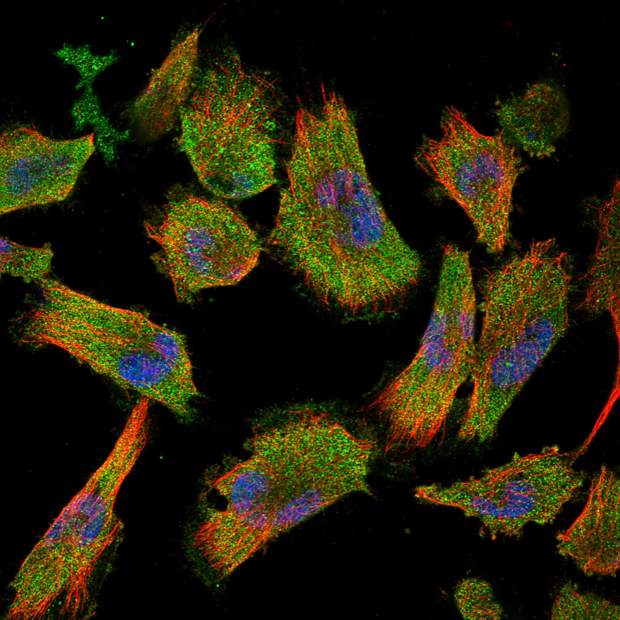
| IF/ICC cross-check: what should be seen? | HPA supports cytosol as the main ICC-IF location; nucleoplasm, primary cilium, and cilium tip are uncertain additional locations (HPA subcellular). Use that as compartment context, while interpreting paraffin-section results against the tissue IHC profile (HPA subcellular; HPA tissue IHC). |
| Retrieval and detection choices | Target-specific fixation effects are not established by the supplied assay evidence. Verify with a matched IHC source before attributing a result to fixation. |
| Situation | Likely cause | Next action |
|---|---|---|
| Expected-positive neurons are unstained (HPA tissue IHC: caudate and cortex High). | Weak assay performance, unsuitable retrieval, or lost tissue integrity may explain absent signal; the payload identifies no TOLLIP-specific fixation effect (general IHC practice; HPA tissue IHC). | Verify morphology and positive-control performance, then adjust retrieval and antibody incubation within the assay workflow (general IHC practice). |
| All cells show similar diffuse chromogen, including cells without clear cytoplasmic staining. | Excess detection signal, insufficient blocking, or nonspecific primary binding may overwhelm the cell pattern (general IHC practice; HPA tissue IHC: general cytoplasmic profile). | Run a no-primary control, review blocking and detection steps, and reduce assay signal until cell boundaries can be scored (general IHC practice). |
| Color remains in a no-primary control. | Endogenous detection activity or reagent background can produce color without primary antibody binding (general IHC practice). | Apply the detection system's appropriate blocking control and reassess the no-primary section before scoring TOLLIP (general IHC practice). |
| Nuclear staining dominates an IHC section. | It conflicts with general cytoplasmic tissue staining; HPA's uncertain nucleoplasmic ICC-IF observation does not establish a dominant nuclear IHC pattern (HPA tissue IHC; HPA subcellular). | Check positive-cell cytoplasm and no-primary controls; confirm the compartment pattern with another IHC-validated antibody if available (HPA antibodies: two Enhanced IHC reagents; general IHC practice). |
| A smooth muscle cell population stains as strongly as a High reference population (HPA tissue IHC). | HPA reports smooth muscle cells as Not detected; nonspecific binding or endogenous activity is possible, though specimen variation remains possible (HPA tissue IHC; general IHC practice). | Compare the same run with an HPA High population and the no-primary control; investigate discordance before calling the smooth muscle signal specific (HPA tissue IHC; general IHC practice). |
| A low-reference population appears faint beside a strong positive population (HPA tissue IHC). | Low neuronal staining in hippocampus or low staining in liver cholangiocytes is reported and need not indicate assay failure (HPA tissue IHC: Low). | Judge the run using an HPA High population and control sections; record the faint signal by cell type and compartment (HPA tissue IHC; general IHC practice). |


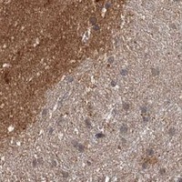
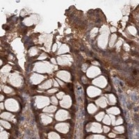

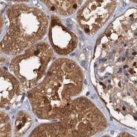

Comprehensive Human Protein Atlas IHC scoring per tissue (reliability: Enhanced — Medium consistency between antibody staining and RNA expression data. Paired antibodies with high similarity supports the protein expression profile.). Rows are taken directly from the HPA tissue chart — click any row's HPA link to view the source.
| Tissue | Cell type | Level | Evidence | Source |
|---|---|---|---|---|
| Caudate | Neuronal cells | High | Protein (IHC) | HPA → |
| Cerebral cortex | Neuronal cells | High | Protein (IHC) | HPA → |
| Epididymis | Glandular cells | High | Protein (IHC) | HPA → |
| Pancreas | Pancreatic endocrine cells | High | Protein (IHC) | HPA → |
| Skin | Fibrohistiocytic cells | High | Protein (IHC) | HPA → |
Troubleshoot TOLLIP staining in paraffin sections by checking retrieval, cell type, subcellular pattern, and controls before comparing signal across samples.
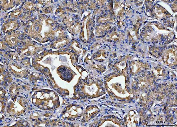
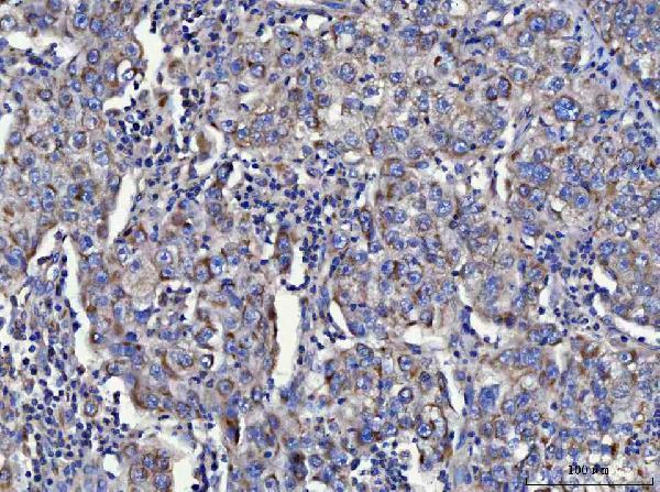
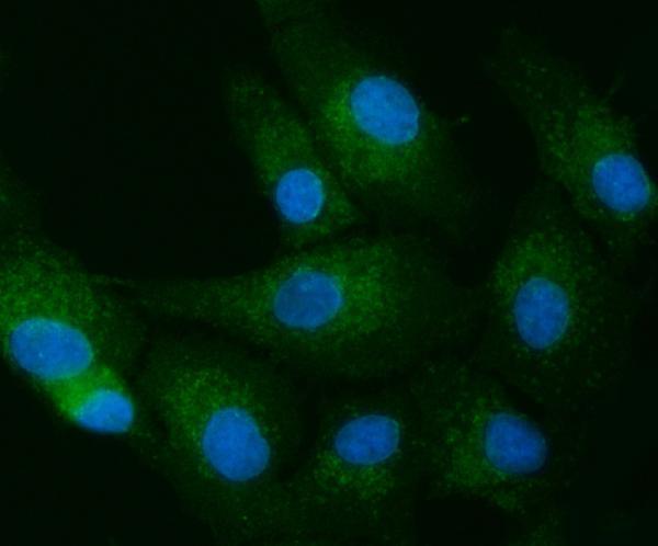
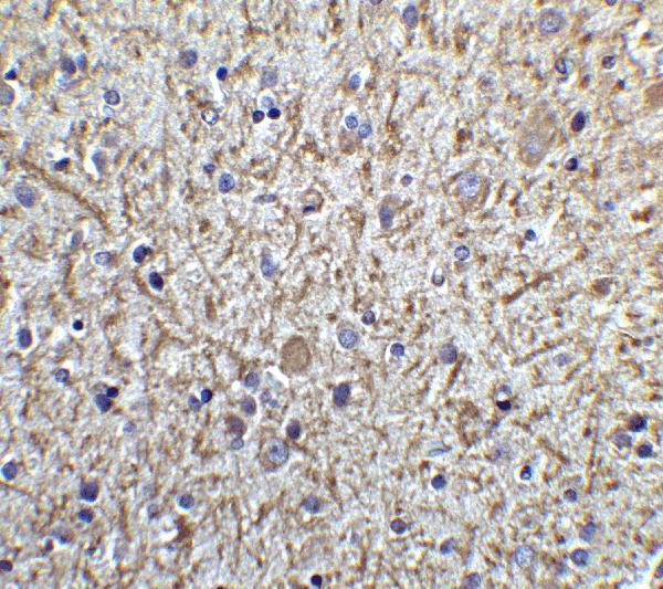
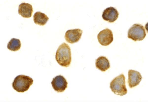
Three anti-TOLLIP antibodies have tissue IHC images from human samples, with mouse and rat tissue examples for selected products; each also has IF or ICC images (catalog IHC and IF captions).
PA2005 has paraffin-section IHC images from human lung and ovarian cancers and mouse and rat brain, plus IF in U2OS cells (PA2005 captions). A02039-1 has paraffin-section IHC images from human prostate and cervical cancers and rat brain, plus IF in A549 cells; A02039 has IHC in human brain and IF or ICC in human brain and THP-1 cells (A02039-1 and A02039 captions).
Which to pick: For paraffin-section IHC, choose PA2005 for the broadest illustrated species coverage, or A02039-1 for its human and rat examples; A02039 lists IHC-P, but its IHC caption identifies human brain tissue without describing processing (PA2005, A02039-1 and A02039 captions; A02039 application list). For IF/ICC, choose by the illustrated sample: PA2005 for U2OS cells, A02039-1 for A549 cells, or A02039 for THP-1 cells or human brain; all three list human, mouse and rat reactivity, while clonality is unreported (catalog IF captions; reactivity and clone fields). The PA2005 and A02039-1 IHC captions specify paraffin sections and EDTA retrieval at pH 8.0, but none of the three IHC captions reports the fixative (catalog IHC captions).