This website uses cookies to ensure you get the best experience on our website.


- Table of Contents
Plan chromogenic TOMM20 IHC in paraffin sections using the catalog antibody at 2–5 μg/ml (datasheet A04039-2). Assess granular cytoplasmic staining, with lower signal expected in adipocytes and smooth muscle cells (HPA tissue IHC).

Expected localisation, validated protocols, controls and antibodies — the at-a-glance facts below, then the full design guide.
| Expected localisation | Granular cytoplasm (HPA tissue IHC); mitochondrial outer membrane expected (UniProt) | |
| Staining pattern | Granular cytoplasmic staining across tissues (HPA tissue IHC) | |
| Antigen retrieval | EDTA pH 8.0 HIER, heat-mediated (datasheet A04039-2) | |
| Positive control | Adrenal gland+4 more · see all | |
| Negative control | None in HPA (detected in all 45 tissues); use no-primary + isotype controls |
| Fixation | Keep fixation consistent across sections (standard IHC practice; not target-specific) | |
| Caveat | Low staining in adipocytes and smooth muscle cells (HPA tissue IHC) | |
| Regulation | No expression regulator specified (UniProt) | |
| Isoform / epitope | No isoforms or cleavage annotated; residues 25–145 face cytoplasm (UniProt) |
The catalog antibody’s IHC-P protocol is accompanied by 3 published TOMM20 IHC protocols for thyroid and mouse liver sections (PMC4909595; PMC7297855; PMC4663018).
| Sample | Paraffin-embedded human lung cancer tissue; fixative not specified (datasheet A04039-2) |
| Fixation | Image fixative and duration unreported (datasheet A04039-2); verify before use. |
| Sectioning | 4–5 µm sections on charged slides (standard) |
| Deparaffinisation | Xylene, graded ethanol series to water (standard) |
| Antigen retrieval | Heat retrieval: EDTA pH 8.0 (datasheet A04039-2); 20 min, 95–100 °C (standard) |
| Peroxidase block | 3% H2O2, 10 min, room temperature (standard) |
| Blocking | 10% goat serum (datasheet A04039-2) |
| Primary antibody | Rabbit anti-TOMM20, 2-5 μg/ml (datasheet A04039-2) |
| Primary incubation | Overnight at 4 °C (datasheet A04039-2) |
| Detection | HRP-conjugated secondary, DAB chromogen (datasheet A04039-2) |
| Counterstain | Hematoxylin, blue, dehydrate and mount (standard) |
| Expected result | TOMM20-positive staining in glandular cells of adrenal gland (HPA tissue IHC: High). HPA tissue profile: Cytoplasmic expression with a granular pattern in all tissues. No signal in the no-primary control. |
TOMM20 is anchored in the mitochondrial outer membrane, with residues 25–145 facing the cytoplasm (UniProt Q15388 topology). In paraffin-section IHC, expect granular cytoplasmic staining across tissues, including strong staining in several glandular, neuronal and hematopoietic cell populations (HPA tissue IHC). HPA rates tissue staining “Supported” but reports very low consistency between antibody staining and RNA expression; interpret intensity with that limitation in mind (HPA tissue IHC).
| Discrete granular cytoplasmic staining in the expected cells. | This matches the reported tissue pattern and mitochondrial location (HPA tissue IHC; UniProt Q15388 subcellular location). Judge the pattern within identifiable cells rather than treating an overall dark section as a positive result (general IHC practice). |
| Predominantly nuclear, surface-only or uniformly diffuse staining. | These patterns do not match the reported granular cytoplasmic distribution (HPA tissue IHC). Review staining specificity and section quality before scoring them as TOMM20; compartment mismatch alone does not identify the precise artefact (general IHC practice). |
| Strong staining in a cell population expected to stain weakly. | HPA reports low staining in smooth muscle cells and adipocytes (HPA tissue IHC). Check cell identification, then consider cross-reactivity or endogenous detection activity if staining persists; neither cause is established by this observation alone (general IHC practice). |
| Diffuse colour across tissue or the slide, obscuring cell boundaries. | Background prevents a reliable assessment of the granular cytoplasmic pattern (HPA tissue IHC; general IHC practice). Inspect the negative detection control and compare background inside and outside tissue before assigning cellular positivity (general IHC practice). |
| No signal in a documented high-staining tissue. | High staining is reported in adrenal gland glandular cells, bronchial respiratory epithelial cells and bone marrow hematopoietic cells (HPA tissue IHC). Confirm the sampled cells are present, then review the IHC run; absence of signal alone cannot establish biological loss (general IHC practice). |
| Epitope location | TOMM20 has one membrane-spanning segment at residues 7–24 and a cytoplasm-facing region at 25–145 (UniProt Q15388 topology). Antibody epitope information is needed to predict whether a particular reagent recognizes either region. |
| Tissue and cell context | HPA reports granular cytoplasmic staining across tissues, with high staining in several listed cell populations and low staining in smooth muscle cells and adipocytes (HPA tissue IHC). Compare like cell types when judging intensity. |
| Strength of tissue evidence | Tissue IHC is rated Supported, yet HPA notes very low consistency between antibody staining and RNA expression (HPA tissue IHC). Treat the observed pattern as a guide, not a guarantee of intensity in every specimen. |
| Antibody validation | HPA lists HPA011562 and CAB005585 as Supported for IHC (HPA antibodies). This supports using their reported staining patterns for comparison, but does not establish the performance of a different catalog antibody. |
| IF/ICC Q: What should a positive cell look like? | A: Mitochondrial localization is reported, with ICC-IF images listed for A-431, U-251MG and U2OS (HPA subcellular). This localization informs pattern interpretation; the IF/ICC guide covers its own experimental workflow. |
| Target-specific fixation effect | No TOMM20-specific fixation sensitivity is reported in the supplied UniProt or HPA records. Standard retrieval optimization may be needed for paraffin-section IHC, but these sources cannot predict its effect on TOMM20 staining (general IHC practice). |
| Situation | Likely cause | Next action |
|---|---|---|
| Known high-staining cells remain blank. | The run, detection steps or cell identification may need review; HPA documents high staining in the listed populations (HPA tissue IHC; general IHC practice). | Verify the expected cells in the section and compare a documented high-staining control processed in the same run; review retrieval and detection settings as general IHC variables (general IHC practice). |
| The entire section looks brown, with no clear granules. | Broad background can obscure the reported granular cytoplasmic pattern (HPA tissue IHC; general IHC practice). | Compare with a negative detection control, then review blocking, antibody concentration, washing and chromogen development as general IHC variables (general IHC practice). |
| Nuclei dominate the apparent signal. | A nuclear-dominant pattern conflicts with mitochondrial outer-membrane localization and HPA’s granular cytoplasmic pattern (UniProt Q15388 subcellular location; HPA tissue IHC). | Check counterstain and detection controls, and require a convincing cytoplasmic granular component before scoring TOMM20 positivity (general IHC practice). |
| Smooth muscle cells or adipocytes appear as strong as adjacent high-staining cells. | Those cell types are listed as low-staining by HPA; misidentification, cross-reactivity or endogenous detection activity are possibilities, not diagnoses (HPA tissue IHC; general IHC practice). | Confirm cell boundaries and identity, compare another section, and inspect negative detection controls before interpreting the strong colour (general IHC practice). |
| A high-staining reference tissue gives uneven results between sections. | Target-specific fixation effects are not established by the supplied assay evidence. Verify with a matched IHC source before attributing a result to fixation. | Check whether the same cell population is present and compare section quality and run conditions; avoid assigning the difference to fixation without separate evidence (general IHC practice). |
| The study antibody gives a different pattern from HPA images. | HPA’s Supported IHC status applies to HPA011562 and CAB005585; it does not validate another antibody’s staining pattern (HPA antibodies). | Compare cell types and controls, then assess the study antibody’s own IHC validation before using HPA images to interpret its signal (general IHC practice). |
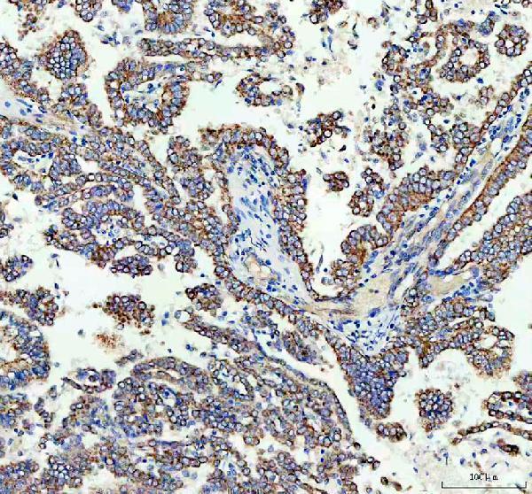
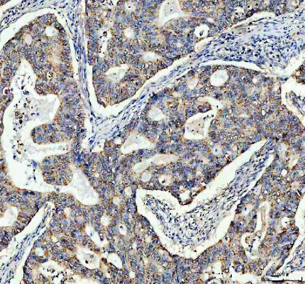


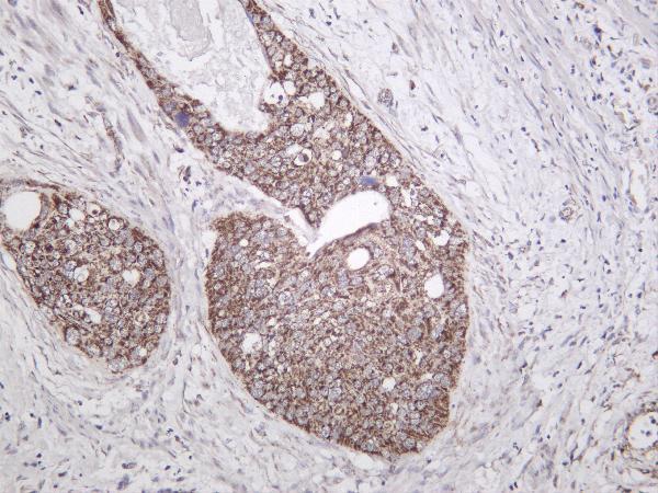


Comprehensive Human Protein Atlas IHC scoring per tissue (reliability: Supported — Very low consistency between antibody staining and RNA expression data.). Rows are taken directly from the HPA tissue chart — click any row's HPA link to view the source.
| Tissue | Cell type | Level | Evidence | Source |
|---|---|---|---|---|
| Adrenal gland | Glandular cells | High | Protein (IHC) | HPA → |
| Appendix | Glandular cells | High | Protein (IHC) | HPA → |
| Bone marrow | Hematopoietic cells | High | Protein (IHC) | HPA → |
| Breast | Glandular cells | High | Protein (IHC) | HPA → |
| Bronchus | Respiratory epithelial cells | High | Protein (IHC) | HPA → |
| Tissue | Cell type | Level | Evidence | Source |
|---|---|---|---|---|
| None in HPA: TOMM20 is detected in all 45 scored tissues. Use a no-primary (secondary-only) and an isotype control instead. | ||||
Troubleshoot TOMM20 staining in paraffin sections by checking retrieval, compartment pattern, controls, and scoring before interpreting biological differences.
These anti-TOMM20 antibodies have human tissue IHC images; A04039-2 also has cell and tissue IF images. All list human, mouse, and rat reactivity (catalog captions/reactivity).
A04039-2 shows IHC in human lung cancer, colon cancer, and liver paraffin sections and IF in HeLa cells and human colon cancer paraffin sections (A04039-2 captions); M04039 shows IHC in human colorectal adenocarcinoma and lung cancer paraffin sections (M04039 IHC captions). M04039-1 shows IHC staining of human cervical carcinoma; its caption does not report section processing (M04039-1 IHC caption).
Which to pick: For paraffin tissue IHC, choose A04039-2 for its three documented human tissues or M04039 for a rabbit monoclonal with two documented human tissues (A04039-2 and M04039 IHC captions; catalog clonality). For IF/ICC, A04039-2 has documented HeLa cell and paraffin tissue IF images; M04039 also lists ICC/IF, while M04039-1 lists IF without an IF image in this payload (catalog applications and IF captions). All three list human, mouse, and rat reactivity, but their shown IHC samples are human; the IHC captions do not report a fixative, and M04039-1 does not report whether its section was paraffin embedded (catalog reactivity; IHC captions).