This website uses cookies to ensure you get the best experience on our website.


- Table of Contents
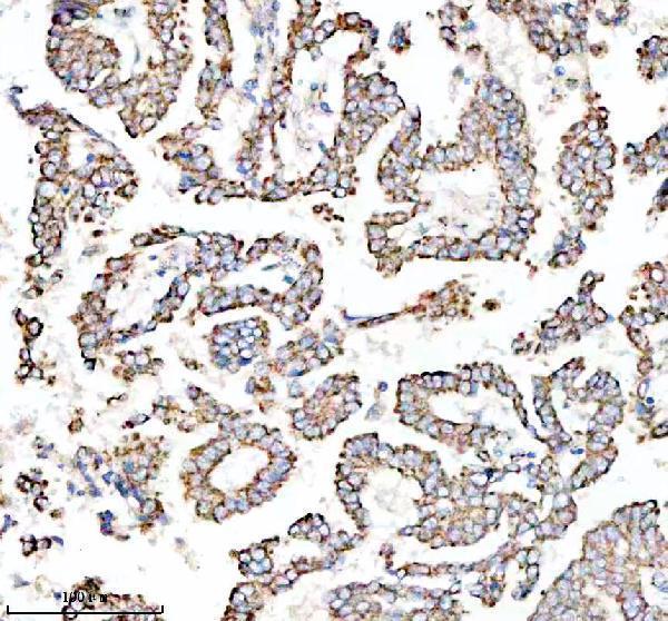
Plan paraffin-section TOMM22 IHC around the granular cytoplasmic tissue pattern (HPA tissue IHC). The catalog antibody was used at 1:100 overnight at 4°C, followed by HRP/DAB detection (datasheet A08668-1).

Expected localisation, validated protocols, controls and antibodies — the at-a-glance facts below, then the full design guide.
| Expected localisation | Granular cytoplasm (HPA tissue IHC); mitochondrial outer membrane (UniProt) | |
| Staining pattern | Ubiquitous granular cytoplasmic staining (HPA tissue IHC) | |
| Antigen retrieval | EDTA pH 8.0 HIER, heat-mediated (datasheet A08668-1) | |
| Positive control | Adrenal gland+4 more · see all | |
| Negative control | None in HPA (detected in all 45 tissues); use no-primary + isotype controls |
| Fixation | Keep fixation consistent across paraffin sections. (standard IHC practice; not target-specific) | |
| Caveat | Bone marrow hematopoietic cells stain highly; score by cell type (HPA tissue IHC) | |
| Regulation | No regulator reported (UniProt) | |
| Isoform / epitope | No isoforms listed; epitope side unspecified (UniProt) |
The catalog antibody’s IHC-P protocol (datasheet: A08668-1) is followed by published TOMM22 IHC methods for brain sections (PMC8215754) and pancreatic tissue (PMC11673509).
| Sample | Paraffin-embedded human lung cancer tissue; fixative not specified (datasheet A08668-1) |
| Fixation | Image fixative and duration unreported (datasheet A08668-1); verify before use. |
| Sectioning | 4–5 µm sections on charged slides (standard) |
| Deparaffinisation | Xylene, graded ethanol series to water (standard) |
| Antigen retrieval | Heat retrieval: EDTA pH 8.0 (datasheet A08668-1); 20 min, 95–100 °C (standard) |
| Peroxidase block | 3% H2O2, 10 min, room temperature (standard) |
| Blocking | 10% goat serum (datasheet A08668-1) |
| Primary antibody | Rabbit anti-TOMM22, 1:50 recommended; image 1:100 (datasheet A08668-1) |
| Primary incubation | Overnight at 4 °C (datasheet A08668-1) |
| Detection | HRP-conjugated secondary, DAB chromogen (datasheet A08668-1) |
| Counterstain | Hematoxylin, blue, dehydrate and mount (standard) |
| Expected result | TOMM22-positive staining in glandular cells of adrenal gland (HPA tissue IHC: High). HPA tissue profile: Ubiquitous cytoplasmic expression in a granular pattern. No signal in the no-primary control. |
TOMM22 resides in the mitochondrial outer membrane, with residues 2–83 facing the cytoplasm and 104–142 facing the intermembrane space (UniProt Q9NS69 topology). In paraffin section IHC, expect granular cytoplasmic staining across many cell types, consistent with the HPA tissue profile and its Supported reliability rating (HPA: ubiquitous granular cytoplasmic expression; IHC Supported).
| Granular cytoplasmic chromogen in glandular, respiratory epithelial, hematopoietic, or glial cells. | This fits the mitochondrial location and reported tissue pattern (UniProt Q9NS69 location; HPA: ubiquitous granular cytoplasmic expression). HPA reports High staining in these cell types in the listed tissues, including adrenal gland, bronchus, bone marrow, and cerebral cortex (HPA: tissue IHC). |
| Predominantly nuclear or cell surface staining, without a convincing granular cytoplasmic component. | The dominant compartment conflicts with the reported mitochondrial location (UniProt Q9NS69 location; HPA: tissue IHC). Treat it as a possible artefact and review morphology, controls, and detection conditions before assigning TOMM22 expression (general IHC practice). |
| Strong signal confined to cells that do not match the expected staining within the sampled tissue. | Check cell identity against the tissue's HPA image and report; unexpected staining can reflect cross-reactivity or endogenous detection activity (HPA: tissue IHC; general IHC practice). Ubiquitous expression means an unlisted cell type alone is not evidence of a false positive (UniProt Q9NS69 tissue specificity). |
| Diffuse color fills tissue spaces or obscures cellular granules. | That appearance is less interpretable than the reported granular cytoplasmic pattern (HPA: tissue IHC). Assess background with appropriate detection controls and review blocking, washing, and chromogen development as general IHC variables (general IHC practice). |
| No signal in a section containing a documented High staining cell population. | A negative result in, for example, adrenal gland glandular cells or bone marrow hematopoietic cells warrants an assay check (HPA: High in adrenal gland glandular cells; High in bone marrow hematopoietic cells). It does not by itself show that TOMM22 is absent (general IHC practice). |
| Membrane topology | TOMM22 spans the mitochondrial outer membrane at residues 84–103 (UniProt Q9NS69 topology). The antibody epitope is not supplied, so topology alone cannot predict whether a particular retrieval condition will expose it (UniProt Q9NS69 topology; supplied evidence). |
| Tissue distribution | TOMM22 is described as ubiquitous, while HPA reports low RNA tissue specificity and ubiquitous granular cytoplasmic IHC (UniProt Q9NS69 tissue specificity; HPA: tissue IHC). Compare compartments and cell identity before treating intensity differences as expression differences (general IHC practice). |
| Reference staining | HPA reports High staining in adrenal gland and appendix glandular cells, bronchus respiratory epithelial cells, and caudate glial cells (HPA: tissue IHC). These are observed examples, not prescribed positive controls for every run (HPA: tissue IHC). |
| Strength of IHC evidence | The tissue IHC profile is Supported because antibody staining is consistent with RNA data (HPA: tissue IHC reliability). HPA003037 is IHC Supported; HPA074642 has no stated IHC status in the supplied antibody record (HPA: antibodies). |
| IF/ICC Q: Where should signal appear? | A: In mitochondria (HPA: ICC-IF, mitochondria Enhanced; UniProt Q9NS69 location). This localization supports interpretation of the IHC pattern; IF/ICC assay design belongs in its separate guide (HPA: subcellular ICC-IF). |
| Situation | Likely cause | Next action |
|---|---|---|
| No chromogen in a known High staining cell population. | An assay or detection failure is possible; absence cannot be assigned from one negative section (HPA: tissue IHC; general IHC practice). | Check a documented High staining population, section integrity, primary antibody use, and detection controls; review the catalog antibody's IHC-P instructions for its specified conditions (HPA: tissue IHC; general IHC practice). |
| Weak granules are difficult to distinguish from background. | Low contrast can obscure the reported granular cytoplasmic pattern (HPA: tissue IHC; general IHC practice). | Compare stained cells with a detection control and inspect at higher magnification; adjust general IHC detection conditions only against controls (general IHC practice). |
| Nuclei carry the dominant signal. | A dominant nuclear pattern conflicts with mitochondrial TOMM22 localization (UniProt Q9NS69 location; HPA: ICC-IF mitochondria). | Verify the counterstain and chromogen separately, then inspect controls and repeat interpretation by compartment rather than counting all dark cells as positive (general IHC practice). |
| Only an unexpected cell population stains strongly. | Cell identification may be wrong, or cross-reactivity or endogenous detection activity may contribute (general IHC practice). | Confirm morphology and compare the same tissue with HPA staining; use appropriate detection controls. Do not label unlisted cells negative solely because TOMM22 is widespread (HPA: tissue IHC; UniProt Q9NS69 tissue specificity; general IHC practice). |
| Broad, smooth color replaces discrete cytoplasmic granules. | Diffuse background can mask the HPA reported granular pattern (HPA: tissue IHC; general IHC practice). | Review blocking, washes, and chromogen development with a detection control; score cellular granules only when they remain distinguishable from background (general IHC practice). |
| A proposed negative tissue control stains. | No negative tissue is supplied, and TOMM22 is described as ubiquitous (HPA: tissue IHC negative list empty; UniProt Q9NS69 tissue specificity). | Use a detection control to assess nonspecific color and compare compartment patterns in documented HPA tissues; do not treat an unlisted tissue as a validated negative (HPA: tissue IHC; general IHC practice). |


Comprehensive Human Protein Atlas IHC scoring per tissue (reliability: Supported — High consistency between antibody staining and RNA expression data.). Rows are taken directly from the HPA tissue chart — click any row's HPA link to view the source.
| Tissue | Cell type | Level | Evidence | Source |
|---|---|---|---|---|
| Adrenal gland | Glandular cells | High | Protein (IHC) | HPA → |
| Appendix | Glandular cells | High | Protein (IHC) | HPA → |
| Bone marrow | Hematopoietic cells | High | Protein (IHC) | HPA → |
| Breast | Glandular cells | High | Protein (IHC) | HPA → |
| Bronchus | Respiratory epithelial cells | High | Protein (IHC) | HPA → |
| Tissue | Cell type | Level | Evidence | Source |
|---|---|---|---|---|
| None in HPA: TOMM22 is detected in all 45 scored tissues. Use a no-primary (secondary-only) and an isotype control instead. | ||||
TOMM22 IHC should show granular cytoplasmic staining consistent with its mitochondrial outer membrane location (HPA: tissue IHC profile; UniProt Q9NS69 localization).
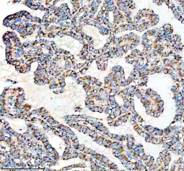
Anti-TOMM22 options include human paraffin-section IHC images for A08668-1 (A08668-1 IHC captions) and an IF image for M08668 with no sample identified (M08668 IF caption).
A08668-1 will render with human lung cancer paraffin-section IHC (A08668-1 IHC caption); its catalog lists IHC and IF use and Human, Mouse, and Rat reactivity (A08668-1 catalog applications/reactivity). M08668 will render with an IF image at 1:50, with no sample identified (M08668 IF caption); its catalog lists IHC and ICC/IF use and Human reactivity (M08668 catalog applications/reactivity).
Which to pick: For tissue IHC, choose A08668-1: its own captions show human lung and thyroid cancer paraffin sections with EDTA retrieval at pH 8.0 and DAB detection; the fixative is unreported (A08668-1 IHC captions). For IF/ICC, M08668 is the rabbit monoclonal option with an IF image and listed ICC/IF use (M08668 catalog clone/applications; M08668 IF caption). For cross-species work, choose rabbit polyclonal A08668-1, whose listed reactivity includes Human, Mouse, and Rat; M08668 lists Human only (catalog clonality/reactivity).