This website uses cookies to ensure you get the best experience on our website.


- Table of Contents
Plan TOMM34 paraffin IHC around cytoplasmic staining and high expression in spermatogonia (HPA tissue IHC). This guide uses the catalog antibody’s 2–5 μg/ml IHC range (datasheet A06775-1) and flags the medium consistency between staining and RNA data (HPA tissue IHC).

Expected localisation, validated protocols, controls and antibodies — the at-a-glance facts below, then the full design guide.
| Expected localisation | Cytoplasmic staining (HPA tissue IHC); outer membrane pool (UniProt) | |
| Staining pattern | Cytoplasmic in spermatogonia, ovarian follicles and immune subsets (HPA tissue IHC) | |
| Antigen retrieval | EDTA pH 8.0 HIER, heat-mediated (datasheet A06775-1) | |
| Positive control | Testis+4 more · see all | |
| Negative control | Adipose tissue+4 more · see all |
| Fixation | Keep paraffin-section fixation consistent (standard IHC practice; not target-specific) | |
| Caveat | Medium staining–RNA consistency; verify independently (HPA tissue IHC) | |
| Regulation | No expression regulator reported (UniProt) | |
| Isoform / epitope | No isoforms; one 1–309 chain with no signal peptide (UniProt) |
The catalog antibody’s IHC-P protocol (datasheet A06775-1) is followed by published colon cancer and oral squamous cell carcinoma protocols (PMC9948080; PMC8262227).
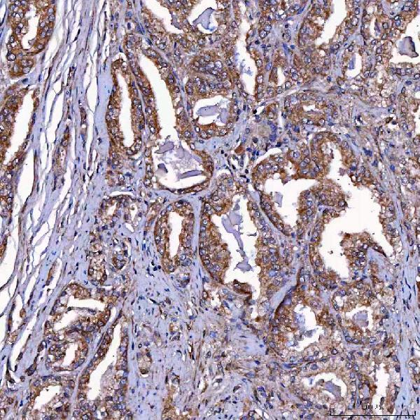
| Sample | Paraffin-embedded human prostate cancer tissue; fixative not specified (datasheet A06775-1) |
| Fixation | Image fixative and duration unreported (datasheet A06775-1); verify before use. |
| Sectioning | 4–5 µm sections on charged slides (standard) |
| Deparaffinisation | Xylene, graded ethanol series to water (standard) |
| Antigen retrieval | Heat retrieval: EDTA pH 8.0 (datasheet A06775-1); 20 min, 95–100 °C (standard) |
| Peroxidase block | 3% H2O2, 10 min, room temperature (standard) |
| Blocking | 10% goat serum (datasheet A06775-1) |
| Primary antibody | Rabbit anti-TOMM34, 2-5 μg/ml (datasheet A06775-1) |
| Primary incubation | Overnight at 4 °C (datasheet A06775-1) |
| Detection | HRP-conjugated secondary, DAB chromogen (datasheet A06775-1) |
| Counterstain | Hematoxylin, blue, dehydrate and mount (standard) |
| Expected result | TOMM34-positive staining in spermatogonia cells of testis (HPA tissue IHC: High). HPA tissue profile: Cytoplasmic expression in several tissues, highly abundant in spermatogonia in testis, ovarian follicles and subsets of immune cells. No signal in the no-primary control. |
TOMM34 should appear mainly cytoplasmic, with a possible mitochondrial component: UniProt places it in the cytoplasm and mitochondrial outer membrane and reports no transmembrane segment (UniProt Q15785 localization and topology). In tissue IHC, the strongest listed signal is in testicular spermatogonia; several glandular and immune cell populations also stain (HPA: tissue IHC). HPA rates the tissue profile Enhanced, with medium RNA–staining consistency and external verification pending (HPA: tissue reliability).
| Strong cytoplasmic staining in spermatogonia, with some other cell populations showing weaker staining. | This fits the listed tissue distribution: spermatogonia are High, while glandular cells in colon and adrenal gland and hematopoietic cells in bone marrow are Medium (HPA: tissue IHC). Compare intensity within identified cell types; a medium tissue need not match testis. |
| Signal appears cytoplasmic, sometimes with a more punctate component. | A mainly cytoplasmic result agrees with tissue IHC (HPA: tissue profile). A punctate component can be compatible with mitochondrial localization (UniProt Q15785 localization; HPA: ICC-IF), but chromogenic puncta alone do not identify mitochondria. |
| Staining is predominantly nuclear, with little cytoplasmic signal. | That compartment conflicts with the reported cytoplasm and mitochondrial outer membrane locations (UniProt Q15785 localization) and cytosolic ICC-IF pattern (HPA: subcellular). Treat it as suspect; review morphology, controls, and staining conditions before assigning TOMM34 expression. |
| Adipocytes or kidney glomerular cells stain strongly. | Those specific cell populations are listed as Not detected (HPA: adipose tissue; HPA: kidney). Check cell identity and no-primary detection controls for cross-reactivity or endogenous detection activity. The HPA calls apply to those cells, not every cell in either organ. |
| Spermatogonia show no convincing signal. | This conflicts with the High spermatogonial staining reported for testis (HPA: testis). Confirm that spermatogonia are present and assess the full IHC run with suitable controls before interpreting the sample as biologically negative. |
| Cell type and tissue selection | Use spermatogonia in testis as the clearest listed positive reference (HPA: High in spermatogonia). Colon glandular cells or bone marrow hematopoietic cells offer Medium comparators (HPA: tissue IHC). Adipocytes and kidney glomerular cells are listed as Not detected in their respective tissues (HPA: tissue IHC). |
| Compartment and topology | The expected emphasis is cytoplasmic (HPA: tissue profile). TOMM34 is also assigned to the mitochondrial outer membrane, with no transmembrane segment (UniProt Q15785 localization and topology). This supports checking compartment, but does not make mitochondrial morphology alone a specificity test. |
| Strength of tissue evidence | HPA labels the tissue IHC profile Enhanced, while describing medium consistency with RNA and pending external verification (HPA: tissue reliability). Its two listed rabbit polyclonal antibodies each have Enhanced IHC validation (HPA: HPA018845; HPA056875). Use the cell-resolved pattern and controls when interpreting a new stain. |
| Fixation and antigen retrieval | Target-specific fixation sensitivity and retrieval requirements are unreported in the supplied UniProt and HPA records. As general IHC practice, assess retrieval using the chosen antibody's documented IHC-P conditions and positive tissue; do not infer a TOMM34-specific retrieval setting from expression or topology. |
| IF/ICC: what pattern is expected? | HPA reports mainly cytosolic signal, with additional vesicular and mitochondrial localization; cytosol and mitochondria are Supported, while vesicles are Approved (HPA: subcellular ICC-IF). That cell-based pattern can inform compartment interpretation, but does not establish an IHC-P dilution or retrieval condition. |
| Situation | Likely cause | Next action |
|---|---|---|
| No signal in testicular spermatogonia | A known High population is absent from the stain, or the run failed (HPA: testis; general IHC practice). | Verify the cells on the section, then review the IHC-validated antibody's documented conditions, retrieval, detection reagents, and a concurrent positive control. Do not call the specimen negative until the run performs. |
| Uniform brown deposit across tissue and empty areas | Nonspecific background or detection chemistry can obscure cell boundaries (general chromogenic IHC practice). | Inspect a no-primary control; review blocking, washing, antibody concentration, and chromogen development. Judge TOMM34 only where signal remains cell associated and cytoplasmic (HPA: tissue profile). |
| Strong staining in adipocytes or glomerular cells | The result conflicts with their Not detected calls (HPA: adipose tissue; HPA: kidney); cross-reactivity or endogenous detection activity is possible. | Confirm the cell type and compare no-primary and appropriate detection controls. Reassess specificity before treating staining in those cells as TOMM34 expression. |
| Predominantly nuclear staining | The dominant compartment disagrees with cytoplasmic tissue staining and cytosolic ICC-IF localization (HPA: tissue profile; HPA: subcellular). | Check counterstain and cellular boundaries, review controls and staining conditions, and compare with a cytoplasmic positive reference. Do not score nuclear-only signal as the expected pattern. |
| Medium comparator looks much weaker than testis | That difference may match the reported levels: spermatogonia are High, while selected glandular and hematopoietic cells are Medium (HPA: tissue IHC). | Score the identified cell population against its own expected level and the same run's controls. Avoid requiring colon glandular cells or bone marrow hematopoietic cells to match spermatogonia. |
| Puncta dominate and diffuse cytoplasm is unclear | Mitochondrial localization is supported, but tissue IHC is described as cytoplasmic and ICC-IF as mainly cytosolic (UniProt Q15785 localization; HPA: tissue profile; HPA: subcellular). | Review morphology and controls for deposits or background. Interpret puncta cautiously in chromogenic IHC; use the separate IF/ICC guide for a cell-based compartment assessment. |


Comprehensive Human Protein Atlas IHC scoring per tissue (reliability: Enhanced — Medium consistency between antibody staining and RNA expression data. Pending external verification.). Rows are taken directly from the HPA tissue chart — click any row's HPA link to view the source.
| Tissue | Cell type | Level | Evidence | Source |
|---|---|---|---|---|
| Testis | Spermatogonia cells | High | Protein (IHC) | HPA → |
| Adrenal gland | Glandular cells | Medium | Protein (IHC) | HPA → |
| Appendix | Glandular cells | Medium | Protein (IHC) | HPA → |
| Bone marrow | Hematopoietic cells | Medium | Protein (IHC) | HPA → |
| Bronchus | Respiratory epithelial cells | Medium | Protein (IHC) | HPA → |
| Tissue | Cell type | Level | Evidence | Source |
|---|---|---|---|---|
| Adipose tissue | Adipocytes | Not detected | Protein (IHC) | HPA → |
| Breast | Adipocytes | Not detected | Protein (IHC) | HPA → |
| Cervix | Glandular cells | Not detected | Protein (IHC) | HPA → |
| Kidney | Cells in glomeruli | Not detected | Protein (IHC) | HPA → |
| Liver | Cholangiocytes | Not detected | Protein (IHC) | HPA → |
Use the catalog antibody’s paraffin-section IHC conditions as a starting point, then judge TOMM34 staining by cell type, compartment and controls.
A06775-1 has real IHC images from paraffin-embedded human prostate, cervical, liver and lung cancer sections (catalog image captions). The antibody lists human and rat IHC use; no IF data are supplied (catalog applications).
A06775-1 is the only SKU that will render, with IHC images from paraffin-embedded human prostate, cervical, liver and lung cancer sections (catalog image captions). Its application data list IHC for human and rat samples; the broader reactivity list also includes mouse, but no mouse IHC image or IF validation is supplied (catalog application and reactivity data).
Which to pick: For paraffin-section tissue IHC, choose A06775-1: its own images document heat retrieval in EDTA at pH 8.0 and primary antibody at 2 μg/ml (catalog image captions). A06775-1 is the only listed cross-species option, with human, mouse and rat reactivity, though IHC is listed for human and rat only (catalog application and reactivity data). No SKU is listed for IF/ICC (catalog applications); the paraffin-section captions do not report the fixative (catalog image captions).