This website uses cookies to ensure you get the best experience on our website.


- Table of Contents
Plan TOMM40 paraffin IHC around the granular cytoplasmic tissue pattern (HPA tissue IHC). Use strongly stained glandular or epithelial cells as positive references and interpret the pattern alongside TOMM40’s outer mitochondrial membrane location (HPA tissue IHC; UniProt).

Expected localisation, validated protocols, controls and antibodies — the at-a-glance facts below, then the full design guide.
| Expected localisation | Granular cytoplasm (HPA tissue IHC); mitochondrial outer membrane (UniProt) | |
| Staining pattern | Granular cytoplasmic staining in glandular and epithelial cells (HPA tissue IHC) | |
| Antigen retrieval | EDTA pH 8.0 HIER, heat-mediated (datasheet A03166-2) | |
| Positive control | Adrenal gland+4 more · see all | |
| Negative control | Soft tissue |
| Fixation | Keep fixation conditions consistent across sections. (standard IHC practice; not target-specific) | |
| Caveat | Low macrophage staining may be missed (HPA tissue IHC) | |
| Regulation | Specific regulation is not annotated (UniProt) | |
| Isoform / epitope | 2 isoforms; epitope coverage is unspecified (UniProt) |
The catalog antibody uses EDTA pH 8.0 heat retrieval (datasheet A03166-2). The published tissue microarray protocol below uses citrate retrieval (PMC13380028).
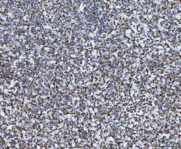
| Sample | Paraffin-embedded human diffuse large B cell lymphoma tissue; fixative not specified (datasheet A03166-2) |
| Fixation | Image fixative and duration unreported (datasheet A03166-2); verify before use. |
| Sectioning | 4–5 µm sections on charged slides (standard) |
| Deparaffinisation | Xylene, graded ethanol series to water (standard) |
| Antigen retrieval | Heat retrieval: EDTA pH 8.0 (datasheet A03166-2); 20 min, 95–100 °C (standard) |
| Peroxidase block | 3% H2O2, 10 min, room temperature (standard) |
| Blocking | 10% goat serum (datasheet A03166-2) |
| Primary antibody | Rabbit anti-TOMM40, 2-5 μg/ml (datasheet A03166-2) |
| Primary incubation | Overnight at 4 °C (datasheet A03166-2) |
| Detection | HRP-conjugated secondary, DAB chromogen (datasheet A03166-2) |
| Counterstain | Hematoxylin, blue, dehydrate and mount (standard) |
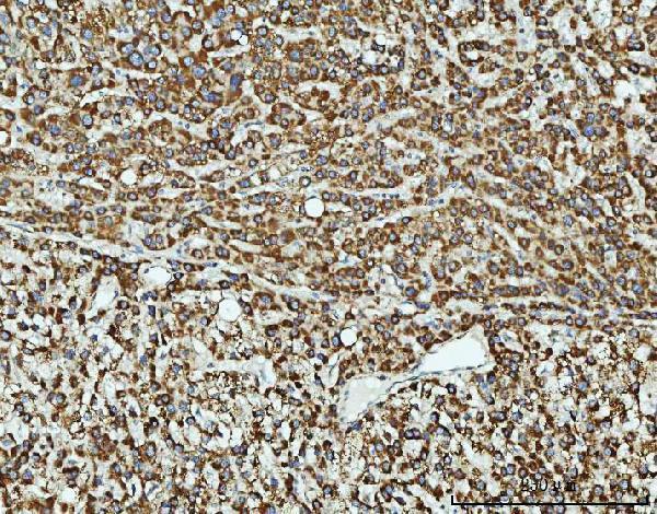
| Expected result | TOMM40-positive staining in glandular cells of adrenal gland (HPA tissue IHC: High). HPA tissue profile: General cytoplasmic expression with a granular pattern. No signal in the no-primary control. |
TOMM40 resides in the mitochondrial outer membrane, where UniProt annotates multiple membrane-spanning segments (UniProt O96008 topology). In paraffin-section IHC, expect granular cytoplasmic staining, especially in glandular cells of the colon, appendix, rectum and small intestine, among other HPA high-staining cells (HPA: tissue IHC). HPA rates the tissue staining pattern Supported, with high consistency between antibody staining and RNA expression (HPA: reliability Supported).
| Granular cytoplasmic staining in colon or small-intestine glandular cells (HPA: High in glandular cells). | This fits the reported tissue pattern and mitochondrial outer-membrane location (HPA: general granular cytoplasmic pattern; UniProt O96008: outer membrane). Judge signal within cells against a counterstain; a brown deposit alone does not establish its compartment (standard IHC practice). |
| Predominantly nuclear, cell-surface or extracellular staining, with little granular cytoplasmic signal. | Those compartments do not match the expected IHC pattern (HPA: granular cytoplasmic pattern; UniProt O96008: outer membrane). Consider nonspecific binding, detection artefact or a localization error; the slide alone cannot identify which. Review morphology and appropriate controls before assigning TOMM40 expression (standard IHC practice). |
| Strong staining in chondrocytes while expected positive glandular cells remain faint or unstained (HPA: chondrocytes Not detected; glandular cells High). | That reversal warrants a specificity check, including possible antibody cross-reactivity or endogenous detection activity (standard IHC practice). HPA's chondrocyte result is an observed reference pattern, not proof that every chondrocyte in every specimen must be negative (HPA: soft-tissue chondrocytes Not detected). |
| Diffuse chromogen across tissue, stroma and cell-free areas obscures cytoplasmic granules. | Uniform background is difficult to reconcile with the reported cell-associated granular pattern (HPA: tissue IHC profile). Review blocking, washes and detection controls as general IHC checks; this appearance cannot by itself establish whether TOMM40 is present (standard IHC practice). |
| No convincing signal in colon or appendix glandular cells (HPA: High in both). | A negative result in an HPA high-staining reference population merits a technical check before biological interpretation (HPA: tissue IHC). Confirm tissue morphology, control staining, antibody dilution and detection performance; HPA's observed intensity does not guarantee the same intensity in every preparation (standard IHC practice). |
| Compartment and topology (UniProt O96008 topology; HPA: tissue IHC profile). | TOMM40 is an outer-mitochondrial-membrane protein, while tissue IHC reports a granular cytoplasmic appearance. Read granules within the cytoplasm rather than expecting a continuous outline of each mitochondrion in a chromogenic section (UniProt O96008: outer membrane; HPA: granular cytoplasmic pattern). |
| Reference-cell selection (HPA: tissue IHC). | HPA reports High staining in several glandular populations and skin basal-layer cells, but Low staining in lung macrophages, smooth-muscle cells, skeletal myocytes and adipocytes. These levels guide comparison between cell types; they do not set a universal intensity cutoff for individual slides (HPA: tissue IHC). |
| Antibody evidence (HPA: antibody validation). | HPA lists IHC as Supported for HPA036231 and HPA036232, both rabbit polyclonal antibodies (HPA: antibody records). That supports comparison with the reported tissue pattern; it does not establish that an unlisted catalog antibody has identical specificity or staining intensity (HPA: antibody records). |
| Isoforms and processing (UniProt O96008). | UniProt records two isoforms and a chain spanning residues 1–361, with no signal peptide, propeptide or annotated glycosylation sites (UniProt O96008: isoforms and processing). The supplied evidence does not identify the IHC antibody epitope or establish isoform-specific staining, so avoid assigning a granule to either isoform. |
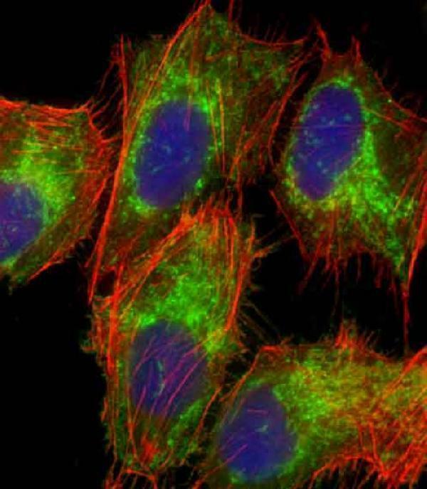
| IF/ICC Q&A: What localization is expected? (HPA: subcellular ICC-IF). | Mainly mitochondrial signal, with an additional cytosolic location reported by HPA (HPA: mitochondria enhanced; cytosol approved). This provides a localization cross-check for IF/ICC; it does not specify an IF/ICC protocol or change the granular cytoplasmic interpretation of paraffin-section IHC (HPA: subcellular ICC-IF; tissue IHC). |
| Situation | Likely cause | Next action |
|---|---|---|
| Known positive glandular cells show no stain (HPA: colon and appendix glandular cells High). | The stain or detection run may have failed; absence on one slide cannot establish TOMM40 loss (standard IHC practice). | Check a positive control and the detection run, then verify antibody dilution against its supplied IHC instructions (standard IHC practice). |
| Signal is weak but confined to cytoplasmic granules (HPA: granular cytoplasmic pattern). | Expression varies by cell population: HPA reports Low staining in several cell types (HPA: tissue IHC). | Compare the same cell type with an HPA high-staining reference and assess morphology before calling the run negative (HPA: tissue IHC; standard IHC practice). |
| Most nuclei are stained more strongly than cytoplasm. | A predominantly nuclear pattern conflicts with the expected mitochondrial and granular cytoplasmic localization; its precise cause remains undetermined (UniProt O96008: outer membrane; HPA: tissue IHC). | Check counterstain and detection controls, then reassess localization with the IHC-validated antibody if needed (standard IHC practice; HPA: IHC Supported). |
| Chondrocytes stain strongly (HPA: chondrocytes Not detected). | Possible nonspecific antibody binding or endogenous detection activity; HPA's reference result alone cannot distinguish them (HPA: tissue IHC; standard IHC practice). | Review a reagent-omission control and the staining of expected positive cells in the same run (standard IHC practice; HPA: glandular cells High). |
| Diffuse deposit obscures cell borders and granules. | Background from blocking, washing or detection can conceal the reported granular cytoplasmic pattern (standard IHC practice; HPA: tissue IHC profile). | Inspect a detection control and review blocking and wash steps before scoring cell-associated signal (standard IHC practice). |
| Two antibodies give different staining patterns (HPA: HPA036231 and HPA036232 IHC Supported). | Supported IHC status does not guarantee identical staining in every preparation (HPA: antibody records). | Compare their cell-type and compartment patterns against HPA references, using matched controls; treat discordant localization as unresolved (HPA: tissue IHC; standard IHC practice). |


Comprehensive Human Protein Atlas IHC scoring per tissue (reliability: Supported — High consistency between antibody staining and RNA expression data.). Rows are taken directly from the HPA tissue chart — click any row's HPA link to view the source.
| Tissue | Cell type | Level | Evidence | Source |
|---|---|---|---|---|
| Adrenal gland | Glandular cells | High | Protein (IHC) | HPA → |
| Appendix | Glandular cells | High | Protein (IHC) | HPA → |
| Colon | Glandular cells | High | Protein (IHC) | HPA → |
| Esophagus | Squamous epithelial cells | High | Protein (IHC) | HPA → |
| Parathyroid gland | Glandular cells | High | Protein (IHC) | HPA → |
| Tissue | Cell type | Level | Evidence | Source |
|---|---|---|---|---|
| Soft tissue | Chondrocytes | Not detected | Protein (IHC) | HPA → |
Troubleshoot TOMM40 staining in paraffin sections by checking retrieval, compartment, controls, and scoring against the documented tissue pattern.
The catalog includes human-reactive TOMM40 antibodies with IHC images from paraffin tissue sections and IF images from cultured cells and a paraffin tissue section (catalog image captions).
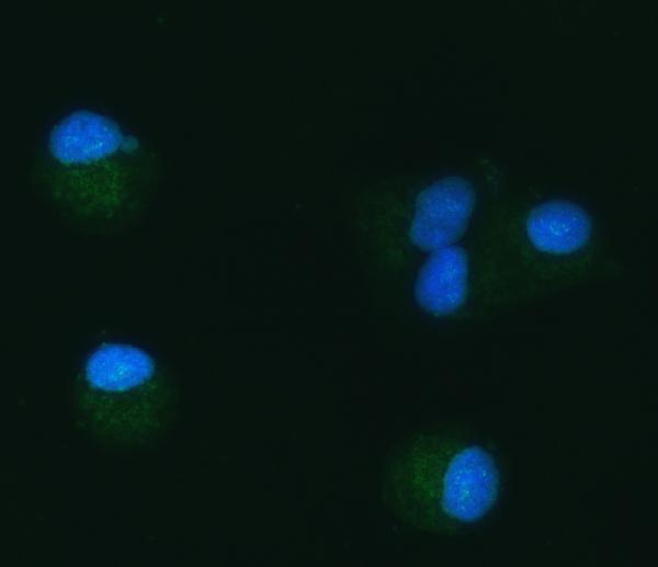
A03166-2 has IHC images from human paraffin sections of diffuse large B cell lymphoma, liver cancer, lung adenocarcinoma, and ovarian cancer, plus IF images from A549 cells and a colon cancer section (A03166-2 image captions). M03166 has IF data from U-2 OS cells and lists no IHC application (M03166 image caption; catalog applications).
Which to pick: Choose A03166-2 for human paraffin-section IHC: its own caption documents EDTA retrieval at pH 8.0 and 2 μg/ml primary antibody, but does not report the fixative (A03166-2 IHC caption). For IF/ICC, A03166-2 lists both applications and has A549 cell IF data; M03166 lists IF and shows mitochondrial staining in U-2 OS cells, but does not list ICC (catalog applications; A03166-2 and M03166 IF captions). Neither SKU has documented cross-species reactivity; both list human reactivity, and M03166 is described as polyclonal while A03166-2 has no clonality specified (catalog reactivity; M03166 dilution notes; A03166-2 catalog entry).