This website uses cookies to ensure you get the best experience on our website.


- Table of Contents
Plan paraffin-section TOP2A IHC around the nuclear staining seen mainly in highly proliferative cells (HPA tissue IHC). This guide covers fixation consistency, controls, chromogenic detection and interpretation of staining across cell populations.

Expected localisation, validated protocols, controls and antibodies — the at-a-glance facts below, then the full design guide.
| Expected localisation | Nuclear staining in proliferative cells (HPA tissue IHC) | |
| Staining pattern | Nuclear signal mainly in highly proliferative cells (HPA tissue IHC) | |
| Antigen retrieval | Tris-EDTA pH 9.0 HIER, 95–98 °C, 20 min (rule: nuclear antigen) | |
| Positive control | Bone marrow+4 more · see all | |
| Negative control | Adipose tissue+4 more · see all |
| Fixation | Keep fixation consistent across sections. (standard IHC practice; not target-specific) | |
| Caveat | Low-proliferation areas may show little staining (HPA tissue IHC) | |
| Regulation | Enriched in proliferative cells (HPA tissue IHC) | |
| Isoform / epitope | 4 isoforms; check whether the epitope is shared (UniProt) |
Compare the catalog antibody’s IHC-P protocol (datasheet) with four published TOP2A IHC methods (PMC10011289; PMC2613415; PMC3140260; PMC8761301).
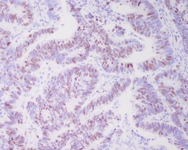
| Sample | Paraffin-embedded human colon cancer tissue; fixative not specified (datasheet M00953-1) |
| Fixation | Image fixative and duration unreported (datasheet M00953-1); verify before use. |
| Sectioning | 4–5 µm sections on charged slides (standard) |
| Deparaffinisation | Xylene, graded ethanol series to water (standard) |
| Antigen retrieval | Heat-induced epitope retrieval in Tris-EDTA buffer, pH 9.0, 20 min at 95–98 °C (standard rule: nuclear antigen) |
| Peroxidase block | 3% H2O2, 10 min, room temperature (standard) |
| Blocking | 10% normal serum of the secondary host, 30 min, room temperature (standard) |
| Primary antibody | Rabbit monoclonal (clone FB-20) anti-TOP2A, 1:50-1:200 (datasheet M00953-1) |
| Primary incubation | Overnight at 4 °C (standard) |
| Detection | HRP-polymer secondary, DAB chromogen 5–10 min (standard) |
| Counterstain | Hematoxylin, blue, dehydrate and mount (standard) |
| Expected result | TOP2A-positive staining in hematopoietic cells of bone marrow (HPA tissue IHC: High). HPA tissue profile: Nuclear expression mainly observed in highly proliferative cells. No signal in the no-primary control. |
TOP2A staining is mainly nuclear in highly proliferative cells (HPA: Enhanced IHC; nuclear profile). Tonsil germinal-center cells and bone-marrow hematopoietic cells stain strongly (HPA: High). TOP2A has no transmembrane segment (UniProt P11388 topology).
| Strong nuclear stain in tonsil germinal-center cells (HPA: High). | Expected pattern in proliferative cells (HPA: nuclear profile). |
| Predominantly membranous stain. | Unexpected compartment; assess nonspecific staining (UniProt P11388: no transmembrane segment). |
| Strong adipocyte stain. | Unexpected; assess cross-reactivity or endogenous activity (HPA: adipocytes not detected). |
| Diffuse stain across the section. | Possible background; check blocking and washes (standard IHC practice). |
| No nuclear stain in tonsil germinal centers. | Unexpected in a known-positive site; check assay controls (HPA: High). |
| Cell context | Nuclear staining favors highly proliferative cells (HPA: tissue IHC profile). |
| IF/ICC: where is signal? | Mainly nucleoplasm; also nucleoli (HPA: supported ICC-IF localization). |
| IHC antibody validation | 3 listed antibodies have Enhanced IHC validation (HPA: antibodies). |
| Isoforms | 4 isoforms are listed; antibody epitope coverage is unreported (UniProt P11388). |
| Situation | Likely cause | Next action |
|---|---|---|
| No nuclear stain in tonsil germinal centers. | Signal is expected there (HPA: High); assay failure is possible. | Check positive control, retrieval and detection (standard IHC practice). |
| Diffuse background. | Blocking or washing may be inadequate (standard IHC practice). | Optimize blocking and washes; run a no-primary control (standard IHC practice). |
| Membranous stain dominates. | Unexpected for TOP2A (UniProt P11388: no transmembrane segment). | Check controls and antibody specificity (standard IHC practice). |
| Strong adipocyte stain. | Unexpected cell type (HPA: adipocytes not detected); cross-reactivity is possible. | Compare with a no-primary control and an IHC-validated antibody (HPA: Enhanced IHC). |
| Only some tonsil cells stain. | Signal favors proliferative germinal-center cells (HPA: nuclear profile; High). | Assess germinal-center nuclei separately (HPA: tonsil germinal-center cells High). |
| Stain persists without primary antibody. | Endogenous detection activity is possible (standard IHC practice). | Check the detection control and block endogenous activity (standard IHC practice). |


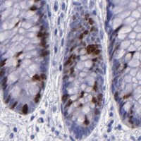


Comprehensive Human Protein Atlas IHC scoring per tissue (reliability: Enhanced — High consistency between antibody staining and RNA expression data.). Rows are taken directly from the HPA tissue chart — click any row's HPA link to view the source.
| Tissue | Cell type | Level | Evidence | Source |
|---|---|---|---|---|
| Bone marrow | Hematopoietic cells | High | Protein (IHC) | HPA → |
| Lymph node | Germinal center cells | High | Protein (IHC) | HPA → |
| Placenta | Cytotrophoblasts | High | Protein (IHC) | HPA → |
| Skin | Cells in basal layer | Medium | Protein (IHC) | HPA → |
| Testis | Pachytene spermatocytes | High | Protein (IHC) | HPA → |
| Tissue | Cell type | Level | Evidence | Source |
|---|---|---|---|---|
| Adipose tissue | Adipocytes | Not detected | Protein (IHC) | HPA → |
| Caudate | Glial cells | Not detected | Protein (IHC) | HPA → |
| Cerebellum | Cells in granular layer | Not detected | Protein (IHC) | HPA → |
| Cerebral cortex | Endothelial cells | Not detected | Protein (IHC) | HPA → |
| Epididymis | Glandular cells | Not detected | Protein (IHC) | HPA → |
Troubleshoot TOP2A staining in paraffin sections by checking retrieval, nuclear localisation, controls, and scoring before interpreting differences between samples.
The catalog includes TOP2A antibodies with IHC images from human paraffin sections and an IF/ICC image from U20S cells (catalog image captions). Both list human, mouse and rat reactivity (catalog reactivity).
The rendered card is M00953-1, with an IHC image from paraffin-embedded human colon cancer (M00953-1 IHC image caption). M00953-1 lists IHC, IP and WB applications and human, mouse and rat reactivity (M00953-1 catalog applications and reactivity).
Which to pick: For tissue IHC, choose M00953-1: it is a rabbit monoclonal with an image from a human paraffin section; the caption does not report the fixative (M00953-1 catalog clone and IHC image caption). For IF/ICC, choose A00953, which lists both applications and has an IF/ICC image from U20S cells (A00953 catalog applications and IF image caption). For mouse or rat tissue IHC, both SKUs list reactivity with those species, while their IHC image captions show human paraffin sections (A00953 and M00953-1 catalog reactivity and IHC image captions).