This website uses cookies to ensure you get the best experience on our website.


- Table of Contents
Plan TOX IHC-P around nuclear staining in subsets of immune cells (HPA tissue IHC). Use the catalog antibody at 2–5 μg/ml (datasheet A08441-2), with cell-type-aware controls and nuclear scoring (standard IHC practice).

Expected localisation, validated protocols, controls and antibodies — the at-a-glance facts below, then the full design guide.
| Expected localisation | Nuclear staining in subsets of immune cells (HPA tissue IHC) | |
| Staining pattern | A subset of immune cells shows nuclear staining (HPA tissue IHC) | |
| Antigen retrieval | EDTA pH 8.0 HIER, heat-mediated (datasheet A08441-2) | |
| Positive control | Appendix+4 more · see all | |
| Negative control | Adipose tissue+4 more · see all |
| Fixation | Keep fixation consistent across sections (standard IHC practice; not target-specific) | |
| Caveat | Collecting ducts and Sertoli cells also stain (HPA tissue IHC) | |
| Regulation | High in tumor-infiltrating CD8+ T cells (UniProt) | |
| Isoform / epitope | No isoforms or processing annotated; one 1–526 chain (UniProt) |
The catalog antibody’s IHC-P protocol (datasheet: A08441-2) is accompanied by three published TOX IHC protocols (PMC10518132; PMC8774984; PMC4374776).
| Sample | Paraffin-embedded human intestinal cancer tissue; fixative not specified (datasheet A08441-2) |
| Fixation | Image fixative and duration unreported (datasheet A08441-2); verify before use. |
| Sectioning | 4–5 µm sections on charged slides (standard) |
| Deparaffinisation | Xylene, graded ethanol series to water (standard) |
| Antigen retrieval | Heat retrieval: EDTA pH 8.0 (datasheet A08441-2); 20 min, 95–100 °C (standard) |
| Peroxidase block | 3% H2O2, 10 min, room temperature (standard) |
| Blocking | 10% goat serum (datasheet A08441-2) |
| Primary antibody | Rabbit anti-TOX, 2-5 μg/ml (datasheet A08441-2) |
| Primary incubation | Overnight at 4 °C (datasheet A08441-2) |
| Detection | HRP-conjugated secondary, DAB chromogen (datasheet A08441-2) |
| Counterstain | Hematoxylin, blue, dehydrate and mount (standard) |
| Expected result | TOX-positive staining in lymphoid tissue of appendix (HPA tissue IHC: High). HPA tissue profile: Nuclear expression mainly in subset of immune cells. No signal in the no-primary control. |
TOX is a nuclear protein with no transmembrane segment (UniProt O94900). In paraffin-section IHC, expect staining mainly in a subset of immune cells (HPA tissue IHC). HPA also reports high staining in kidney collecting ducts and testis Sertoli cells (HPA tissue IHC). The tissue IHC profile is rated Enhanced, with medium consistency between antibody staining and RNA expression (HPA tissue IHC).
| Distinct nuclear staining in some lymphoid cells, with adjacent cells unstained. | This fits the reported subset pattern (HPA tissue IHC) and nuclear location (UniProt O94900). Compare cells within the same section; a mixed positive and negative population can be expected. |
| Predominantly cytoplasmic or membranous staining. | This conflicts with the reported nuclear location (UniProt O94900). Treat it as a possible artefact; check morphology, background and detection controls before interpreting it as TOX. |
| Strong staining across a cell population reported as undetected. | Adipocytes and cardiomyocytes are reported as not detected (HPA tissue IHC). Check cell identity and consider cross-reactivity or endogenous detection activity (general IHC practice); these controls do not prove antibody specificity. |
| Diffuse colour over tissue and spaces between cells, without clear nuclear borders. | This is difficult to score as nuclear TOX (UniProt O94900). Check blocking, washes and the detection-only control for nonspecific colour development (general IHC practice). |
| No nuclear signal in a known-positive compartment. | Germinal center cells in tonsil and lymph node are reported High (HPA tissue IHC). Review the run controls and staining workflow (general IHC practice) before calling that compartment TOX-negative. |
| Cell identity and tissue compartment | High staining is reported in tonsil and lymph-node germinal center cells, appendix lymphoid tissue, kidney collecting ducts and testis Sertoli cells (HPA tissue IHC). Score the named compartment, not the whole tissue. |
| Reported low or absent staining | Hippocampal neuronal cells are Low, while adipocytes are Not detected (HPA tissue IHC). These are cell-specific reference patterns; an unstained field elsewhere is not automatically a valid negative control. |
| Protein location and processing | TOX is nuclear, has no transmembrane segment or signal peptide, and is annotated as one 1–526 chain (UniProt O94900). An extracellular staining pattern has no support from those annotations. |
| IHC evidence and its limit | HPA rates tissue IHC Enhanced and describes medium staining–RNA consistency (HPA tissue IHC). HPA018322 is IHC Enhanced (HPA antibodies); neither rating establishes target-specific fixation sensitivity. |
| IF/ICC Q&A: where should signal appear? | Nucleoplasm and nucleoli fibrillar center are approved IF/ICC locations (HPA subcellular). Those finer locations are IF/ICC observations; assess chromogenic IHC primarily for nuclear staining (HPA tissue IHC). |
| Situation | Likely cause | Next action |
|---|---|---|
| Tonsil germinal center cells show no signal. | This conflicts with their reported High staining (HPA tissue IHC); the cause cannot be assigned from the slide alone. | Confirm the compartment, then review retrieval, antibody dilution, detection and run controls (general IHC practice). |
| Only faint nuclear staining appears in a positive compartment. | Signal may be hard to separate from counterstain (general IHC practice); HPA levels describe observed cells, not a required intensity for every run (HPA tissue IHC). | Compare the positive compartment with adjacent cells and a matched control; review detection and counterstain (general IHC practice). |
| Colour is widespread, including areas without recognizable nuclei. | Diffuse staining does not match nuclear TOX (UniProt O94900) and may reflect background (general IHC practice). | Inspect a detection-only control; review blocking, washes and colour-development time (general IHC practice). |
| Cytoplasmic staining dominates while nuclei remain clear. | The compartment conflicts with UniProt's nuclear assignment (UniProt O94900); morphology or nonspecific binding may complicate interpretation (general IHC practice). | Verify nuclear boundaries with the counterstain and compare with a positive control before scoring (general IHC practice). |
| Adipocytes or cardiomyocytes stain strongly. | Both cell types are reported as Not detected (HPA tissue IHC); cross-reactivity or endogenous detection activity is possible (general IHC practice). | Confirm cell identity and examine the detection-only control; use an independent antibody if available (general IHC practice). |
| A lymphoid section shows scattered positive nuclei rather than uniform staining. | HPA describes nuclear expression mainly in a subset of immune cells (HPA tissue IHC); a patchy pattern alone need not indicate a failed run. | Score identifiable cells and compartments, and compare with a reported positive control such as tonsil germinal center cells (HPA tissue IHC). |
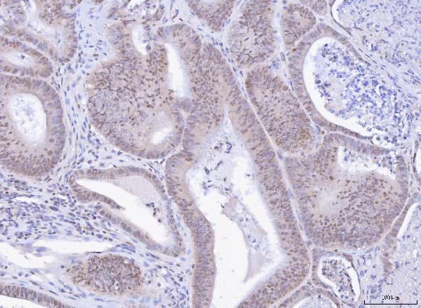
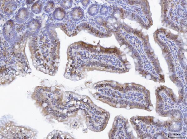
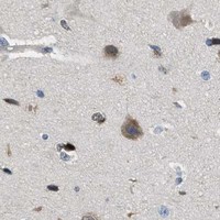


Comprehensive Human Protein Atlas IHC scoring per tissue (reliability: Enhanced — Medium consistency between antibody staining and RNA expression data.). Rows are taken directly from the HPA tissue chart — click any row's HPA link to view the source.
| Tissue | Cell type | Level | Evidence | Source |
|---|---|---|---|---|
| Appendix | Lymphoid tissue | High | Protein (IHC) | HPA → |
| Kidney | Collecting ducts | High | Protein (IHC) | HPA → |
| Lymph node | Germinal center cells | High | Protein (IHC) | HPA → |
| Testis | Sertoli cells | High | Protein (IHC) | HPA → |
| Tonsil | Germinal center cells | High | Protein (IHC) | HPA → |
| Tissue | Cell type | Level | Evidence | Source |
|---|---|---|---|---|
| Adipose tissue | Adipocytes | Not detected | Protein (IHC) | HPA → |
| Caudate | Glial cells | Not detected | Protein (IHC) | HPA → |
| Cerebellum | Cells in granular layer | Not detected | Protein (IHC) | HPA → |
| Epididymis | Glandular cells | Not detected | Protein (IHC) | HPA → |
| Fallopian tube | Glandular cells | Not detected | Protein (IHC) | HPA → |
Troubleshoot TOX staining in paraffin section IHC by checking retrieval, nuclear localisation, background, and cell specific scoring (datasheet A08441-2; UniProt O94900: nucleus).
A08441-2 has IHC images from paraffin-embedded human intestinal cancer and mouse lymphaden tissue (IHC image captions). No IF/ICC image is provided (IF image alts).
A08441-2 is listed for IHC and has paraffin-section images from human intestinal cancer and mouse lymphaden tissue (catalog applications; IHC image captions). Its listed reactivity is human and mouse, while IF/ICC is absent from its application list (catalog reactivity; catalog applications).
Which to pick: Choose A08441-2 for paraffin-section tissue IHC; its images show human and mouse samples stained with 2 μg/ml antibody after EDTA pH 8.0 retrieval (IHC image captions). It is also the cross-species choice on the evidence provided, although its clone and the samples’ fixative are unreported (catalog reactivity; catalog clone; IHC image captions). No listed SKU is validated for IF/ICC, so an IF/ICC choice cannot be made from this payload (catalog applications; IF image alts).