This website uses cookies to ensure you get the best experience on our website.


- Table of Contents
Use this guide to plan TPBG chromogenic IHC on paraffin sections, with kidney tubules as a high-staining control and hippocampal glia as a no-signal comparison (HPA tissue IHC). Assess cytoplasmic staining with a membranous pattern (HPA tissue IHC) alongside TPBG’s cell membrane topology (UniProt).

Expected localisation, validated protocols, controls and antibodies — the at-a-glance facts below, then the full design guide.
| Expected localisation | Cytoplasmic staining with a membranous pattern (HPA tissue IHC) | |
| Staining pattern | Glandular cells: cytoplasmic with a membranous pattern (HPA tissue IHC) | |
| Antigen retrieval | Citrate pH 6.0 HIER, 95–98 °C, 20 min (rule: cytoplasmic / membrane antigen) | |
| Positive control | Adrenal gland+4 more · see all | |
| Negative control | Hippocampus+1 more · see all |
| Fixation | Keep fixation consistent across paraffin sections. (standard IHC practice; not target-specific) | |
| Caveat | Signal varies by cell type; glia and chondrocytes are undetected (HPA tissue IHC) | |
| Regulation | No expression regulator annotated (UniProt) | |
| Isoform / epitope | No isoforms listed; extracellular versus cytoplasmic epitope matters (UniProt) |
The catalog antibody protocol is paired with one published TPBG protocol for chromogenic IHC on paraffin sections (PMC12208316).
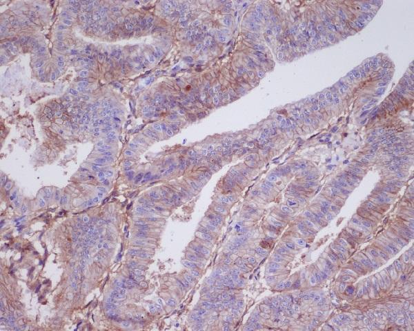
| Sample | Paraffin-embedded human uters cancer tissue; fixative not specified (datasheet M07442) |
| Fixation | Image fixative and duration unreported (datasheet M07442); verify before use. |
| Sectioning | 4–5 µm sections on charged slides (standard) |
| Deparaffinisation | Xylene, graded ethanol series to water (standard) |
| Antigen retrieval | Heat-induced epitope retrieval in citrate buffer, pH 6.0, 20 min at 95–98 °C (standard rule: cytoplasmic / membrane antigen) |
| Peroxidase block | 3% H2O2, 10 min, room temperature (standard) |
| Blocking | 10% normal serum of the secondary host, 30 min, room temperature (standard) |
| Primary antibody | Rabbit monoclonal (clone EHE-20) anti-TPBG, 1:50 (datasheet M07442) |
| Primary incubation | Overnight at 4 °C (standard) |
| Detection | HRP-polymer secondary, DAB chromogen 5–10 min (standard) |
| Counterstain | Hematoxylin, blue, dehydrate and mount (standard) |
| Expected result | TPBG-positive staining in glandular cells of adrenal gland (HPA tissue IHC: High). HPA tissue profile: Cytoplasmic expression with a membranous pattern in several tissues. No signal in the no-primary control. |
TPBG is a cell-membrane protein with an extracellular region spanning residues 32–355 and a transmembrane segment at 356–376 (UniProt Q13641 topology). In paraffin-section IHC, expect cytoplasmic staining with a membranous pattern in selected cells (HPA tissue IHC: Enhanced reliability). HPA reports high staining in kidney tubule cells, lung type I alveolar cells, and several glandular cell populations; expression varies by cell type (HPA tissue IHC).
| Cell edges are outlined, with accompanying cytoplasmic stain in the expected cells. | This fits the reported tissue IHC pattern (HPA: cytoplasmic expression with a membranous pattern). Membrane-associated signal is consistent with TPBG topology (UniProt Q13641: cell membrane; extracellular residues 32–355). Score the identified cell population, because an adjacent unstained population does not negate a positive result. |
| Kidney tubule cells or lung type I alveolar cells stain strongly, while neighboring cells differ. | These are useful positive-reference populations (HPA: High in kidney tubule cells; High in lung type I alveolar cells). Compare staining within the named population and against local background. HPA describes tissue IHC staining, so a strong result in one population does not establish that every cell in that tissue must stain. |
| Staining is exclusively nuclear, without a convincing cytoplasmic or membranous IHC pattern. | Investigate the compartment assignment: nuclear-only staining differs from the tissue IHC profile (HPA: cytoplasmic expression with a membranous pattern). Interpret cautiously because the separate ICC-IF record reports nucleoplasm as an approved location, while this antibody's ICC validation is Uncertain (HPA subcellular; HPA antibody HPA010554). |
| Unexpected cells stain, including glial cells in hippocampus or chondrocytes in soft tissue. | HPA reports TPBG as Not detected in those specific cell populations (HPA tissue IHC). Recheck cell identification and compare with controls; unexpected chromogen could reflect cross-reactivity or endogenous detection activity (general IHC practice). A result in those cells warrants investigation, not an automatic conclusion about the whole tissue. |
| The section shows broad haze, or a known high-staining population has no signal. | Diffuse staining that obscures cell boundaries does not establish TPBG localisation; assess background controls (general IHC practice). Absence of signal in a high-staining reference population calls for a run check before interpreting the study section as negative (HPA: High in kidney tubule cells and other listed populations). |
| Cell population and tissue | HPA grades selected glandular cells, kidney tubule cells, lung type I alveolar cells, and skin eccrine glands High; other populations are Low or Not detected (HPA tissue IHC). Choose and score controls by cell population rather than assuming uniform staining across a section. |
| Protein topology | TPBG has an extracellular region at 32–355, a transmembrane segment at 356–376, and a cytoplasmic tail at 377–420 (UniProt Q13641 topology). This supports a membrane-associated interpretation, but no antibody epitope is supplied; topology alone cannot predict retrieval requirements. |
| Evidence strength and antibody scope | The tissue IHC profile has Enhanced reliability, described as medium consistency with RNA data (HPA tissue IHC). Antibody HPA010554 is IHC Enhanced but ICC Uncertain (HPA antibodies). These labels support use of the tissue IHC pattern while preserving uncertainty about results in a different application. |
| IF/ICC Q&A: should nuclear IF determine the IHC score? | No. The separate ICC-IF record lists nucleoplasm as an approved location (HPA subcellular), whereas tissue IHC reports a cytoplasmic pattern with membranous staining (HPA tissue IHC). The listed antibody's ICC status is Uncertain (HPA antibodies); resolve an IHC score using the IHC slide and controls. |
| Situation | Likely cause | Next action |
|---|---|---|
| No staining in a reference population expected to stain strongly. | A run-level detection or tissue problem is possible; the result alone cannot identify its cause (general IHC practice). | Check section integrity, primary-antibody addition, retrieval and detection records, then compare with a concurrently stained positive control. Kidney tubule cells are one documented High population (HPA tissue IHC). |
| Cytoplasmic colour is present, but cell borders cannot be distinguished. | High background, excess detection signal, or poor morphological resolution can obscure the reported pattern (general IHC practice; HPA tissue IHC: cytoplasmic with membranous pattern). | Review a negative control and tissue morphology; adjust general IHC blocking, washing, antibody concentration, or detection development as the control findings indicate (general IHC practice). |
| Colour appears broadly across the section, including cell-free areas. | Nonspecific reagent deposition or endogenous detection activity may contribute (general IHC practice). | Inspect no-primary and detection controls; address the implicated detection chemistry and washing before scoring TPBG (general IHC practice). |
| Only nuclei stain in an IHC section. | The compartment differs from the reported tissue IHC pattern; the cause cannot be determined from this appearance alone (HPA tissue IHC). | Check the counterstain and detection controls, then reassess localisation at adequate magnification (general IHC practice). Keep the ICC-IF nucleoplasm report separate because ICC validation is Uncertain (HPA subcellular; HPA antibodies). |
| Unexpected cell populations stain more strongly than the intended population. | Cell misidentification, cross-reactivity, or endogenous activity are possibilities (general IHC practice). HPA grades hippocampal glial cells and soft-tissue chondrocytes Not detected (HPA tissue IHC). | Confirm cell identity from morphology, compare with the HPA cell-specific profile, and inspect negative controls before treating the signal as TPBG (HPA tissue IHC; general IHC practice). |
| A study section appears negative while the control stains appropriately. | TPBG staining differs across cell populations (HPA tissue IHC), and an appropriate run control does not establish expression in the study population (general IHC practice). | Record the specific cells assessed and their background; report absence of detectable IHC staining in that population under these conditions. Avoid extending that finding to other cells or tissues (general IHC practice). |


Comprehensive Human Protein Atlas IHC scoring per tissue (reliability: Enhanced — Medium consistency between antibody staining and RNA expression data.). Rows are taken directly from the HPA tissue chart — click any row's HPA link to view the source.
| Tissue | Cell type | Level | Evidence | Source |
|---|---|---|---|---|
| Adrenal gland | Glandular cells | High | Protein (IHC) | HPA → |
| Epididymis | Glandular cells | High | Protein (IHC) | HPA → |
| Gallbladder | Glandular cells | High | Protein (IHC) | HPA → |
| Kidney | Cells in tubules | High | Protein (IHC) | HPA → |
| Lung | Alveolar cells type I | High | Protein (IHC) | HPA → |
Use the documented paraffin section image and TPBG localisation data to plan controls, then optimise staining and scoring within each specimen series.
The catalog antibody has a human paraffin-section IHC image (M07442 image caption; catalog: Human, IHC). No IF image or IF application is listed (catalog: applications and image captions).
M07442 will render with an IHC image captioned as paraffin-embedded human “uters cancer” (M07442 image caption). Its listed reactivity is Human and its applications include IHC, with no IF application listed (catalog: M07442 applications and reactivity).
Which to pick: Choose M07442 for human tissue IHC: it is a rabbit monoclonal with a paraffin-section IHC image (catalog: M07442 host and clone; M07442 image caption). The caption does not report the fixative (M07442 image caption). No SKU in the payload is listed for IF/ICC or cross-species use (catalog: M07442 applications and reactivity).