This website uses cookies to ensure you get the best experience on our website.


- Table of Contents
Plan TPH1 staining in paraffin sections around the granular cytoplasmic pattern reported in glandular cells (HPA tissue IHC). This guide covers the catalog antibody’s IHC conditions (datasheet A01626-4) and controls for uncertain staining specificity (HPA tissue IHC).

Expected localisation, validated protocols, controls and antibodies — the at-a-glance facts below, then the full design guide.
| Expected localisation | Granular cytoplasmic tissue staining (HPA tissue IHC) | |
| Staining pattern | Granular cytoplasm in glandular cells (HPA tissue IHC) | |
| Antigen retrieval | EDTA pH 8.0 HIER, heat-mediated (datasheet A01626-4) | |
| Positive control | Colon+4 more · see all | |
| Negative control | Adipose tissue+1 more · see all |
| Fixation | Keep fixation consistent across sections. (standard IHC practice; not target-specific) | |
| Caveat | Presumed off-target binding can mimic TPH1 staining (HPA tissue IHC) | |
| Regulation | Signal varies by tissue (HPA tissue IHC) | |
| Isoform / epitope | 2 isoforms; epitope coverage is unresolved (UniProt) |
The catalog antibody protocol is accompanied by published TPH1 IHC protocols for rat organs, human liver, and human placenta (PMC5609917; PMC7156786; PMC6724713).
| Sample | Paraffin-embedded human bladder epithelial carcinoma tissue; fixative not specified (datasheet A01626-4) |
| Fixation | Image fixative and duration unreported (datasheet A01626-4); verify before use. |
| Sectioning | 4–5 µm sections on charged slides (standard) |
| Deparaffinisation | Xylene, graded ethanol series to water (standard) |
| Antigen retrieval | Heat retrieval: EDTA pH 8.0 (datasheet A01626-4); 20 min, 95–100 °C (standard) |
| Peroxidase block | 3% H2O2, 10 min, room temperature (standard) |
| Blocking | 10% goat serum (datasheet A01626-4) |
| Primary antibody | Rabbit anti-TPH1, 2-5 μg/ml (datasheet A01626-4) |
| Primary incubation | Overnight at 4 °C (datasheet A01626-4) |
| Detection | Streptavidin-biotin complex (SABC), DAB chromogen (datasheet A01626-4) |
| Counterstain | Hematoxylin, blue, dehydrate and mount (standard) |
| Expected result | TPH1-positive staining in glandular cells of colon (HPA tissue IHC: High). HPA tissue profile: General cytoplasmic expression with a granular pattern. No signal in the no-primary control. |
TPH1 should appear mainly as granular cytoplasmic staining in glandular cells of colon, duodenum, rectum, small intestine, and stomach (HPA: tissue IHC profile; High in these cells). Cytosolic localization is supported separately by ICC-IF (HPA: Cytosol approved). TPH1 has no transmembrane segment (UniProt P17752 topology). Interpret tissue staining cautiously: HPA rates its IHC evidence Uncertain and reports presumed off-target binding (HPA: tissue IHC reliability).
| Granular cytoplasmic stain in gastrointestinal glandular cells, with little stain outside those cells. | This fits the reported tissue pattern and the High staining recorded in colon, duodenum, rectum, small intestine, and stomach (HPA: tissue IHC). It is a plausible positive result, subject to the Uncertain IHC reliability rating (HPA: tissue IHC reliability). |
| Predominantly nuclear or sharply membrane-bound signal, with little cytoplasmic staining. | The compartment conflicts with granular cytoplasmic tissue staining and approved cytosolic ICC-IF localization (HPA: tissue IHC profile; Cytosol approved). Review morphology, counterstain, and controls before assigning it to TPH1; UniProt reports no transmembrane segment (UniProt P17752 topology). |
| Strong staining in adipocytes or soft-tissue fibroblasts, especially if gastrointestinal glandular cells are weak. | HPA reports TPH1 as Not detected in those cells (HPA: adipocytes; soft-tissue fibroblasts). Unexpected staining may reflect cross-reactivity or detection background; HPA reports presumed off-target binding in its tissue IHC assessment (HPA: tissue IHC reliability). |
| Uniform stain across cells, stroma, and empty spaces, without a discernible granular cell pattern. | That distribution does not match the reported granular cytoplasmic profile (HPA: tissue IHC profile). As general IHC practice, examine the reagent-only control and review blocking, washes, and chromogen exposure before scoring cells as positive. |
| No discernible stain in gastrointestinal glandular cells on a run expected to show TPH1. | A negative result needs a run check because HPA records High staining in several gastrointestinal glandular populations (HPA: tissue IHC). Confirm tissue morphology and positive-control performance, then review retrieval, primary incubation, and detection as general IHC checks; the HPA IHC rating remains Uncertain. |
| Tissue evidence and confidence | HPA records High glandular-cell staining in colon, duodenum, rectum, small intestine, and stomach, but rates tissue IHC Uncertain and notes presumed off-target binding (HPA: tissue IHC). Treat the distribution as a guide to interpretation, with controls alongside it. |
| Compartment evidence | The granular cytoplasmic tissue profile and approved cytosolic ICC-IF assignment agree on a cytoplasmic expectation (HPA: tissue IHC profile; Cytosol approved). The ICC-IF approval does not change the separate Uncertain rating for tissue IHC (HPA: antibody validation). |
| Comparison tissue and cell type | Adipocytes and soft-tissue fibroblasts are reported Not detected, while adrenal glandular cells and bone-marrow hematopoietic cells are Medium (HPA: tissue IHC). Score the named cell population within each tissue; whole-section darkness cannot substitute for cell-level assessment. |
| Protein form and epitope | UniProt lists two isoforms, no signal peptide or propeptide, and a chain spanning residues 1–444 (UniProt P17752: isoforms; processing). The supplied record gives no antibody epitope, so it cannot establish whether the catalog antibody distinguishes isoforms. |
| IF/ICC Q&A: where is the expected signal? | Cytosol is the approved ICC-IF location, with images listed for A-431, U-251MG, and U2OS (HPA: subcellular). Use that as compartment context only; this section makes no IF/ICC protocol recommendation. |
| Situation | Likely cause | Next action |
|---|---|---|
| Gastrointestinal glandular cells are negative, and the run control is also weak. | The run may have insufficient detectable signal; HPA reports High staining in these glandular cells, but its tissue IHC rating is Uncertain (HPA: tissue IHC). | As general IHC practice, verify control tissue, retrieval and primary-antibody conditions, detection reagents, and chromogen development before interpreting the sample as negative. |
| A section is strongly stained throughout, including areas without intact cells. | Nonspecific detection or overdevelopment can obscure the granular cytoplasmic pattern reported by HPA (HPA: tissue IHC profile). | Inspect the reagent-only control; review blocking, washes, and development time as general IHC checks. Score only signal that can be assigned to intact cells. |
| Nuclear stain dominates in an otherwise well-preserved section. | That location disagrees with HPA's granular cytoplasmic tissue profile and approved cytosolic ICC-IF location (HPA: tissue IHC; subcellular). | Check the counterstain and reagent-only control, then reassess compartment assignment. Do not use nuclear intensity alone as evidence of TPH1. |
| Adipocytes or soft-tissue fibroblasts stain as strongly as gastrointestinal glandular cells. | These cell types are reported Not detected, and HPA flags presumed off-target binding in tissue IHC (HPA: tissue IHC). | Compare staining with the control and with glandular-cell morphology; treat the unexpected positive cells as unresolved until specificity is supported. |
| Only weak, scattered stain appears in cerebral cortical neurons, lung alveolar cells, or ovarian follicle cells. | HPA lists these populations as Low, so weak signal alone does not establish a failed run or a strong positive (HPA: tissue IHC low-expression entries). | Use the gastrointestinal glandular-cell comparison and run controls to judge assay performance; record the observed cell type and intensity separately. |
| Two antibody results disagree on which cells are positive. | The listed tissue IHC assessments are Uncertain for both HPA022483 and CAB010767 (HPA: antibody validation); HPA also reports presumed off-target binding (HPA: tissue IHC reliability). | Compare matched sections, cell morphology, and controls before interpreting either pattern as TPH1. Report the disagreement when scoring the result. |
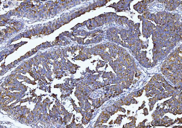
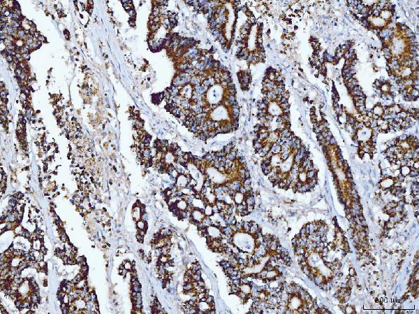
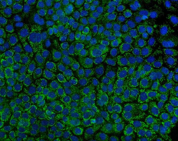


Comprehensive Human Protein Atlas IHC scoring per tissue (reliability: Uncertain — Pending external verification. Presumed off target binding observed and disregarded.). Rows are taken directly from the HPA tissue chart — click any row's HPA link to view the source.
| Tissue | Cell type | Level | Evidence | Source |
|---|---|---|---|---|
| Colon | Glandular cells | High | Protein (IHC) | HPA → |
| Duodenum | Glandular cells | High | Protein (IHC) | HPA → |
| Rectum | Glandular cells | High | Protein (IHC) | HPA → |
| Small intestine | Glandular cells | High | Protein (IHC) | HPA → |
| Stomach | Glandular cells | High | Protein (IHC) | HPA → |
Troubleshoot TPH1 staining in paraffin sections by checking retrieval, controls, cell identity and cytoplasmic localisation before interpreting chromogenic signal.
A01626-4 has human paraffin-section IHC images and an A431-cell IF image (catalog image captions). Its listed reactivity is human (catalog reactivity).
A01626-4 is the only SKU with a card; its IHC captions show human bladder epithelial carcinoma, colonic adenocarcinoma, ovarian cancer, and small intestine paraffin sections (catalog IHC captions). Its IF caption shows A431 cells (catalog IF caption); M01626 has no IHC or IF figure, so it has no card (catalog image fields).
Which to pick: Choose A01626-4 for human paraffin-section IHC: its captions document that preparation, but do not report the fixative (catalog IHC captions). Choose A01626-4 for IF/ICC because its A431-cell IF image supports that use (catalog IF caption). For human, mouse, or rat IHC, M01626 is a rabbit monoclonal option, clone EDH-20, with those species and IHC listed; it has no IHC image caption documenting sample processing (catalog clone/applications/reactivity/image fields).