This website uses cookies to ensure you get the best experience on our website.


- Table of Contents
Plan paraffin-section IHC for TPP1 using its lysosomal tissue staining profile and cell-type controls (HPA tissue IHC). The guide also flags how maturation can affect epitope selection (UniProt).

Expected localisation, validated protocols, controls and antibodies — the at-a-glance facts below, then the full design guide.
| Expected localisation | Lysosomal staining in tissue (HPA tissue IHC) | |
| Staining pattern | Lysosomal staining in glandular, ciliated and glial cells (HPA tissue IHC) | |
| Antigen retrieval | EDTA pH 8.0 HIER, heat-mediated (datasheet PB9899) | |
| Positive control | Adrenal gland+4 more · see all | |
| Negative control | Adipose tissue+4 more · see all |
| Fixation | Keep fixation consistent across sections (standard IHC practice; not target-specific); Matched tissue-IHC evidence does not establish the fixation claim. Validate the specimen-specific method before use. (selected-SKU IHC image PB9899) | |
| Caveat | Staining varies by cell type and tissue (HPA tissue IHC) | |
| Regulation | Regulation not specified (UniProt) | |
| Isoform / epitope | 2 isoforms; maturation removes residues 1–195, so check the epitope (UniProt) |
The catalog antibody’s IHC-P protocol (datasheet: PB9899) and published chromogenic TPP1 IHC methods for endometrial sections (PMC7734757) and mouse brain sections (PMC4268109) provide starting conditions.
| Sample | Paraffin-embedded human lung cancer tissue; fixative not specified (datasheet PB9899) |
| Fixation | Image fixative and duration unreported (datasheet PB9899); verify before use. |
| Sectioning | 4–5 µm sections on charged slides (standard) |
| Deparaffinisation | Xylene, graded ethanol series to water (standard) |
| Antigen retrieval | Heat retrieval: EDTA pH 8.0 (datasheet PB9899); 20 min, 95–100 °C (standard) |
| Peroxidase block | 3% H2O2, 10 min, room temperature (standard) |
| Blocking | 10% goat serum (datasheet PB9899) |
| Primary antibody | Rabbit anti-TPP1, 0.5-1μg/ml (datasheet PB9899) |
| Primary incubation | Overnight at 4 °C (datasheet PB9899) |
| Detection | Streptavidin-biotin complex (SABC), DAB chromogen (datasheet PB9899) |
| Counterstain | Hematoxylin, blue, dehydrate and mount (standard) |
| Expected result | TPP1-positive staining in glandular cells of adrenal gland (HPA tissue IHC: High). HPA tissue profile: Ubiquitous expression in lysosomes. No signal in the no-primary control. |
TPP1 is a soluble lysosomal protease with no transmembrane segment (UniProt O14773: function and topology). In paraffin sections, expect cytoplasmic puncta in many cell types, including glandular cells and glia with high observed staining (HPA: ubiquitous lysosomal expression; high in adrenal glandular cells and caudate glia). HPA rates the tissue IHC pattern Enhanced, indicating consistency with RNA expression (HPA: tissue IHC reliability).
| Discrete cytoplasmic puncta in adrenal glandular cells or caudate glia, with little nuclear signal (HPA: high in both cell types). | This fits the reported lysosomal pattern (HPA: ubiquitous expression in lysosomes; UniProt O14773: lysosome). Score the cells and puncta seen on the slide; HPA's High category does not set a universal staining intensity for every section (HPA: tissue IHC levels). |
| Predominantly uniform nuclear, surface-rim, or extracellular staining, without convincing cytoplasmic puncta. | The dominant compartment conflicts with the reported lysosomal pattern and lack of a transmembrane segment (HPA: lysosomal profile; UniProt O14773: topology). Treat it as suspect and review morphology, controls, and detection background before assigning TPP1 positivity (general IHC practice). |
| Strong staining confined to adipocytes or soft-tissue fibroblasts while expected positive cells remain unstained. | HPA reports TPP1 as Not detected in those cell types (HPA: adipocytes; soft-tissue fibroblasts). Cross-reactivity or endogenous detection activity is possible; a Not detected reference is a comparison point, not proof that every specimen must be negative (general IHC interpretation). |
| Widespread diffuse color obscures cell boundaries and puncta across the section. | The distribution cannot establish lysosomal localization (HPA: lysosomal profile). Consider nonspecific antibody binding, endogenous enzyme activity, or chromogen background, and compare with the appropriate detection control before scoring (general chromogenic IHC practice). |
| No signal in a section containing adrenal glandular cells or caudate glia. | This conflicts with HPA's High observations in those cell types (HPA: adrenal glandular cells; caudate glia). First assess tissue preservation, retrieval, antibody application, and detection with a known-positive control; absence in one run does not establish biological loss (general IHC practice). |
| Lysosomal localization and topology (UniProt O14773; HPA: tissue IHC profile) | Interpret granular cytoplasmic staining in cell context; a membrane-only pattern has weak support because UniProt lists no transmembrane segment (UniProt O14773: topology). |
| Cell-specific reference levels (HPA: tissue IHC) | High staining is reported in bronchial and fallopian-tube ciliated cell bodies, while adipocytes are Not detected (HPA: named cell types). Use cell identity when comparing regions; these categories do not prescribe an exact optical density. |
| Precursor processing (UniProt O14773: chains) | TPP1 has a signal peptide at residues 1–19, propeptide at 20–195, and mature chain at 196–563 (UniProt O14773: processing). Antibody epitope location is unspecified here, so this record cannot predict which processed form the IHC-validated antibody detects. |
| Glycosylation and isoforms (UniProt O14773) | Five glycosylation sites and two isoforms are listed (UniProt O14773: features). Their effects on this antibody's section staining cannot be inferred without an epitope map or comparative validation; do not explain a negative slide from these features alone. |
| Melanosome annotation (UniProt O14773: subcellular location) | TPP1 was identified by mass spectrometry in stage I–IV melanosome fractions (UniProt O14773: melanosome note). This does not establish a distinctive melanosomal chromogenic pattern in the supplied tissue IHC observations (HPA: lysosomal tissue profile). |
| IF/ICC Q: Is the IHC pattern an IF localization control? | A: No IF localization is provided: HPA lists no main ICC-IF location or cell-line images, and neither listed antibody has ICC validation (HPA: subcellular record; HPA037709 and HPA044868 validation). Assess IF on its own guide page. |
| Situation | Likely cause | Next action |
|---|---|---|
| Positive tissue lacks visible puncta despite intact morphology. | The result disagrees with HPA's High examples, but the supplied sources do not identify a TPP1-specific fixation or retrieval failure (HPA: adrenal glandular cells; caudate glia). | Check antibody and detection steps against their validated instructions; review retrieval and a known-positive section as general paraffin-IHC controls (general IHC practice). |
| Color is concentrated in nuclei or along cell surfaces. | Dominant localization differs from the lysosomal profile and soluble topology (HPA: lysosomal expression; UniProt O14773: no transmembrane segment). | Compare a detection-only control and inspect whether cytoplasmic puncta persist after background is excluded (general IHC practice); withhold a positive call if the discordant pattern dominates. |
| Adipocytes or soft-tissue fibroblasts appear strongly positive. | Both are listed as Not detected, so the result may reflect cross-reactivity or detection background (HPA: adipocytes; soft-tissue fibroblasts; general IHC interpretation). | Check staining in an HPA-high cell type on the same run and review detection controls before treating the unexpected cells as TPP1-positive (HPA: high in adrenal glandular cells; general IHC practice). |
| Brown deposits cover tissue-free areas or obscure cell boundaries. | A deposit outside interpretable cells cannot support the reported lysosomal distribution (HPA: lysosomal profile); chromogen precipitation or nonspecific detection is possible (general chromogenic IHC practice). | Review reagent cleanliness, washes, and a detection-only control, then score only clearly cell-associated staining (general IHC practice). |
| Glial staining varies between hippocampus and caudate. | HPA reports Low in hippocampal glia and High in caudate glia (HPA: tissue IHC). Regional differences alone therefore do not establish assay failure. | Record region and cell type separately, and compare matching regions across slides before interpreting intensity differences (HPA: glial levels; general IHC scoring practice). |
| Puncta are suggested in IF, but their identity is uncertain. | The supplied HPA subcellular record has no ICC-IF images or assigned main location; listed antibodies lack ICC validation (HPA: subcellular record; HPA037709 and HPA044868 validation). | Treat IF localization as unverified by these data and consult the separate IF/ICC guide for its controls and assessment (HPA: ICC-IF record). |

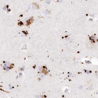


Comprehensive Human Protein Atlas IHC scoring per tissue (reliability: Enhanced — High consistency between antibody staining and RNA expression data.). Rows are taken directly from the HPA tissue chart — click any row's HPA link to view the source.
| Tissue | Cell type | Level | Evidence | Source |
|---|---|---|---|---|
| Adrenal gland | Glandular cells | High | Protein (IHC) | HPA → |
| Bronchus | Ciliated cells (cell body) | High | Protein (IHC) | HPA → |
| Caudate | Glial cells | High | Protein (IHC) | HPA → |
| Cerebellum | Bergmann glia - cytoplasm/membrane | High | Protein (IHC) | HPA → |
| Duodenum | Glandular cells | High | Protein (IHC) | HPA → |
| Tissue | Cell type | Level | Evidence | Source |
|---|---|---|---|---|
| Adipose tissue | Adipocytes | Not detected | Protein (IHC) | HPA → |
| Cervix | Glandular cells | Not detected | Protein (IHC) | HPA → |
| Ovary | Ovarian stroma cells | Not detected | Protein (IHC) | HPA → |
| Soft tissue | Fibroblasts | Not detected | Protein (IHC) | HPA → |
| Vagina | Squamous epithelial cells | Not detected | Protein (IHC) | HPA → |
Use the catalog antibody’s paraffin section result as the starting point, then judge staining against TPP1’s lysosomal localisation and documented cell patterns.
PB9899 is a human-reactive IHC antibody with an image showing TPP1 staining in a paraffin-embedded human lung cancer section (catalog applications/reactivity; PB9899 IHC caption).
PB9899 lists IHC for human samples (catalog applications/reactivity). Its IHC image shows staining in a paraffin-embedded human lung cancer section; no IF image is supplied (PB9899 IHC caption; catalog IF image list).
Which to pick: Choose PB9899 for human paraffin-section IHC: its image documents EDTA retrieval at pH 8.0 and primary antibody at 1 μg/ml (PB9899 IHC caption). The fixative is unreported, so the caption does not establish fixation (PB9899 IHC caption). No SKU in this payload is listed for IF/ICC or species beyond human, and PB9899 has no clone designation (catalog applications/reactivity/clone field). The selected PB9899 tissue-IHC caption documents paraffin sections, but does not specify the fixative (selected-SKU IHC image PB9899).