TPR / Nucleoprotein TPR · IHC design guide

Design Immunohistochemistry for TPR

TPR shows ubiquitous nuclear and nuclear membranous staining in tissue IHC (HPA tissue IHC). This guide covers paraffin-section staining with the catalog antibody at 0.5–1 μg/mL and interpretation of mitotic redistribution (datasheet A00695-1; UniProt).

Evidence assembled Oct 2026 · For research use; verify linked source records and product datasheet before use
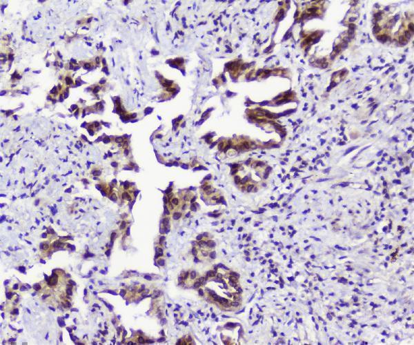
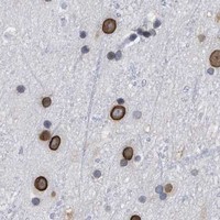
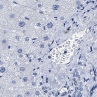
Immunohistochemistry protocol sheet for TPR (IHC for TPR): expected localisation Nuclear and nuclear membranous staining (HPA tissue IHC), antibody A00695-1, validated IHC image, and IHC protocol steps
Printable TPR IHC protocol sheet — expected localisation Nuclear and nuclear membranous staining (HPA tissue IHC), antibody A00695-1, controls and protocol steps. Open the full TPR IHC guide →

TPR Immunohistochemistry Experimental Design Guide

Expected localisation, validated protocols, controls and antibodies — the at-a-glance facts below, then the full design guide.

Must know before staining
Expected localisation Nuclear and nuclear membranous staining (HPA tissue IHC)
Staining pattern Ubiquitous nuclear and nuclear membranous staining (HPA tissue IHC)
Antigen retrieval Citrate pH 6 HIER, heat-mediated (datasheet A00695-1)
Positive control ⓘ Adrenal gland+4 more · see all
Negative control ⓘ None in HPA (detected in all 45 tissues); use no-primary + isotype controls
Important caveats
Reasons your staining may differ from the expected pattern.
Fixation Keep fixation consistent across paraffin sections. (standard IHC practice; not target-specific)
Caveat Mitotic TPR can redistribute to the spindle (UniProt)
Regulation Expression regulation is unreported (UniProt)
Isoform / epitope 2 isoforms; epitope differences are unspecified (UniProt)
Section 1

Recommended TPR IHC & IF Protocols

Compare the catalog antibody’s IHC-P protocol (datasheet: A00695-1) with four published TPR tissue-staining protocols (PMC13109368; PMC5862582; PMC8078762; PMC8225803).

Recommended immunohistochemistry (IHC-P) protocol parameters
SampleParaffin-embedded human lung cancer tissues; fixative not specified (datasheet A00695-1)
FixationImage fixative and duration unreported (datasheet A00695-1); verify before use.
Sectioning4–5 µm sections on charged slides (standard)
DeparaffinisationXylene, graded ethanol series to water (standard)
Antigen retrievalHeat retrieval: Citrate pH 6, 20 min (datasheet A00695-1)
Peroxidase block3% H2O2, 10 min, room temperature (standard)
Blocking10% goat serum (datasheet A00695-1)
Primary antibodyRabbit anti-TPR, 0.5-1μg/ml (datasheet A00695-1)
Primary incubationOvernight at 4 °C (datasheet A00695-1)
DetectionStreptavidin-biotin complex (SABC), DAB chromogen (datasheet A00695-1)
CounterstainHematoxylin, blue, dehydrate and mount (standard)
Expected resultTPR-positive staining in glandular cells of adrenal gland (HPA tissue IHC: High). HPA tissue profile: Ubiquitous nuclear and nuclear membranous expression. No signal in the no-primary control.
💡Decision noteStart with heat-mediated citrate retrieval at pH 6 (datasheet: A00695-1); the published protocols also use heat retrieval near pH 6 (PMC13109368; PMC5862582; PMC8078762; PMC8225803).
Section 2

What Is the Expected TPR Staining Pattern?

TPR is a nuclear pore complex protein with no transmembrane segment; in interphase it occupies the nucleoplasmic side of the nuclear envelope (UniProt P12270). Expect predominantly nuclear membranous staining across many cell types, with some nuclear staining (HPA: ubiquitous nuclear and nuclear membranous expression). HPA rates the tissue IHC pattern Enhanced, citing high consistency between antibody staining and RNA expression (HPA: tissue IHC reliability).

What am I looking at on my slide?
Nuclear rim staining appears in glandular cells, hematopoietic cells or glia, with some nuclear signal.This fits TPR at the nuclear basket (UniProt P12270) and the broad nuclear and nuclear membranous IHC pattern (HPA: tissue IHC). HPA reports High staining in adrenal and colon glandular cells, bone marrow hematopoietic cells, and cerebral cortex glial cells (HPA: tissue IHC).
Strong, widespread plasma membrane or cytoplasmic staining dominates interphase cells, without a nuclear rim.Treat this as a questionable IHC pattern: HPA's dominant tissue pattern is nuclear and nuclear membranous (HPA: tissue IHC). Review morphology and controls before calling it TPR. UniProt also lists cytoplasmic and spindle localization, particularly during mitosis, so isolated mitotic profiles need separate assessment (UniProt P12270).
A cell population expected to stain weakly shows intense signal while nearby expected positive cells are blank.For example, HPA lists liver cholangiocytes as Low, while adrenal glandular cells are High (HPA: tissue IHC). An inverted pattern raises concern for nonspecific antibody binding or endogenous chromogen activity (standard IHC practice); it does not establish cross-reactivity on appearance alone.
Brown haze covers nuclei and surrounding tissue without a discernible nuclear rim.Diffuse signal cannot support a compartment call. Compare a no-primary control, inspect background in the same section, and review blocking, antibody concentration and detection conditions (standard IHC practice). A genuine interphase result should be judged against nuclear membranous localization (HPA: tissue IHC; UniProt P12270).
An adequately sampled known-positive tissue shows no nuclear or nuclear rim signal.First confirm that the expected cell population is present: HPA reports High staining in adrenal glandular and bone marrow hematopoietic cells (HPA: tissue IHC). A blank result there calls for checking section quality, retrieval, antibody and detection controls (standard IHC practice); it alone cannot establish absent TPR expression.
💡Expected TPR appearanceCall an interphase IHC section positive when many expected cells show clear nuclear rim staining, with possible nuclear signal and readily visible strong staining in HPA High populations such as adrenal glandular cells (HPA: tissue IHC; UniProt P12270); isolated diffuse cytoplasmic or surface staining without a rim is a suspect positive (HPA: tissue IHC).
How each factor affects the staining
Compartment and cell-cycle stateTPR anchors to the nucleoplasmic side of the nuclear pore complex in interphase, but associates with the spindle from late prometaphase through anaphase and returns to the reforming nuclear envelope (UniProt P12270). Score interphase nuclear rims separately from mitotic figures; mitotic redistribution alone does not invalidate an otherwise matching section.
Choice of tissue controlHPA describes ubiquitous nuclear and nuclear membranous expression and low tissue specificity (HPA: tissue IHC). Adrenal glandular cells or bone marrow hematopoietic cells have reported High staining; liver cholangiocytes are reported Low (HPA: tissue IHC). Use the High populations to check that a staining run can reveal the expected pattern, without treating Low as necessarily absent.
Antibody and assay evidenceThree listed rabbit polyclonal antibodies, HPA019661, HPA019663 and HPA024336, each have Enhanced IHC and ICC status (HPA: antibodies). HPA describes Enhanced IHC validation as agreement between independent antibodies or orthogonal data (HPA: antibody validation). This supports the reported pattern, but the antibody used on the current section still needs appropriate controls (standard IHC practice).
Isoforms and epitope interpretationUniProt records two TPR isoforms but supplies no antibody epitope mapping in this evidence (UniProt P12270; HPA: antibodies). A difference between antibodies could reflect what they recognize, yet the supplied records cannot assign an isoform to a staining pattern. Check the catalog antibody's documented immunogen before making isoform-specific claims (standard IHC practice).
Retrieval and target-specific sensitivityAntigen retrieval is a routine variable in paraffin IHC (standard IHC practice). Neither the supplied HPA tissue profile nor UniProt topology establishes a TPR-specific fixation or retrieval effect (HPA: tissue IHC; UniProt P12270). Interpret a changed signal through matched positive and negative controls rather than assuming a particular retrieval condition reveals TPR.
Why is my staining missing, weak or wrong?
SituationLikely causeNext action
Nuclear rims are weak in a section expected to stain strongly.The run may have inadequate retrieval, antibody incubation or detection performance (standard IHC practice); HPA reports High staining in adrenal glandular cells (HPA: tissue IHC).Check a known-positive section in the same run, then review the catalog antibody's IHC-P instructions and detection controls (standard IHC practice).
The whole section has diffuse brown background.Excess antibody, insufficient blocking or endogenous enzyme activity can obscure localization in chromogenic IHC (standard IHC practice).Inspect no-primary and detection controls; adjust blocking, washing or antibody concentration according to the assay controls (standard IHC practice).
Signal is prominent at tissue surfaces or in cytoplasm, with no interphase nuclear rim.The distribution conflicts with HPA's predominant nuclear membranous pattern (HPA: tissue IHC); nonspecific binding or detection background is possible (standard IHC practice).Recheck morphology and controls; compare nuclear rim staining in a known-positive population before scoring the section (HPA: tissue IHC; standard IHC practice).
Cholangiocytes appear much stronger than the chosen positive control.HPA reports Low staining in cholangiocytes, so this result needs scrutiny (HPA: tissue IHC); differential background or technical failure is possible (standard IHC practice).Confirm cell identity, inspect no-primary staining, and repeat with a High HPA population as a run control (HPA: tissue IHC; standard IHC practice).
A known-positive section is blank, including its expected cells.A failed staining or detection step is possible (standard IHC practice); blank staining alone does not overturn HPA's High report for that cell population (HPA: tissue IHC).Verify that expected cells are present, then check reagent performance, retrieval and the assay's positive control (standard IHC practice).
What should a separate IF/ICC assessment show?HPA reports an Enhanced nuclear membrane location in ICC-IF images (HPA: subcellular ICC-IF).Look for a nuclear rim in interphase cells and assess any spindle-associated mitotic signal against cell-cycle morphology (HPA: subcellular ICC-IF; UniProt P12270).

Sample controls for TPR IHC & IF

🧪Run bone marrow first: hematopoietic cells show high TPR staining (HPA: High in bone marrow hematopoietic cells). HPA detects TPR in all 45 scored tissues, so no negative tissue is available; use the no-primary and isotype controls, and expect any anucleate erythrocytes on the positive slide to lack nuclear-rim staining (HPA: no negative tissues; UniProt P12270 subcellular localization).
Positive control tissue: Adrenal gland (Glandular cells, HPA High)
Negative control tissue: None in HPA: TPR is detected in all 45 scored tissues. Use a no-primary (secondary-only) and an isotype control instead.
ICC-IF cell lines (HPA subcellular resource): HPA ICC-IF images show TPR in A-431, U-251MG, U2OS, KOLF2.1J, with annotated localisation: Nuclear membrane (enhanced) (HPA subcellular).
Technical controls: Include a no-primary, secondary-only slide and a host- and clonality-matched rabbit IgG isotype control, plus an identically processed TPR-knockout cell pellet as a biological negative (selected A00695-1 IHC caption: rabbit primary). Quench endogenous peroxidase in bone marrow and check the control slides for residual chromogenic background (standard IHC practice).
⚠️Feasibility: The selected A00695-1 paraffin-section caption reports citrate pH 6 heat retrieval for 20 min, but the fixative is unreported; no target-specific fixation window or fixation effect is supplied (selected A00695-1 IHC caption). Nuclear-envelope signal may be easier to resolve by IF/ICC, consistent with the HPA nuclear-membrane images, but the supplied evidence does not establish that frozen sections are easier (HPA: subcellular localization; UniProt P12270 subcellular localization). In chromogenic bone-marrow sections, residual endogenous peroxidase can obscure specific staining, making the no-primary control especially useful (standard IHC practice).

HPA tissue IHC evidence for TPR

Comprehensive Human Protein Atlas IHC scoring per tissue (reliability: Enhanced — High consistency between antibody staining and RNA expression data.). Rows are taken directly from the HPA tissue chart — click any row's HPA link to view the source.

Positive expression · recommended positive controls

TissueCell typeLevelEvidenceSource
Adrenal gland Glandular cells High Protein (IHC) HPA →
Bone marrow Hematopoietic cells High Protein (IHC) HPA →
Bronchus Ciliated cells (cell body) High Protein (IHC) HPA →
Caudate Neuronal cells High Protein (IHC) HPA →
Cerebellum Bergmann glia - nucleus High Protein (IHC) HPA →

Undetected expression · recommended negative controls

TissueCell typeLevelEvidenceSource
None in HPA: TPR is detected in all 45 scored tissues. Use a no-primary (secondary-only) and an isotype control instead.
Section 3

Advanced TPR IHC Tips

Troubleshoot paraffin-section TPR IHC by checking nuclear-envelope localisation, retrieval conditions, and cell-level scoring (UniProt P12270 localisation; HPA tissue IHC).

Which retrieval conditions should I try when TPR staining is weak?
Start with heat-mediated citrate retrieval at pH 6 for 20 minutes (datasheet A00695-1). The selected paraffin-section image used that treatment before overnight incubation with 1 μg/mL primary antibody at 4°C (datasheet A00695-1). Run a known staining section alongside the test section so a failed retrieval step can be distinguished from low tissue signal (standard IHC practice; HPA tissue IHC). If staining remains weak, compare a second retrieval condition on adjacent sections while keeping detection and exposure conditions matched (standard IHC practice). Assess whether signal resolves at the nuclear boundary, the expected interphase location, before accepting stronger diffuse staining as improvement (UniProt P12270 localisation).
Could fixation explain weak or uneven TPR staining?
The selected paraffin-section caption does not state its fixative, so target-specific fixation sensitivity is unknown (datasheet A00695-1). Record the specimen's fixative, fixation duration, section thickness, and processing history before comparing staining across cases (standard IHC practice). If fixation varies, process adjacent sections with the same citrate pH 6, 20-minute retrieval and matched detection conditions (datasheet A00695-1; standard IHC practice). Inspect tissue preservation and nuclear morphology before attributing absent signal to TPR expression (standard IHC practice). Use similarly processed reference tissue to distinguish a batch effect from a biological difference, without inferring fixation sensitivity from the reported tissue pattern (HPA tissue IHC; standard IHC practice).
Should TPR appear throughout the nucleus or mainly at its edge?
Expect prominent nuclear-envelope or nuclear-membrane staining in interphase sections, consistent with TPR at the nucleoplasmic face of the nuclear pore (HPA subcellular; UniProt P12270 localisation). Nuclear or discrete intranuclear signal can also occur, so classify its pattern separately from the envelope signal (HPA tissue IHC; UniProt P12270 localisation). UniProt also describes spindle and kinetochore association during mitosis, which should be evaluated in cells with matching morphology (UniProt P12270 localisation). Compare the DAB distribution with a nuclear counterstain and intact cell boundaries at high magnification (standard IHC practice). Treat widespread cytoplasmic DAB in interphase cells as a prompt to check background and morphology before assigning it to TPR (UniProt P12270 localisation; standard IHC practice).
Can this antibody distinguish TPR isoforms or modified epitopes?
TPR has 2 listed isoforms, but the supplied antibody evidence does not map its epitope or establish isoform selectivity (UniProt P12270 isoforms; datasheet A00695-1). The record lists modified residues, including acetylated lysines and phosphoserine 379; their effects on this antibody's staining are untested here (UniProt P12270 modified residues; datasheet A00695-1). TPR has no annotated transmembrane segment, so an apparent nuclear rim should be interpreted through its nuclear-pore association (UniProt P12270 topology and localisation). If isoform or modification specificity matters, seek independent epitope and validation evidence before assigning a staining difference to either feature (standard IHC practice). Score the observed compartment and cell population without labeling the DAB signal as a particular isoform (standard IHC practice).
How can I assess TPR by IF alongside a cell-type marker?
Use this as a separate IF optimisation: the selected antibody evidence describes paraffin-section chromogenic IHC, not an IF procedure (datasheet A00695-1). Pair TPR with a validated marker for the cell type being assessed and inspect their signals in separate channels before interpreting overlap (standard IF practice). Choose fluorophores after checking the specimen's autofluorescence, and include single-stain controls to assess spectral bleed-through (standard IF practice). Because interphase TPR sits on the nucleoplasmic side of the nuclear pore, optimise permeabilisation to expose that side while preserving the nuclear rim (UniProt P12270 localisation; standard IF practice). Confirm that the resulting pattern agrees with nuclear-membrane localisation rather than relying on channel overlap alone (HPA subcellular; standard IF practice).
How do I reduce diffuse or edge-heavy DAB staining?
The selected paraffin-section method used 10% goat-serum blocking, a biotinylated secondary, and DAB development (datasheet A00695-1). Include a no-primary control to locate signal from detection reagents or tissue before changing the primary concentration (standard IHC practice). With biotin-based detection, assess whether endogenous biotin contributes to staining and apply an appropriate block if needed (datasheet A00695-1; standard IHC practice). Apply a peroxidase block for DAB workflows, then compare wash stringency and development time across matched sections (standard IHC practice). Judge background against TPR's expected nuclear-envelope distribution; broad staining at tissue edges or damaged regions needs separate scrutiny (HPA subcellular; standard IHC practice).
What should I score when comparing TPR across tissue sections? ⚠ ANSWER MARKED FOR VERIFICATION
Score nuclear-envelope staining in morphologically identified cells, and record intranuclear staining separately when present (HPA tissue IHC; UniProt P12270 localisation). An H-score can combine the percentage of cells at each intensity, while the percentage of positive cells offers a simpler measure (standard IHC practice). Define positivity and intensity thresholds on control sections before evaluating the study set (standard IHC practice). Normalise counts to the number of eligible intact cells; if reporting stained-cell density, use a measured area in mm² and state which tissue regions were included (standard IHC practice). Keep acquisition and scoring rules consistent, and stratify cell populations because TPR staining varies among reported tissue cell types (HPA tissue IHC; standard IHC practice).
How can I distinguish genuine TPR staining from artefact?
A convincing interphase result follows the nuclear membrane or envelope in intact cells, with possible additional nuclear signal (HPA tissue IHC; HPA subcellular; UniProt P12270 localisation). Compare stained cells with tissue morphology because the reported IHC profile is widespread, while cholangiocytes are listed at a low level (HPA tissue IHC). Reject an isolated bright edge, necrotic focus, or poorly preserved region as evidence until adjacent intact cells show a coherent pattern (standard IHC practice). Check a no-primary control and the peroxidase block when DAB persists outside the expected compartment (standard IHC practice). Interpret spindle-associated staining only where mitotic morphology supports that phase-specific localisation (UniProt P12270 localisation).
Boster reagents

Best TPR / Nucleoprotein TPR IHC Antibodies

Two anti-TPR antibodies have IHC images from human, mouse and rat paraffin sections (catalog IHC captions); IF images cover paraffin sections and cultured cells (catalog IF captions).

Real IHC data IHC analysis of TPR using anti-TPR antibody (A00695-1). TPR was detected in paraffin-embedded section of human lung cancer tissues. Heat mediated antigen retrieval was performed in citrate buffer (pH6, epitope retrieval solution) for 20 mins. The tissue section was blocked with 10% goat serum. The tissue section was then incubated with 1μg/ml rabbit anti-TPR Antibody (A00695-1) overnight at 4°C. Biotinylated goat anti-rabbit IgG was used as secondary antibody and incubated for 30 minutes at 37°C. The tissue section was developed using Strepavidin-Biotin-Complex (SABC)(Catalog # SA1022) with DAB as the chromogen.
Anti-TPR Antibody ®
Cat # A00695-1
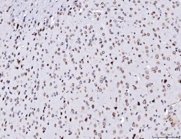
Real IHC data IHC analysis of TPR using anti-TPR antibody (A00695-2). TPR was detected in a paraffin-embedded section of human colorectal cancer tissue. Heat mediated antigen retrieval was performed in EDTA buffer (pH 8.0, epitope retrieval solution). The tissue section was blocked with 10% goat serum. The tissue section was then incubated with 2 μg/ml rabbit anti-TPR Antibody (A00695-2) overnight at 4°C. Peroxidase Conjugated Goat Anti-rabbit IgG was used as secondary antibody and incubated for 30 minutes at 37°C. The tissue section was developed using HRP Conjugated Rabbit IgG Super Vision Assay Kit (Catalog # SV0002) with DAB as the chromogen.
Anti-TPR Antibody ®
Cat # A00695-2

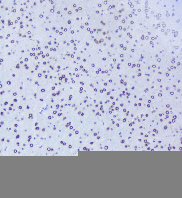
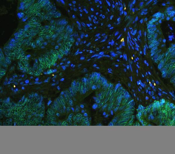
A00695-1 has IHC images from human lung cancer, mouse brain and kidney, and rat spleen, plus IF images from human intestinal cancer and mouse brain sections (A00695-1 image captions). A00695-2 has IHC images from human colorectal cancer, mouse brain and rat brain, plus IF/ICC images from SiHa and CACO-2 cells (A00695-2 image captions).

Which to pick: For paraffin-section IHC, choose A00695-1 for a citrate pH 6 retrieval starting point or A00695-2 for EDTA pH 8 retrieval (each SKU’s IHC caption); fixation is unreported in both captions. For IF, A00695-1 has tissue-section images, while A00695-2 has ICC cell images (catalog IF captions). Both offer human, mouse and rat reactivity and IHC and IF applications (catalog listings), with IHC images across all three species (catalog IHC captions); both are rabbit antibodies, and clonality is unreported (catalog listings).

Each figure is that product's own IHC / IF validation image from its datasheet.

References

  1. UniProt Consortium. UniProt entry P12270 (TPR_HUMAN, Nucleoprotein TPR).
  2. Human Protein Atlas. TPR tissue IHC expression (reliability: Enhanced).
  3. Human Protein Atlas. TPR subcellular location (ICC-IF): Localized to the nuclear membrane..
  4. Human Protein Atlas. TPR antibody validation summary (3 antibodies).
  5. Nucleoporin TPR integrates MAPK signaling with mitogen-induced transcriptional programs. Cell death & disease 2026 — PMC13109368.
  6. Colorectal cancer cells require glycogen synthase kinase-3β for sustaining mitosis via translocated promoter region (TPR)-dynein interaction. Oncotarget 2018 — PMC5862582.
  7. Nucleoporin TPR (translocated promoter region, nuclear basket protein) upregulation alters MTOR-HSF1 trails and suppresses autophagy induction in ependymoma. Autophagy 2021 — PMC8078762.
  8. The human nucleoporin Tpr protects cells from RNA-mediated replication stress. Nature communications 2021 — PMC8225803.
  9. PubMed PMID:1549355 — UniProt-cited evidence.
  10. PubMed PMID:1437155 — UniProt-cited evidence.
  11. PubMed PMID:7798308 — UniProt-cited evidence.