This website uses cookies to ensure you get the best experience on our website.


- Table of Contents
Plan chromogenic TPSAB1 IHC in paraffin sections with the catalog antibody at 0.5–1 μg/ml (datasheet: PB10016). Expect cytoplasmic staining in leukocyte subsets, probably mast cells (HPA tissue IHC), and account for possible detection of protein from more than one gene (HPA tissue IHC).

Expected localisation, validated protocols, controls and antibodies — the at-a-glance facts below, then the full design guide.
| Expected localisation | Cytoplasmic staining in leukocyte subsets (HPA tissue IHC) | |
| Staining pattern | Subset of leukocytes, probably mast cells: cytoplasmic (HPA tissue IHC) | |
| Antigen retrieval | EDTA pH 8.0 HIER, heat-mediated (datasheet PB10016) | |
| Positive control | Bone marrow+2 more · see all | |
| Negative control | Adipose tissue+4 more · see all |
| Fixation | Keep fixation consistent across sections. (standard IHC practice; not target-specific) | |
| Caveat | Antibody may detect protein from more than one gene (HPA tissue IHC) | |
| Regulation | Degranulation releases tryptase (UniProt) | |
| Isoform / epitope | Two isoforms; mature chain starts at residue 31 (UniProt) |
The catalog antibody’s IHC-P protocol is presented alongside published chromogenic TPSAB1 protocols for mouse ovary and rat synovium (PMC3790711; PMC13460010).
| Sample | Paraffin-embedded human lung cancer tissue; fixative not specified (datasheet PB10016) |
| Fixation | Image fixative and duration unreported (datasheet PB10016); verify before use. |
| Sectioning | 4–5 µm sections on charged slides (standard) |
| Deparaffinisation | Xylene, graded ethanol series to water (standard) |
| Antigen retrieval | Heat retrieval: EDTA pH 8.0 (datasheet PB10016); 20 min, 95–100 °C (standard) |
| Peroxidase block | 3% H2O2, 10 min, room temperature (standard) |
| Blocking | 10% goat serum (datasheet PB10016) |
| Primary antibody | Rabbit anti-TPSAB1, 0.5-1μg/ml (datasheet PB10016) |
| Primary incubation | Overnight at 4 °C (datasheet PB10016) |
| Detection | Streptavidin-biotin complex (SABC), DAB chromogen (datasheet PB10016) |
| Counterstain | Hematoxylin, blue, dehydrate and mount (standard) |
| Expected result | TPSAB1-positive staining in hematopoietic cells of bone marrow (HPA tissue IHC: Medium). HPA tissue profile: Selective cytoplasmic expression in subsets of leukocytes, probably mast cells. No signal in the no-primary control. |
TPSAB1 is a secreted protease released from mast-cell granules and has no transmembrane segment (UniProt Q15661). In tissue IHC, expect selective cytoplasmic staining in subsets of leukocytes, probably mast cells (HPA: tissue IHC profile). HPA rates the tissue pattern Supported, while reporting very low agreement with RNA expression and warning that the antibodies may recognize proteins from more than one gene (HPA: tissue IHC reliability).
| Discrete cytoplasmic staining in scattered leukocytes, with surrounding cells largely unstained. | This matches the selective pattern HPA describes in leukocyte subsets, probably mast cells (HPA: tissue IHC profile). Granule-associated signal is plausible because tryptase is released from mast-cell secretory granules (UniProt Q15661). The pattern supports interpretation but does not identify every positive cell as a mast cell (HPA: tissue IHC profile). |
| Strong, uniform nuclear or membrane outlining signal replaces the selective cytoplasmic pattern. | Treat this as a pattern mismatch and check the assay before scoring it as TPSAB1. HPA reports cytoplasmic staining, while UniProt describes a secreted protein without a transmembrane segment (HPA: tissue IHC profile; UniProt Q15661). Review morphology and controls for staining artifact (general IHC practice). |
| Broad staining appears in adipocytes, respiratory epithelium, or other unexpected cells. | HPA reports no detection in adipocytes of adipose tissue or breast, or in bronchial respiratory epithelial cells (HPA: tissue IHC). Consider antibody cross-reactivity or endogenous chromogenic activity, and compare a no-primary control (HPA: multigene-target caution; general IHC practice). Do not apply those negatives to every cell in those tissues. |
| Diffuse color covers stroma, lumens, and many cell types without clear cell borders. | Background or extracellular protein can obscure selective cellular staining (general IHC practice; UniProt Q15661: secreted protein). Because secretion can separate protein location from its RNA source, do not assign diffuse staining to a cell type solely from nearby RNA expression (HPA: tissue IHC reliability). |
| No convincing staining appears in a bone marrow, lymph-node, or tonsil section. | First confirm the relevant cell population is present: HPA reports Medium staining in bone-marrow hematopoietic cells and non-germinal-center cells of lymph node and tonsil (HPA: tissue IHC). A negative run calls for control and assay review; HPA's reported levels do not guarantee every section will contain positive cells. |
| Cell identity and tissue context | HPA's positive calls concern specific populations: bone-marrow hematopoietic cells and non-germinal-center cells in lymph node and tonsil, each at Medium level (HPA: tissue IHC). Its negative calls likewise name cell populations, such as adipocytes and bronchial respiratory epithelial cells (HPA: tissue IHC). Score cells, not whole organs. |
| Secretion and processing | UniProt lists a signal peptide at residues 1–18, a propeptide at 19–30, and the mature chain at 31–275; tryptase is released on mast-cell activation (UniProt Q15661). Extracellular signal therefore needs morphological context. The supplied record does not locate the catalog antibody's epitope, so processing cannot predict its staining strength. |
| Isoforms and endothelial staining | UniProt reports two isoforms and describes isoform 2 in endothelial cells of some vessels around the aorta and breast tumor, with little if any mast-cell protein there (UniProt Q15661). Do not automatically label every endothelial signal an artifact. HPA separately reports no detection in cerebral-cortex endothelial cells (HPA: tissue IHC); site matters. |
| Antibody specificity and evidence | Four listed HPA antibodies have Supported IHC status, but HPA warns that staining may reflect proteins from more than one gene and shows very low agreement with RNA expression (HPA: antibody validation; tissue IHC reliability). Interpret a matching pattern as evidence of tryptase-associated staining, not proof that every signal is uniquely TPSAB1. |
| IF/ICC: what should a positive cell show? | HPA reports vesicular localization in ICC-IF, with the main location approved as vesicles (HPA: subcellular). This is consistent with UniProt's secretory-granule description (UniProt Q15661). Use the separate IF/ICC guide for that application; these IHC tissue levels do not set an IF intensity threshold. |
| Situation | Likely cause | Next action |
|---|---|---|
| The reported positive tissue is blank. | The sampled section may lack the relevant cells, or the staining run may have failed; HPA's Medium calls apply to named cell populations (HPA: tissue IHC; general IHC practice). | Check tissue morphology and a positive-run control, then review antibody dilution, retrieval, detection, and counterstain settings (general IHC practice). No TPSAB1-specific retrieval or fixation sensitivity is established by the supplied sources. |
| The entire section develops color. | Excess background or endogenous chromogenic activity may overwhelm selective staining (general IHC practice). HPA describes selective cytoplasmic leukocyte staining (HPA: tissue IHC profile). | Compare a no-primary control; review blocking, washes, detection exposure, and counterstain (general IHC practice). Assess whether discrete cytoplasmic positive cells remain visible. |
| Adipocytes or bronchial respiratory epithelial cells stain prominently. | Those named cell types were not detected in HPA tissue IHC; antibody cross-reactivity or detection background is possible (HPA: tissue IHC and multigene-target caution; general IHC practice). | Review cell identity and no-primary staining, then compare the suspect pattern with a Supported IHC antibody where available (HPA: antibody validation; general IHC practice). |
| Nuclear or continuous membrane staining dominates. | The compartment conflicts with HPA's cytoplasmic pattern and UniProt's secreted, non-transmembrane annotation (HPA: tissue IHC profile; UniProt Q15661). | Check morphology, no-primary background, and detection settings before assigning a positive score (general IHC practice). |
| Vascular endothelial cells stain. | Some endothelial staining may reflect isoform 2 near the aorta or breast tumor, although HPA reports no detection in cerebral-cortex endothelial cells (UniProt Q15661; HPA: tissue IHC). | Record the vessel site and cell identity. Compare with the expected local pattern and controls; avoid a blanket positive or artifact call across tissues (UniProt Q15661; HPA: tissue IHC; general IHC practice). |
| Protein staining and local RNA expression disagree. | HPA reports very low staining–RNA consistency and notes that secreted protein can lie away from the cells producing it (HPA: tissue IHC reliability). | Score the observed cellular and extracellular pattern with tissue morphology and staining controls; report the RNA discrepancy as a limitation rather than using RNA alone to override the slide (HPA: tissue IHC reliability; general IHC practice). |





Comprehensive Human Protein Atlas IHC scoring per tissue (reliability: Supported — Very low consistency between antibody staining and RNA expression data. Caution, targets protein from more than one gene. Secreted protein, tissue location of RNA and protein is expected to differ. Antibody staining in cells/structures not annotated, view images.). Rows are taken directly from the HPA tissue chart — click any row's HPA link to view the source.
| Tissue | Cell type | Level | Evidence | Source |
|---|---|---|---|---|
| Bone marrow | Hematopoietic cells | Medium | Protein (IHC) | HPA → |
| Lymph node | Non-germinal center cells | Medium | Protein (IHC) | HPA → |
| Tonsil | Non-germinal center cells | Medium | Protein (IHC) | HPA → |
| Tissue | Cell type | Level | Evidence | Source |
|---|---|---|---|---|
| Adipose tissue | Adipocytes | Not detected | Protein (IHC) | HPA → |
| Adrenal gland | Glandular cells | Not detected | Protein (IHC) | HPA → |
| Appendix | Endocrine cells | Not detected | Protein (IHC) | HPA → |
| Breast | Adipocytes | Not detected | Protein (IHC) | HPA → |
| Bronchus | Respiratory epithelial cells | Not detected | Protein (IHC) | HPA → |
Troubleshoot TPSAB1 staining by assessing retrieval, tissue processing, expected localisation and cell identity before comparing signal intensity across sections.
Three anti-TPSAB1 antibodies have human paraffin-section IHC images (catalog image captions). Human reactivity is listed for all three; mouse and rat reactivity is also listed for A08039-1 and M08039 (catalog reactivity).
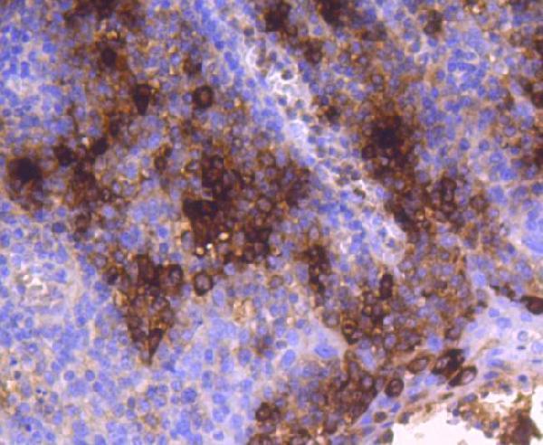
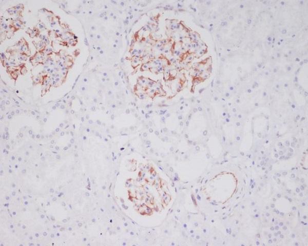
PB10016 has IHC images from paraffin sections of human lung, breast, and intestinal cancer tissue (PB10016 image captions). A08039-1 has a paraffin-section IHC image from human tonsil, and M08039 has one from human kidney (respective image captions).
Which to pick: For human paraffin-section IHC, PB10016 offers a documented starting workflow with EDTA pH 8.0 retrieval and 1 μg/ml primary antibody; the fixative is unreported (PB10016 image caption). For mouse or rat tissue IHC, A08039-1 and monoclonal M08039 list those species, although their paraffin-section images show human tonsil and kidney, respectively; fixation is unreported in both captions (catalog reactivity and clone; respective image captions). No SKU lists IF/ICC or provides an IF image, so these data do not support an IF/ICC pick (catalog applications and IF image fields).