This website uses cookies to ensure you get the best experience on our website.


- Table of Contents
Plan chromogenic IHC on paraffin sections with the catalog antibody at 0.5–1 μg/ml (datasheet: IHC-P). Colon glandular cells provide a high-staining reference (HPA tissue IHC); assess the observed cytoplasmic and nuclear signal alongside TRAM1’s expected ER membrane location (HPA tissue IHC; UniProt).

Expected localisation, validated protocols, controls and antibodies — the at-a-glance facts below, then the full design guide.
| Expected localisation | Cytoplasmic and nuclear staining (HPA tissue IHC); ER membrane expected (UniProt) | |
| Staining pattern | Cells across tissues: variable cytoplasmic and nuclear staining (HPA tissue IHC) | |
| Antigen retrieval | Citrate pH 6.0 HIER, 95–98 °C, 20 min (rule: cytoplasmic / membrane antigen) | |
| Positive control | Cervix+4 more · see all | |
| Negative control | Fallopian tube+1 more · see all |
| Fixation | Keep fixation consistent across sections (standard IHC practice; not target-specific) | |
| Caveat | IHC staining has low consistency with RNA expression (HPA tissue IHC) | |
| Regulation | Variable staining across tissues (HPA tissue IHC) | |
| Isoform / epitope | 2 isoforms; check epitope position against luminal and cytosolic regions (UniProt) |
Compare the catalog antibody’s IHC-P protocol with the published pancreatic tissue microarray protocol (PMC4718295 methods).
| Sample | Tissue sections; selected-image fixative not specified (standard IHC workflow) |
| Fixation | Image fixative and duration unreported (datasheet PA2010); verify before use. |
| Sectioning | 4–5 µm sections on charged slides (standard) |
| Deparaffinisation | Xylene, graded ethanol series to water (standard) |
| Antigen retrieval | Heat-induced epitope retrieval in citrate buffer, pH 6.0, 20 min at 95–98 °C (standard rule: cytoplasmic / membrane antigen) |
| Peroxidase block | 3% H2O2, 10 min, room temperature (standard) |
| Blocking | 10% normal serum of the secondary host, 30 min, room temperature (standard) |
| Primary antibody | Rabbit anti-TRAM1, 0.5-1μg/ml (datasheet PA2010) |
| Primary incubation | Overnight at 4 °C (standard) |
| Detection | HRP-polymer secondary, DAB chromogen 5–10 min (standard) |
| Counterstain | Hematoxylin, blue, dehydrate and mount (standard) |
| Expected result | TRAM1-positive staining in squamous epithelial cells of cervix (HPA tissue IHC: High). HPA tissue profile: Cytoplasmic and nuclear expression at variable levels in all tissues. No signal in the no-primary control. |
TRAM1 is an ER membrane protein (UniProt Q15629 topology). Epithelial staining varies; HPA rates IHC Approved with low RNA concordance (HPA).
| Colon glandular cells stain strongly (HPA: High). | A cytoplasmic component fits ER localisation (UniProt Q15629; HPA: cytoplasmic). |
| Staining is nuclear only (UniProt: ER membrane). | Review specificity; HPA reports variable nuclear staining but does not establish nuclear TRAM1 (HPA). |
| Prostate glands stain strongly (HPA: Not detected). | Check cross-reactivity or endogenous detection activity (HPA; standard IHC practice). |
| Stain is diffuse across the section. | Suspect background; inspect a no-primary control (standard IHC practice). |
| Colon glands show no signal (HPA: High). | Check the positive control, retrieval and detection (HPA; standard IHC practice). |
| Epitope position is unspecified; TRAM1 has 8 membrane spans (UniProt topology). | Antibody access may depend on whether its epitope faces cytoplasm or ER lumen (UniProt topology). |
| HPA IHC is Approved, with low RNA concordance (HPA). | Interpret unexpected tissue staining cautiously (HPA: low consistency). |
| IF/ICC: where should signal appear? | ER membrane is expected (UniProt); HPA says Membrane but provides no ICC images (HPA). |
| Situation | Likely cause | Next action |
|---|---|---|
| No colon gland signal (HPA: High). | Retrieval or detection may be inadequate (standard IHC practice). | Check positive control and retrieval conditions (standard IHC practice). |
| Strong prostate gland signal (HPA: Not detected). | Cross-reactivity or endogenous activity is possible (standard IHC practice). | Run no-primary and detection controls (standard IHC practice). |
| Nuclear-only staining (UniProt: ER membrane). | Specificity is uncertain; HPA reports variable nuclear staining (HPA). | Compare cytoplasmic staining and no-primary control (standard IHC practice). |
| Diffuse section-wide stain. | Blocking or detection background is possible (standard IHC practice). | Inspect no-primary control; adjust blocking and washes (standard IHC practice). |
| Uneven stain across a section. | Reagent coverage may be uneven (standard IHC practice). | Inspect section coverage and wash steps (standard IHC practice). |


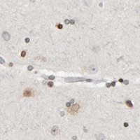


Comprehensive Human Protein Atlas IHC scoring per tissue (reliability: Approved — Low consistency between antibody staining and RNA expression data.). Rows are taken directly from the HPA tissue chart — click any row's HPA link to view the source.
| Tissue | Cell type | Level | Evidence | Source |
|---|---|---|---|---|
| Cervix | Squamous epithelial cells | High | Protein (IHC) | HPA → |
| Colon | Glandular cells | High | Protein (IHC) | HPA → |
| Duodenum | Glandular cells | High | Protein (IHC) | HPA → |
| Esophagus | Squamous epithelial cells | High | Protein (IHC) | HPA → |
| Oral mucosa | Squamous epithelial cells | High | Protein (IHC) | HPA → |
Troubleshoot TRAM1 chromogenic IHC in paraffin sections using its ER membrane topology and tissue staining evidence (UniProt Q15629; HPA tissue IHC).
PA2010 has IHC images from human paraffin sections and rat paraffin and frozen sections, plus an ICC image from HELA cells (PA2010 image captions). Its listed reactivity is human, mouse and rat (PA2010 catalog).
PA2010 has IHC(P) captions for human lung cancer and rat kidney tissue, and frozen-section IHC captions for rat intestine and brain (PA2010 image captions). Its ICC image is labeled HELA cells; IHC, IHC-F and ICC are listed applications (PA2010 image captions; PA2010 catalog).
Which to pick: Choose PA2010 for paraffin-section IHC based on its human lung cancer and rat kidney IHC(P) captions; the fixative is unreported (PA2010 image captions). For cell staining, PA2010 has an ICC image from HELA cells, but IF is not separately listed as an application (PA2010 ICC caption; PA2010 catalog). It is listed as reactive with human, mouse and rat, though the supplied IHC images show human and rat samples only; its host is rabbit and clonality is unreported (PA2010 catalog; PA2010 image captions).