This website uses cookies to ensure you get the best experience on our website.


- Table of Contents
Plan paraffin-section TRIM2 IHC using cerebellar GLUC cells as a high-staining reference and adipocytes as a negative cellular control (HPA tissue IHC). Start the IHC-validated antibody A08957 at 2–5 μg/ml (datasheet A08957), then assess cytoplasmic staining with the reported off-target binding in mind (HPA tissue IHC).

Expected localisation, validated protocols, controls and antibodies — the at-a-glance facts below, then the full design guide.
| Expected localisation | Mainly cytoplasmic staining in the CNS (HPA tissue IHC) | |
| Staining pattern | CNS cytoplasm; cerebellar GLUC cells also stain at the membrane (HPA tissue IHC) | |
| Antigen retrieval | EDTA pH 8.0 HIER, heat-mediated (datasheet A08957) | |
| Positive control | Cerebellum+4 more · see all | |
| Negative control | Adipose tissue+4 more · see all |
| Fixation | Keep fixation consistent across sections (standard IHC practice; not target-specific) | |
| Caveat | Presumed off-target binding may mimic TRIM2 staining (HPA tissue IHC) | |
| Regulation | No stimulus-dependent regulation annotated (UniProt) | |
| Isoform / epitope | 2 isoforms; check epitope coverage for each (UniProt) |
Compare the catalog antibody’s IHC-P protocol (datasheet: A08957) with a published protocol for human breast tumour sections (PMC11171213).
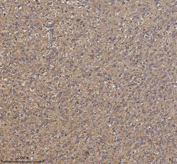
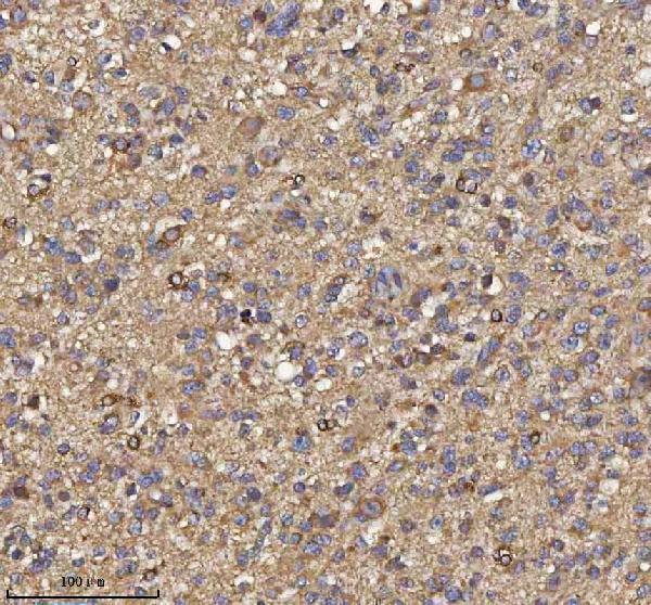
| Sample | Paraffin-embedded human glioblastoma tissue; fixative not specified (datasheet A08957) |
| Fixation | Image fixative and duration unreported (datasheet A08957); verify before use. |
| Sectioning | 4–5 µm sections on charged slides (standard) |
| Deparaffinisation | Xylene, graded ethanol series to water (standard) |
| Antigen retrieval | Heat retrieval: EDTA pH 8.0 (datasheet A08957); 20 min, 95–100 °C (standard) |
| Peroxidase block | 3% H2O2, 10 min, room temperature (standard) |
| Blocking | 10% goat serum (datasheet A08957) |
| Primary antibody | Rabbit anti-TRIM2, 2-5 μg/ml (datasheet A08957) |
| Primary incubation | Overnight at 4 °C (datasheet A08957) |
| Detection | HRP-conjugated secondary, DAB chromogen (datasheet A08957) |
| Counterstain | Hematoxylin, blue, dehydrate and mount (standard) |
| Expected result | TRIM2-positive staining in gLUC cells - cytoplasm/membrane of cerebellum (HPA tissue IHC: High). HPA tissue profile: Cytoplasmic expression mainly in the CNS. No signal in the no-primary control. |
TRIM2 is annotated as cytoplasmic and has no transmembrane segment (UniProt Q9C040). In paraffin-section IHC, expect staining mainly in CNS cells: high staining in cerebellar GLUC cells and medium staining in cerebral cortex and hippocampal glial cells (HPA tissue IHC). HPA rates its tissue evidence Supported, with medium agreement between staining and RNA data; its antibody findings need cautious interpretation (HPA tissue IHC).
| Cerebellar GLUC cells stain strongly in the cytoplasm, with possible membrane-associated staining. | This matches the highest supplied tissue observation: GLUC cells show high cytoplasmic/membrane staining (HPA tissue IHC). The cytoplasmic component agrees with TRIM2's annotated location (UniProt Q9C040). Assess the stained cells and compartment together; intensity alone cannot establish antibody specificity (standard IHC practice). |
| Glial cells in cerebral cortex or hippocampus show moderate staining. | Medium glial-cell staining is reported in both regions (HPA tissue IHC). A weaker result than the high cerebellar GLUC-cell observation can therefore fit the supplied pattern (HPA tissue IHC). Compare like cell populations within the section when scoring, since tissue-wide color can obscure which cells carry the signal (standard IHC practice). |
| Signal is predominantly nuclear, or forms a convincing isolated nuclear pattern. | Treat this as discordant with the annotated cytoplasmic location (UniProt Q9C040). Review morphology and controls before calling it TRIM2; misplaced signal can reflect nonspecific staining or detection artefact (standard IHC practice). The uncertain centrosome call from ICC-IF does not establish an expected nuclear IHC pattern (HPA subcellular ICC-IF). |
| Strong staining appears in cells listed as not detected, such as adipocytes or bone-marrow hematopoietic cells. | Those cell populations are reported as not detected in the supplied tissue observations (HPA tissue IHC). Check for cross-reactivity or endogenous detection activity before interpreting a strong result as TRIM2 (standard IHC practice). HPA also cautions that its tissue antibodies target proteins from more than one gene and that presumed off-target binding was disregarded (HPA tissue IHC). |
| Color is diffuse across tissue structures, or a known-positive region has no discernible signal. | Diffuse color without cell-specific contrast is background rather than a persuasive localisation pattern (standard IHC practice). No signal in cerebellar GLUC cells conflicts with their reported high staining (HPA tissue IHC), but does not by itself prove TRIM2 absence. Compare a positive-control section and detection controls before assigning a biological negative (standard IHC practice). |
| Compartment and topology | TRIM2 is annotated in the cytoplasm and has no transmembrane segment (UniProt Q9C040). Interpret the HPA cerebellar cytoplasmic/membrane observation as a cell staining pattern, without inferring that TRIM2 spans the membrane (HPA tissue IHC; UniProt Q9C040). |
| Tissue and cell selection | Cerebellar GLUC cells provide the supplied high-staining comparison; cortex and hippocampus provide medium glial-cell observations (HPA tissue IHC). Adipocytes and bone-marrow hematopoietic cells are listed as not detected, so use their stated cell identities when evaluating contrast (HPA tissue IHC). |
| Strength of antibody evidence | Two listed antibodies have Supported IHC status, HPA035853 and HPA035854 (HPA antibodies). HPA describes medium consistency with RNA data and cautions about proteins from more than one gene and disregarded presumed off-target binding (HPA tissue IHC). Treat morphology and controls as part of interpretation (standard IHC practice). |
| Isoforms and processing | Two isoforms are listed, with no signal peptide, propeptide, or cleavage into separate annotated chains (UniProt Q9C040). No epitope map is supplied, so these facts cannot predict whether the IHC-validated antibody distinguishes isoforms or whether a particular compartmental signal belongs to one isoform. |
| IF/ICC Q: Should a centrosome spot be expected in tissue IHC? | A: HPA lists centrosome localisation as uncertain in ICC-IF and cautions that the antibodies target proteins from multiple genes (HPA subcellular ICC-IF). Use the cytoplasmic tissue IHC observations for this paraffin-section readout; a centrosome spot alone is insufficient to identify TRIM2 (HPA tissue IHC; HPA subcellular ICC-IF). |
| Situation | Likely cause | Next action |
|---|---|---|
| No signal in cerebellar GLUC cells. | The result conflicts with the reported high staining in those cells (HPA tissue IHC); a failed staining run or missed cell population is possible (standard IHC practice). | Confirm the cells on the counterstained section, then check a known-positive section, antibody application, and detection controls (standard IHC practice). |
| Only nuclei stain strongly. | A predominantly nuclear result differs from the cytoplasmic annotation (UniProt Q9C040); nonspecific signal or detection artefact is possible (standard IHC practice). | Check a no-primary control and review cell boundaries before scoring; repeat with the IHC-validated antibody if controls implicate the staining run (standard IHC practice). |
| Strong signal appears in an HPA not-detected cell population. | Cross-reactivity or endogenous detection activity may explain unexpected staining (standard IHC practice); HPA notes a multi-gene antibody-target caution (HPA tissue IHC). | Verify the precise cell type, inspect a no-primary control, and assess the relevant detection-system control before calling the cells positive (standard IHC practice). |
| Diffuse brown color obscures cell boundaries. | Non-specific binding or detection background can reduce cell-specific contrast (standard IHC practice). | Compare with a no-primary control; review blocking, antibody concentration, washes, and chromogen development using the staining workflow (standard IHC practice). |
| Cortex or hippocampus looks weaker than cerebellum. | This may match medium glial staining versus high cerebellar GLUC-cell staining (HPA tissue IHC). | Score the named cell populations and their compartments separately; retain the region-specific intensity distinction in the result (HPA tissue IHC; standard IHC practice). |
| A punctate IF/ICC result disagrees with tissue IHC. | The centrosome assignment is uncertain and carries a multi-gene antibody caution (HPA subcellular ICC-IF). | Interpret the paraffin-section result against tissue IHC cell and compartment observations; seek independent specificity evidence before assigning the puncta to TRIM2 (HPA tissue IHC; standard IHC practice). |
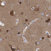
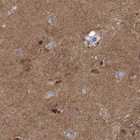
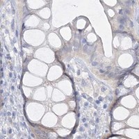
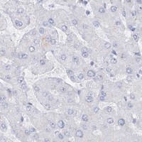


Comprehensive Human Protein Atlas IHC scoring per tissue (reliability: Supported — Medium consistency between antibody staining and RNA expression data. Caution, targets protein from more than one gene. Presumed off target binding observed and disregarded. Pending external verification.). Rows are taken directly from the HPA tissue chart — click any row's HPA link to view the source.
| Tissue | Cell type | Level | Evidence | Source |
|---|---|---|---|---|
| Cerebellum | GLUC cells - cytoplasm/membrane | High | Protein (IHC) | HPA → |
| Cerebral cortex | Glial cells | Medium | Protein (IHC) | HPA → |
| Fallopian tube | Non-ciliated cells | Medium | Protein (IHC) | HPA → |
| Gallbladder | Glandular cells | Medium | Protein (IHC) | HPA → |
| Hippocampus | Glial cells | Medium | Protein (IHC) | HPA → |
| Tissue | Cell type | Level | Evidence | Source |
|---|---|---|---|---|
| Adipose tissue | Adipocytes | Not detected | Protein (IHC) | HPA → |
| Appendix | Glandular cells | Not detected | Protein (IHC) | HPA → |
| Bone marrow | Hematopoietic cells | Not detected | Protein (IHC) | HPA → |
| Breast | Adipocytes | Not detected | Protein (IHC) | HPA → |
| Bronchus | Basal cells | Not detected | Protein (IHC) | HPA → |
Troubleshoot TRIM2 staining in paraffin sections using the catalog antibody’s IHC example and cautious interpretation of tissue and subcellular evidence.
The IHC-validated anti-TRIM2 antibody has paraffin-section images from human glioblastoma and rat brain (A08957 IHC captions); no IF/ICC data are supplied (catalog application list; IF image list).
A08957 is the sole listed SKU and has IHC images from paraffin-embedded human glioblastoma and rat brain sections (A08957 IHC captions). Its listed applications include IHC, WB and ELISA, with human, mouse and rat reactivity; IF/ICC is not listed (A08957 catalog).
Which to pick: Choose A08957 for paraffin-section IHC using its documented 2 μg/ml image condition as a starting point; the catalog IHC range is 2–5 μg/ml (A08957 IHC captions; datasheet). No listed SKU has IF/ICC validation, and A08957’s clonality is unspecified (catalog application list; IF image list; clone field). For cross-species work, A08957 lists human, mouse and rat reactivity, while its IHC images document human and rat sections only; the fixative is unreported (A08957 catalog; IHC captions).