This website uses cookies to ensure you get the best experience on our website.


- Table of Contents
Plan chromogenic IHC for TRMT10C with the IHC-validated antibody at 2–5 μg/ml (datasheet A09612-1). Use placenta decidual cells as a high-staining reference (HPA tissue IHC), and assess nuclear and cytoplasmic staining in light of the reported medium consistency with RNA expression (HPA tissue IHC).

Expected localisation, validated protocols, controls and antibodies — the at-a-glance facts below, then the full design guide.
| Expected localisation | Nuclear and cytoplasmic tissue staining (HPA tissue IHC) | |
| Staining pattern | Nuclear and cytoplasmic staining; high in decidual cells (HPA tissue IHC) | |
| Antigen retrieval | EDTA pH 8.0 HIER, heat-mediated (datasheet A09612-1) | |
| Positive control | Placenta+4 more · see all | |
| Negative control | Adipose tissue+4 more · see all |
| Fixation | Keep fixation consistent across paraffin sections. (standard IHC practice; not target-specific) | |
| Caveat | Staining shows medium consistency with RNA expression (HPA tissue IHC) | |
| Regulation | Low tissue specificity by RNA (HPA tissue RNA) | |
| Isoform / epitope | No isoforms listed; annotated chain spans residues 40–403 (UniProt) |
The catalog antibody's IHC-P protocol (datasheet A09612-1) is accompanied by two published TRMT10C IHC protocols (PMC12080399; PMC13276065).
| Sample | Paraffin-embedded human breast duct carcinoma tissue; fixative not specified (datasheet A09612-1) |
| Fixation | Image fixative and duration unreported (datasheet A09612-1); verify before use. |
| Sectioning | 4–5 µm sections on charged slides (standard) |
| Deparaffinisation | Xylene, graded ethanol series to water (standard) |
| Antigen retrieval | Heat retrieval: EDTA pH 8.0 (datasheet A09612-1); 20 min, 95–100 °C (standard) |
| Peroxidase block | 3% H2O2, 10 min, room temperature (standard) |
| Blocking | 10% goat serum (datasheet A09612-1) |
| Primary antibody | Rabbit anti-TRMT10C, 2-5 μg/ml (datasheet A09612-1) |
| Primary incubation | Overnight at 4 °C (datasheet A09612-1) |
| Detection | HRP-conjugated secondary, DAB chromogen (datasheet A09612-1) |
| Counterstain | Hematoxylin, blue, dehydrate and mount (standard) |
| Expected result | TRMT10C-positive staining in decidual cells of placenta (HPA tissue IHC: High). HPA tissue profile: General nuclear and cytoplasmic expression. No signal in the no-primary control. |
TRMT10C is a soluble mitochondrial matrix and nucleoid protein with no transmembrane segment (UniProt Q7L0Y3). In paraffin section IHC, expect cellular staining in HPA positive populations, strongest in placental decidual cells and moderate in several glandular populations (HPA tissue IHC: High in decidual cells; Medium in glandular cells). HPA describes a general nuclear and cytoplasmic tissue pattern, with Approved reliability and medium consistency with RNA data (HPA tissue IHC).
| Placental decidual cells stain strongly, with cytoplasmic signal; nearby cells vary. | This fits the strongest listed tissue and cell population (HPA tissue IHC: High in placental decidual cells). Mitochondrial enrichment is biologically plausible (UniProt Q7L0Y3: matrix and nucleoid). Chromogenic IHC alone may not resolve individual mitochondria (general IHC practice). |
| Colon, appendix, or cervical glandular cells show moderate staining. | These are listed positive populations (HPA tissue IHC: Medium in glandular cells of colon, appendix, and cervix). Compare the named cell population, not the whole organ. HPA also reports a general nuclear and cytoplasmic tissue pattern, so cytoplasmic staining need not appear as discrete puncta (HPA tissue IHC). |
| Staining is predominantly extracellular or outlines cell membranes. | That distribution conflicts with the reported matrix and nucleoid location and absence of a transmembrane segment (UniProt Q7L0Y3). Treat it as a possible artefact and check controls before scoring. Nuclear signal alone is less decisive because HPA reports nuclear and cytoplasmic tissue staining (HPA tissue IHC). |
| An HPA listed negative cell population stains as strongly as decidual cells. | Check the exact cell type: adipocytes in adipose tissue and cholangiocytes in liver are listed as Not detected, without establishing that every cell in those organs is negative (HPA tissue IHC). Strong staining there raises concern for cross-reactivity or endogenous detection activity; a no-primary control helps distinguish these possibilities (general IHC practice). |
| Color appears broadly across tissue and blank areas, or the positive population has no signal. | Widespread color without cellular definition suggests background from the detection workflow (general IHC practice). Absent signal in placental decidual cells conflicts with their reported High staining (HPA tissue IHC); first assess tissue preservation, retrieval, detection controls, and antibody dilution as general IHC checks. |
| Compartment and optical resolution | TRMT10C is assigned to the mitochondrial matrix and nucleoids and lacks a transmembrane segment (UniProt Q7L0Y3). These facts guide compartment checks, while a chromogenic paraffin section may not resolve mitochondria (general IHC practice). Avoid requiring visible puncta for every positive cell. |
| Cell population and staining strength | Decidual cells are High; listed glandular, hematopoietic, basal, and neuronal populations are Medium (HPA tissue IHC). Other listed populations are Low or Not detected (HPA tissue IHC). Select and score the specified cell type rather than using an organ-wide positive or negative label. |
| Evidence behind a nuclear signal | HPA tissue IHC describes general nuclear and cytoplasmic expression (HPA tissue IHC). Its ICC-IF record supports mitochondria as the main location but marks additional nucleoplasm localization uncertain (HPA subcellular ICC-IF). A nuclear IHC component therefore needs cautious interpretation; it does not by itself establish nucleoplasmic TRMT10C. |
| Antibody validation and protein processing | The listed antibody HPA036671 is IHC Approved, not reported here as IHC Enhanced (HPA antibodies). UniProt lists a 40–403 chain but supplies no antibody epitope position in this payload (UniProt Q7L0Y3). The record cannot predict whether processing changes this antibody’s staining. |
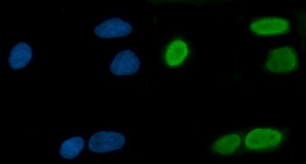
| IF/ICC Q&A: what localization should an IF image support? | Mainly mitochondrial signal; additional nucleoplasmic localization remains uncertain (HPA subcellular ICC-IF). This is localization evidence for interpreting images, not an IF protocol or a reason to demand resolved mitochondrial puncta in chromogenic IHC (general IHC practice). |
| Situation | Likely cause | Next action |
|---|---|---|
| No stain in placental decidual cells. | The result conflicts with the reported High population (HPA tissue IHC); a workflow failure is possible (general IHC practice). | Confirm the correct cells are present, then check the positive control, retrieval conditions, antibody dilution, and detection reagents (general IHC practice). |
| Moderate glandular staining is called negative because it is weaker than placenta. | HPA assigns Medium to glandular cells in colon, appendix, and cervix, versus High in decidual cells (HPA tissue IHC). | Score intensity within the identified cell population and compare with an appropriate positive section processed alongside it (general IHC practice). |
| Color persists in a no-primary control. | Endogenous detection activity or nonspecific detection reagents can create signal without primary antibody (general IHC practice). | Review enzyme blocking and detection steps; repeat the no-primary control before attributing tissue color to TRMT10C (general IHC practice). |
| Most of the section has diffuse haze. | Background can obscure cell-associated staining and its distribution (general IHC practice). | Check blocking, washes, primary antibody dilution, and detection exposure against controls; judge specific signal only after background is reduced (general IHC practice). |
| Only a sharp nuclear signal appears. | Its significance is uncertain: HPA tissue IHC reports nuclear and cytoplasmic staining, while ICC-IF marks nucleoplasm uncertain (HPA tissue IHC; HPA subcellular ICC-IF). | Compare with the expected positive cell population and controls; do not infer a confirmed nucleoplasmic location from nuclear IHC alone (HPA subcellular ICC-IF; general IHC practice). |
| Strong staining appears in adipocytes or liver cholangiocytes. | Those specific populations are listed as Not detected (HPA tissue IHC); cross-reactivity or detection background is possible (general IHC practice). | Verify cell identity, inspect no-primary staining, and compare with placental decidual cells on the same run (HPA tissue IHC: High in decidual cells; general IHC practice). |
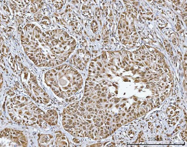
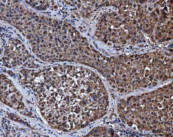


Comprehensive Human Protein Atlas IHC scoring per tissue (reliability: Approved — Medium consistency between antibody staining and RNA expression data.). Rows are taken directly from the HPA tissue chart — click any row's HPA link to view the source.
| Tissue | Cell type | Level | Evidence | Source |
|---|---|---|---|---|
| Placenta | Decidual cells | High | Protein (IHC) | HPA → |
| Adrenal gland | Glandular cells | Medium | Protein (IHC) | HPA → |
| Appendix | Glandular cells | Medium | Protein (IHC) | HPA → |
| Bone marrow | Hematopoietic cells | Medium | Protein (IHC) | HPA → |
| Bronchus | Basal cells | Medium | Protein (IHC) | HPA → |
| Tissue | Cell type | Level | Evidence | Source |
|---|---|---|---|---|
| Adipose tissue | Adipocytes | Not detected | Protein (IHC) | HPA → |
| Breast | Adipocytes | Not detected | Protein (IHC) | HPA → |
| Caudate | Glial cells | Not detected | Protein (IHC) | HPA → |
| Hippocampus | Glial cells | Not detected | Protein (IHC) | HPA → |
| Liver | Cholangiocytes | Not detected | Protein (IHC) | HPA → |
Troubleshoot TRMT10C staining in paraffin sections using the catalog antibody’s tissue IHC conditions, with IF as a complementary localisation check.
A09612-1 has human IHC data from paraffin-embedded carcinoma sections and IF data from A549 cells and paraffin-embedded carcinoma sections (catalog image captions; catalog reactivity: Human).
A09612-1 was tested for IHC in human paraffin-embedded breast duct, liver, lung and lung squamous cell carcinoma sections (A09612-1 IHC image captions). The same SKU has IF images from A549 cells and human paraffin-embedded rectal and ovarian carcinoma sections (A09612-1 IF image captions).
Which to pick: Choose A09612-1 for human tissue IHC; its own captions document paraffin sections, while the fixative and antibody clonality are unreported (A09612-1 IHC image captions; catalog reactivity: Human). Choose A09612-1 for IF/ICC based on its listed applications and IF images (catalog applications; A09612-1 IF image captions). No cross-species option is supported by this catalog payload (catalog reactivity: Human).