This website uses cookies to ensure you get the best experience on our website.


- Table of Contents
Real validated TRPC4 Western blot protocols, expected-band and isoform facts, troubleshooting for weak or shifted signal, and recommended anti-TRPC4 WB antibodies. Everything you need to plan the experiment before you commit precious samples.
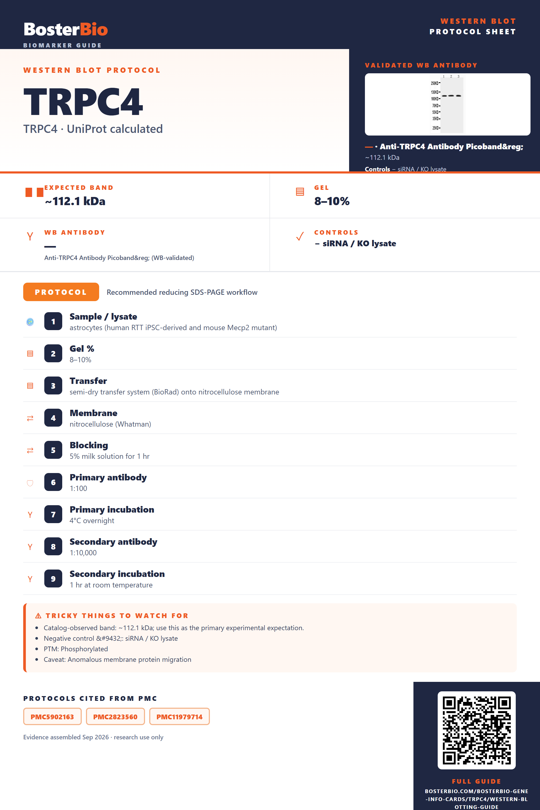
Expected bands, validated protocols, controls and antibodies — the at-a-glance facts below, then the full design guide.
| Expected band | ~112.1 kDa | |
| Observed band | ~150 kDa | |
| Gel | 8–10% | |
| Negative control | siRNA / KO lysate |
| PTM | Phosphorylated | |
| Caveat | Anomalous membrane protein migration | |
| Regulation | IFN-γ-induced | |
| Isoform | 7 isoform(s) |
Literature-validated Western blot parameters for TRPC4 — gel percentage, transfer, blocking, antibody incubation and detection, extracted from published methods.
| Sample / lysate | astrocytes (human RTT iPSC-derived and mouse Mecp2 mutant) |
| Gel % | 8–10% |
| Transfer | semi-dry transfer system (BioRad) onto nitrocellulose membrane |
| Membrane | nitrocellulose (Whatman) |
| Blocking | 5% milk solution for 1 hr |
| Primary antibody | 1:100 |
| Primary incubation | 4°C overnight |
| Secondary antibody | 1:10,000 |
| Secondary incubation | 1 hr at room temperature |
| Detection | infrared dye-conjugated secondary antibody |
| Exposure / imaging | Odyssey infrared imaging system |
TRPC4 has a 112 kDa predicted backbone but typically runs at ~150 kDa on Western blots due to anomalous migration of this multi-pass membrane channel protein.
| Single band around 150 kDa in whole-cell lysate | the mature, membrane-embedded TRPC4 channel migrating well above its 112 kDa predicted mass |
| Band position varies slightly (roughly 110-150 kDa) across cell lines or tissues | reflects which of the seven alternatively spliced TRPC4 isoforms (Alpha, Beta, Delta, Gamma, Epsilon, Zeta, Eta) are expressed |
| No shift to a doubled-size band under non-reducing conditions | the single disulfide (Cys549-Cys554) is a short intrachain loop, not an inter-chain linkage, so it does not generate a dimer-sized species |
| Faint higher-molecular-weight smear above the main ~150 kDa band | residual homotetrameric or TRPC1/TRPC5 heterotetrameric channel assemblies that were not fully dissociated by SDS |
| Band still detected in whole-cell lysate rather than absent | TRPC4 is a cell-membrane multi-pass channel, not a secreted protein, so it is retained when lysates are prepared with adequate detergent |
| Predicted mass from UniProt (112.1 kDa) | sets the theoretical backbone size before any migration or assembly effects are applied |
| Multi-pass transmembrane channel structure | causes anomalous, slower migration on SDS-PAGE so the protein typically runs near 150 kDa instead of its calculated mass |
| Homotetramer/heterotetramer assembly with TRPC1 and/or TRPC5 | incomplete denaturation of this non-covalent oligomer can leave a faint higher-molecular-weight species above the main band |
| Intrachain disulfide bond (Cys549-Cys554) | stabilizes a short loop and can subtly change mobility under non-reducing conditions, but does not create an inter-chain or dimer-sized band |
| Seven named splice isoforms (Alpha, Beta, Delta, Gamma, Epsilon, Zeta, Eta) | can produce additional bands at slightly different apparent sizes depending on which isoforms a sample expresses |
| C-terminal phosphotyrosine sites (Tyr959, Tyr972; FYN) | phosphorylation can add a minor upward mobility shift but is a secondary contributor compared to the transmembrane-driven shift |
| Situation | Likely cause | Next action |
|---|---|---|
| Band higher than expected | multi-pass transmembrane channel proteins migrate anomalously and TRPC4 tetramers may not fully dissociate under standard SDS conditions | compare against the known ~150 kDa apparent size, ensure samples are fully reduced and denatured, and resolve on a lower-percentage gel if needed |
| Multiple bands | co-expression of several TRPC4 splice isoforms or partially denatured oligomeric channel complexes | confirm the specific band with an isoform-aware or validated antibody and a knockdown/knockout control |
| No band in lysate | incomplete solubilization of this multi-pass membrane protein with a lysis buffer lacking sufficient detergent | use a membrane-compatible lysis buffer (e.g., RIPA or NP-40 based) and avoid under-extracting the membrane fraction |
| Weak or no signal | low native TRPC4 expression in the sampled cell type or degradation during lysate preparation | increase protein loading, enrich membrane fractions, and add protease inhibitors during lysis |
| Broad smear instead of sharp band | heterogeneous phosphorylation states or variably denatured oligomeric channel assemblies | ensure fresh lysate, avoid repeated freeze-thaw, and optimize reducing agent and heating conditions |

Comprehensive Human Protein Atlas IHC scoring per tissue. Rows are taken directly from the HPA tissue chart — click any row's HPA link to view the source.
| Tissue | Cell type | Level | Evidence | Source |
|---|
| Tissue | Cell type | Level | Evidence | Source |
|---|
Deeper troubleshooting and optimisation questions for TRPC4, answered from its protein features.
BosterBio's TRPC4 antibodies are among the best-performing WB antibodies on the market — well cited, thoroughly validated, and orthogonally cross-validated against negative tissues and complementary methods.
These Boster anti-TRPC4 antibodies are top-performing, widely cited reagents validated by orthogonal methods including negative-tissue controls and complementary techniques, giving reliable, reproducible Western blot detection of TRPC4 you can trust for publication-quality results.
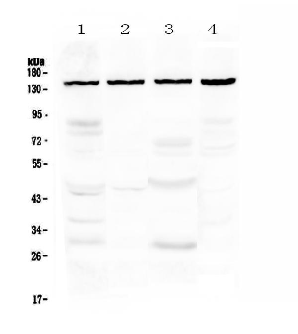
Which to pick: Both PA2307 and A02037-1 include real WB validation images. PA2307 shows detection across COLO320, MCF-7, and PANC lysates; A02037-1 demonstrates standard SDS-PAGE-based WB analysis. Either is a solid choice—pick PA2307 for broader cell-line coverage or A02037-1 if it matches your workflow.