This website uses cookies to ensure you get the best experience on our website.


- Table of Contents
Real validated TRPM8 Western blot protocols, expected-band and isoform facts, troubleshooting for weak or shifted signal, and recommended anti-TRPM8 WB antibodies. Everything you need to plan the experiment before you commit precious samples.
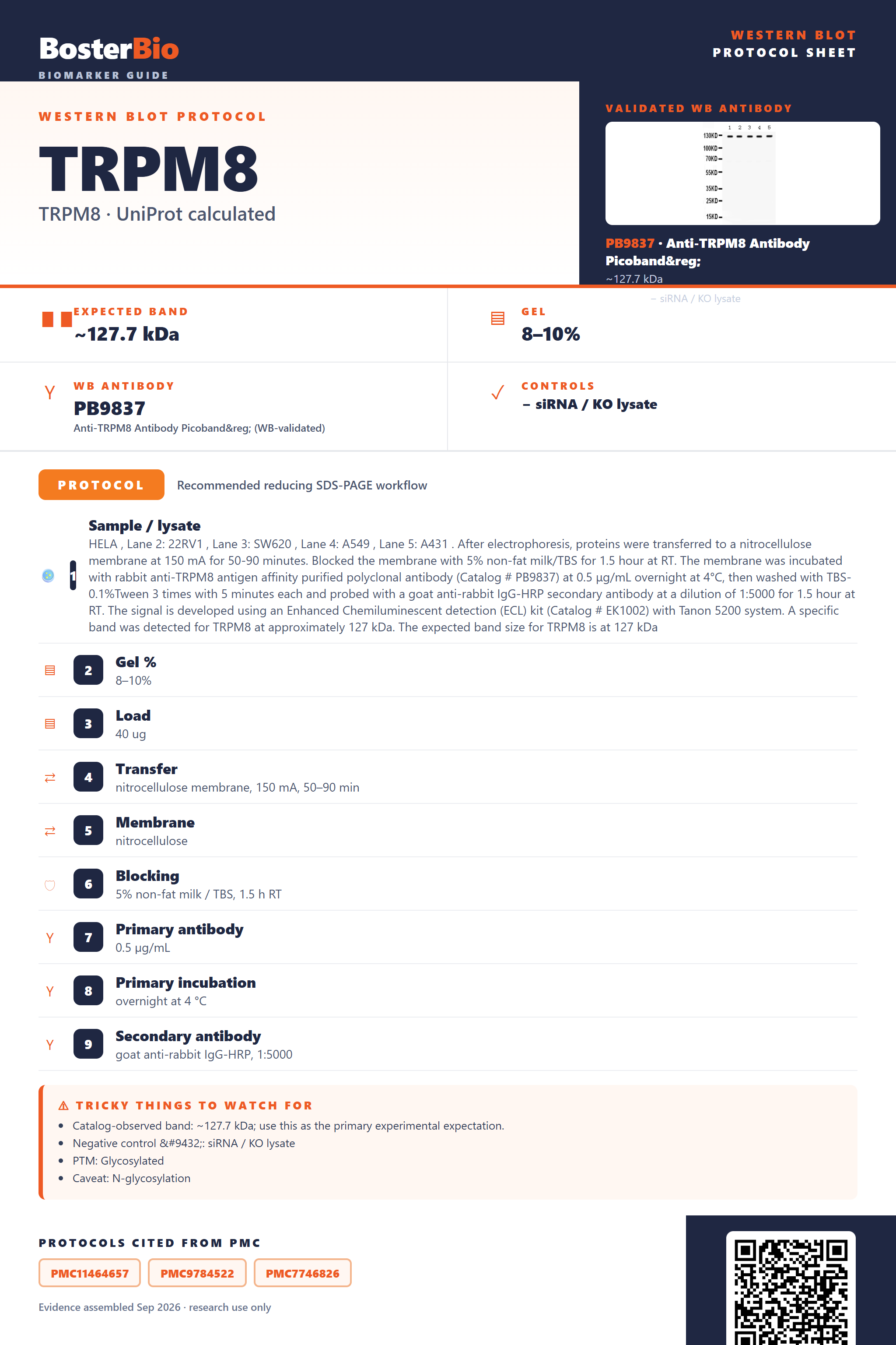
Expected bands, validated protocols, controls and antibodies — the at-a-glance facts below, then the full design guide.
| Expected band | ~127.7 kDa | |
| Observed band | ~127 kDa | |
| Gel | 8–10% | |
| Negative control | siRNA / KO lysate |
| PTM | Glycosylated | |
| Caveat | N-glycosylation | |
| Regulation | LPS-suppressed | |
| Isoform | 4 isoform(s) |
Literature-validated Western blot parameters for TRPM8 — gel percentage, transfer, blocking, antibody incubation and detection, extracted from published methods.
| Sample / lysate | HELA , Lane 2: 22RV1 , Lane 3: SW620 , Lane 4: A549 , Lane 5: A431 . After electrophoresis, proteins were transferred to a nitrocellulose membrane at 150 mA for 50-90 minutes. Blocked the membrane with 5% non-fat milk/TBS for 1.5 hour at RT. The membrane was incubated with rabbit anti-TRPM8 antigen affinity purified polyclonal antibody (Catalog # PB9837) at 0.5 μg/mL overnight at 4°C, then washed with TBS-0.1%Tween 3 times with 5 minutes each and probed with a goat anti-rabbit IgG-HRP secondary antibody at a dilution of 1:5000 for 1.5 hour at RT. The signal is developed using an Enhanced Chemiluminescent detection (ECL) kit (Catalog # EK1002) with Tanon 5200 system. A specific band was detected for TRPM8 at approximately 127 kDa. The expected band size for TRPM8 is at 127 kDa |
| Gel % | 8–10% |
| Load | 40 ug |
| Transfer | nitrocellulose membrane, 150 mA, 50–90 min |
| Membrane | nitrocellulose |
| Blocking | 5% non-fat milk / TBS, 1.5 h RT |
| Primary antibody | 0.5 µg/mL |
| Primary incubation | overnight at 4 °C |
| Secondary antibody | goat anti-rabbit IgG-HRP, 1:5000 |
| Wash | TBS-0.1% Tween, 3 × 5 min |
| Detection | ECL |
| Exposure / imaging | Tanon 5200 |
| Observed band | 127 kDa |
TRPM8 has a 127.7 kDa predicted backbone and runs at the empirically observed ~127 kDa monomer, with minor shifts possible from its single Asn934 N-glycan and its non-covalent homotetramer assembly.
| Single band at ~127 kDa | Matches the full-length TRPM8 monomer; there is no signal peptide or propeptide to trim off, so the observed band reflects the intact predicted mass. |
| Band sitting slightly above the unmodified 127.7 kDa core | The single N-linked complex-type glycan at Asn934 adds a small mass increment to the mature channel. |
| High-molecular-weight band or smear near the top of the gel | TRPM8 assembles as a non-covalent homotetramer; incompletely denatured/reduced samples can retain tetrameric or partial oligomeric complexes. |
| Multiple bands at different apparent masses | Four annotated splice isoforms (1-4), including isoforms that interact with the isoform-1 C-terminus, can contribute additional bands beyond the primary monomer. |
| Fuzzy edge or minor doublet around the main band | Microheterogeneity in occupancy/processing of the single glycosylation site at Asn934 can slightly blur the primary band. |
| Predicted mass (127.7 kDa) | Sets the baseline monomer size, consistent with the empirically observed ~127 kDa band. |
| N-glycosylation at Asn934 (complex-type) | Adds a small mass increment and can slightly broaden the band relative to the unmodified backbone. |
| Homotetramer assembly (non-covalent, no disulfides) | Under incomplete denaturation the tetramer can appear as a high-molecular-weight band or smear, but full reduction/boiling collapses it back to the ~127 kDa monomer. |
| Four splice isoforms (1, 2, 3, 4) | Isoform-specific sequence differences can generate extra bands of slightly different apparent size alongside the primary isoform 1 band. |
| No signal peptide or propeptide | No proteolytic maturation step occurs, so no smaller cleaved-product band is expected relative to the full-length protein. |
| Situation | Likely cause | Next action |
|---|---|---|
| Band higher than expected | Incomplete reduction or denaturation leaves the non-covalent homotetramer partially intact. | Increase SDS and reducing agent (DTT/beta-mercaptoethanol), boil samples longer, and use freshly prepared reducing buffer before loading. |
| Multiple bands | Four splice isoforms with differing C-terminal interactions may be co-expressed or differentially recognized by the antibody. | Confirm which isoform(s) the antibody epitope covers and compare against a sample known to express predominantly isoform 1. |
| Broad smear instead of sharp band | Heterogeneous glycan occupancy or processing at the single N-glycosylation site (Asn934). | Treat lysate with PNGase F before running the gel to collapse the smear toward the core protein mass. |
| Weak or no signal | TRPM8 is a multi-pass membrane protein residing in cell membrane, membrane rafts, and ER membrane, making it resistant to standard lysis conditions. | Use a lysis buffer with stronger membrane-solubilizing detergent and avoid over-heating samples, which can cause membrane-protein aggregation. |
| Band lower than expected | Proteolytic degradation of the large multi-pass membrane protein during lysis or handling. | Add protease inhibitors, keep samples cold throughout preparation, and minimize freeze-thaw cycles. |

Comprehensive Human Protein Atlas IHC scoring per tissue. Rows are taken directly from the HPA tissue chart — click any row's HPA link to view the source.
| Tissue | Cell type | Level | Evidence | Source |
|---|
| Tissue | Cell type | Level | Evidence | Source |
|---|
Deeper troubleshooting and optimisation questions for TRPM8, answered from its protein features.
BosterBio's TRPM8 antibodies are among the best-performing WB antibodies on the market — well cited, thoroughly validated, and orthogonally cross-validated against negative tissues and complementary methods.
The anti-TRPM8 antibody recommended below is Boster's best-performing option for Western blot, widely cited across published studies and thoroughly validated, with specificity orthogonally cross-validated against negative tissue and complementary detection methods for dependable, reproducible results.
Which to pick: Only one TRPM8 antibody is catalogued here, PB9837, and it comes with an actual Western blot validation image demonstrating specific detection—making it the clear default choice, since no alternative SKUs are currently listed for comparison.