This website uses cookies to ensure you get the best experience on our website.


- Table of Contents
Plan TRPV4 IHC on paraffin sections using 1–2 μg/mL primary antibody (datasheet A00565). Assess cytoplasmic and membranous staining (HPA tissue IHC), while accounting for the very low consistency between antibody staining and RNA expression (HPA tissue IHC).

Expected localisation, validated protocols, controls and antibodies — the at-a-glance facts below, then the full design guide.
| Expected localisation | Cytoplasmic and membranous tissue staining (HPA tissue IHC) | |
| Staining pattern | Cytoplasmic and membranous staining in several tissues (HPA tissue IHC) | |
| Antigen retrieval | Citrate pH 6 HIER, heat-mediated (datasheet A00565) | |
| Positive control | Adrenal gland+4 more · see all | |
| Negative control | Adipose tissue+4 more · see all |
| Fixation | Formaldehyde-fixed paraffin sections (selected-SKU IHC image A00565) | |
| Caveat | Very low staining–RNA concordance (HPA tissue IHC) | |
| Regulation | WNK kinases inhibit surface localisation (UniProt) | |
| Isoform / epitope | Six isoforms; check epitope coverage and membrane side (UniProt) |
The catalog antibody protocol is accompanied by published chromogenic TRPV4 IHC methods for lung tissue and intervertebral discs (PMC8740047; PMC5072776).
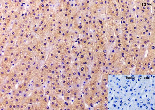
| Sample | Formaldehyde-fixed, paraffin-embedded human liver tissue (datasheet A00565) |
| Fixation | Image formalin-fixed; duration unreported (datasheet A00565); verify before use. |
| Sectioning | 4–5 µm sections on charged slides (standard) |
| Deparaffinisation | Xylene, graded ethanol series to water (standard) |
| Antigen retrieval | Heat retrieval: Citrate pH 6 (datasheet A00565); 20 min, 95–100 °C (standard) |
| Peroxidase block | 3% H2O2, 10 min, room temperature (standard) |
| Blocking | 10% serum (datasheet A00565) |
| Primary antibody | Rabbit anti-TRPV4, 1-2 μg/mL (datasheet A00565) |
| Primary incubation | Overnight at 4 °C (datasheet A00565) |
| Detection | HRP-polymer secondary, DAB chromogen 5–10 min (standard) |
| Counterstain | Hematoxylin, blue, dehydrate and mount (standard) |
| Expected result | TRPV4-positive staining in glandular cells of adrenal gland (HPA tissue IHC: High). HPA tissue profile: Cytoplasmic and membranous expression in several tissues. No signal in the no-primary control. |
TRPV4 is a six-pass membrane protein with cytoplasmic termini; UniProt also places it at apical membranes, adherens junctions, cilia and endoplasmic reticulum (UniProt Q9HBA0 topology; subcellular location). In tissue IHC, expect cytoplasmic and membranous staining in selected cell populations, including adrenal glandular cells, pancreatic endocrine cells and cortical neurons (HPA: tissue IHC). Treat these examples as provisional: HPA rates the tissue staining Uncertain because antibody staining and RNA expression show very low consistency (HPA: reliability).
| Membranous staining, with some cytoplasmic signal, in selected glandular or neuronal cells. | This fits the reported tissue profile and membrane/ER localization (HPA: cytoplasmic and membranous expression; UniProt Q9HBA0 subcellular location). Interpret the pattern alongside controls because HPA rates tissue IHC Uncertain (HPA: reliability). |
| Predominantly nuclear staining, with little signal at the membrane or in cytoplasm. | A nuclear-dominant pattern does not fit the listed TRPV4 locations (UniProt Q9HBA0 subcellular location). Review antibody specificity and detection controls before scoring it as positive (standard IHC practice). |
| Strong signal in a cell population reported as not detected, such as colon glandular cells. | The result conflicts with that HPA tissue example (HPA: colon glandular cells, Not detected). Consider cross-reactivity or endogenous detection activity; HPA's Uncertain rating means the reference pattern is not definitive (HPA: reliability; standard IHC practice). |
| Similar diffuse staining across cells, stroma and the negative control. | This distribution favors background from the staining or detection workflow over cell-restricted TRPV4 signal (standard IHC practice). Compare the primary-antibody omission control and tissue architecture before assigning localization (standard IHC practice). |
| No staining in an adrenal gland section expected to show glandular-cell signal. | HPA reports High staining in adrenal glandular cells, so the run needs review (HPA: adrenal gland, glandular cells, High). Check controls and tissue preservation; absence alone cannot establish TRPV4 loss because HPA rates the IHC evidence Uncertain (HPA: reliability; standard IHC practice). |
| Compartment and epitope orientation | TRPV4 has six transmembrane segments, cytoplasmic N- and C-termini, and extracellular loops (UniProt Q9HBA0 topology). Epitope position is not supplied here, so these data cannot specify an antibody-specific retrieval or permeabilisation condition (UniProt Q9HBA0 topology; standard IHC practice). |
| Intracellular pool | TRPV4 is also listed in the endoplasmic reticulum, where putative tetramers assemble (UniProt Q9HBA0 subcellular location). Cytoplasmic staining can therefore fit the biology, but it requires the same specificity controls as membrane staining (HPA: cytoplasmic and membranous profile; standard IHC practice). |
| Isoforms | UniProt lists six TRPV4 isoforms and reports isoform-specific self-association (UniProt Q9HBA0 isoforms; subunit). Without an antibody epitope or isoform-reactivity record, a tissue pattern cannot be assigned to one isoform (UniProt Q9HBA0 isoforms; standard IHC practice). |
| Reference-pattern confidence | HPA labels antibody HPA007150 IHC Uncertain and reports very low agreement between staining and RNA data (HPA: antibody validation; tissue reliability). Use HPA High and Not detected examples as comparison points, not proof of antibody specificity or universal tissue status (HPA: tissue IHC; reliability). |
| Situation | Likely cause | Next action |
|---|---|---|
| Little or no chromogenic signal in a comparison tissue. | The staining run may have failed, or the chosen section may lack the reported cell population (standard IHC practice). HPA examples remain provisional (HPA: reliability Uncertain). | Verify the detection controls and morphology; inspect the specific HPA-listed cells, such as adrenal glandular cells, before changing IHC conditions (HPA: adrenal gland, glandular cells High; standard IHC practice). |
| Predominantly nuclear signal. | The pattern conflicts with listed membrane, junctional, ciliary and ER locations (UniProt Q9HBA0 subcellular location). | Review primary-antibody omission and antibody-specificity controls; score nuclear-only signal cautiously (standard IHC practice; HPA: IHC Uncertain). |
| Signal appears in colon glandular cells or adipocytes. | HPA lists these cell types as Not detected, though its tissue IHC assessment is Uncertain (HPA: colon glandular cells and adipocytes Not detected; reliability). | Check morphology, antibody specificity and endogenous detection activity before calling the staining TRPV4-positive (standard IHC practice). |
| Diffuse brown background obscures cell boundaries. | Nonspecific binding or endogenous detection activity can obscure compartment assignment (standard IHC practice). | Compare primary-antibody omission controls; review blocking, washes and detection chemistry using standard IHC controls (standard IHC practice). |
| Membrane and cytoplasmic staining vary between sections. | Both compartments are reported for tissue staining, while UniProt also lists an ER pool (HPA: tissue profile; UniProt Q9HBA0 subcellular location). | Score each compartment separately in the same named cell type and compare matched controls; avoid treating membrane-only staining as the sole acceptable pattern (HPA: tissue profile; standard IHC practice). |
| What should an IF/ICC image show? | HPA summarizes TRPV4 as Membrane but provides no ICC-IF image cell lines or main-location detail here (HPA: subcellular summary). | Assess membrane enrichment with compartment and negative controls; use this page's IHC tissue pattern only as context, not an IF/ICC validation claim (HPA: subcellular summary; tissue IHC reliability Uncertain; standard IF practice). |
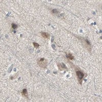


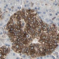
Comprehensive Human Protein Atlas IHC scoring per tissue (reliability: Uncertain — Very low consistency between antibody staining and RNA expression data. Pending external verification.). Rows are taken directly from the HPA tissue chart — click any row's HPA link to view the source.
| Tissue | Cell type | Level | Evidence | Source |
|---|---|---|---|---|
| Adrenal gland | Glandular cells | High | Protein (IHC) | HPA → |
| Cerebral cortex | Neuronal cells | High | Protein (IHC) | HPA → |
| Duodenum | Glandular cells | High | Protein (IHC) | HPA → |
| Hippocampus | Neuronal cells | High | Protein (IHC) | HPA → |
| Pancreas | Pancreatic endocrine cells | High | Protein (IHC) | HPA → |
| Tissue | Cell type | Level | Evidence | Source |
|---|---|---|---|---|
| Adipose tissue | Adipocytes | Not detected | Protein (IHC) | HPA → |
| Bone marrow | Hematopoietic cells | Not detected | Protein (IHC) | HPA → |
| Cervix | Glandular cells | Not detected | Protein (IHC) | HPA → |
| Colon | Glandular cells | Not detected | Protein (IHC) | HPA → |
| Epididymis | Glandular cells | Not detected | Protein (IHC) | HPA → |
Troubleshoot TRPV4 staining in paraffin sections by checking retrieval, tissue controls, cellular localisation and the limits of the available validation evidence.
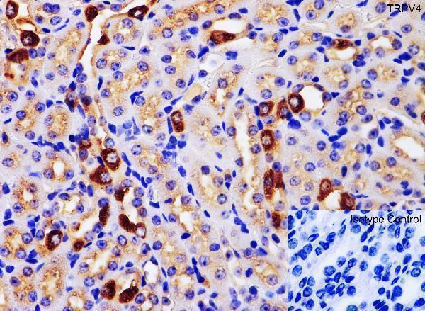
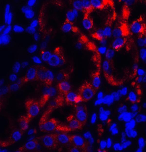
The IHC-validated antibody has paraffin-section images from human liver, mouse kidney and rat kidney, plus an IF image from human kidney (catalog IHC and IF captions).
A00565 is listed for IHC-P and IF in human, mouse and rat samples (catalog applications and reactivity). Its IHC captions show human liver, mouse kidney and rat kidney paraffin sections; its IF caption shows human kidney tissue (catalog image captions).
Which to pick: Choose A00565 for tissue IHC: its own captions document formaldehyde-fixed paraffin sections with citrate pH 6 heat retrieval and 2 μg/mL primary antibody (A00565 IHC captions). Choose A00565 for tissue IF, with human kidney imaged at 5 μg/mL; ICC validation is unreported (A00565 IF caption; catalog applications). For cross-species IHC, A00565 has human, mouse and rat paraffin-section images and is listed as polyclonal (A00565 IHC captions; catalog dilution_raw).