TXN / Thioredoxin · IHC design guide

Design Immunohistochemistry for TXN

Plan paraffin-section TXN IHC around cytoplasmic and nuclear staining, including high signal in kidney tubules and intestinal glandular cells (HPA tissue IHC). Use consistent fixation and score nuclear and cytoplasmic signal separately, since PMA or radiation can shift TXN into nuclei (UniProt).

Evidence assembled Oct 2026 · For research use; verify linked source records and product datasheet before use
Immunohistochemistry protocol sheet for TXN (IHC for TXN): expected localisation Cytoplasmic and nuclear in tissue sections (HPA tissue IHC), antibody A01219, validated IHC image, and IHC protocol steps
Printable TXN IHC protocol sheet — expected localisation Cytoplasmic and nuclear in tissue sections (HPA tissue IHC), antibody A01219, controls and protocol steps. Open the full TXN IHC guide →

TXN Immunohistochemistry Experimental Design Guide

Expected localisation, validated protocols, controls and antibodies — the at-a-glance facts below, then the full design guide.

Must know before staining
Expected localisation Cytoplasmic and nuclear in tissue sections (HPA tissue IHC)
Staining pattern Glandular cells show cytoplasmic and nuclear staining (HPA tissue IHC)
Antigen retrieval Tris-EDTA pH 9.0 HIER, 95–98 °C, 20 min (rule: nuclear antigen)
Positive control ⓘ Duodenum+4 more · see all
Negative control ⓘ Adipose tissue+2 more · see all
Important caveats
Reasons your staining may differ from the expected pattern.
Fixation Keep fixation consistent across sections. (standard IHC practice; not target-specific)
Caveat Leaderless secretion may complicate intracellular scoring (UniProt)
Regulation PMA or radiation shifts TXN toward nuclei (UniProt)
Isoform / epitope 2 isoforms; epitope coverage is unspecified (UniProt)
Section 1

Recommended TXN IHC & IF Protocols

The catalog antibody protocol is followed by published TXN IHC protocols for kidney and salivary adenoid cystic carcinoma sections (PMC11256699; PMC4694848).

Recommended immunohistochemistry (IHC-P) protocol parameters
SampleParaffin-embedded H. esophagus tissue; fixative not specified (datasheet A01219)
FixationImage fixative and duration unreported (datasheet A01219); verify before use.
Sectioning4–5 µm sections on charged slides (standard)
DeparaffinisationXylene, graded ethanol series to water (standard)
Antigen retrievalHeat-induced epitope retrieval in Tris-EDTA buffer, pH 9.0, 20 min at 95–98 °C (standard rule: nuclear antigen)
Peroxidase block3% H2O2, 10 min, room temperature (standard)
Blocking10% normal serum of the secondary host, 30 min, room temperature (standard)
Primary antibodyRabbit anti-TXN, 1:25 (datasheet A01219)
Primary incubationOvernight at 4 °C (standard)
DetectionHRP-polymer secondary, DAB chromogen 5–10 min (standard)
CounterstainHematoxylin, blue, dehydrate and mount (standard)
Expected resultTXN-positive staining in glandular cells of duodenum (HPA tissue IHC: High). HPA tissue profile: General cytoplasmic and nuclear expression. No signal in the no-primary control.
💡Decision noteStart with Tris-EDTA pH 9.0, 95–98 °C for 20 min (page retrieval specification); both published protocols used citrate pH 6.0 (PMC11256699; PMC4694848).
Section 2

What Is the Expected TXN Staining Pattern?

TXN is cytoplasmic and nuclear, with no membrane span (UniProt P10599). Expect strong staining in kidney tubule and intestinal glandular cells (HPA tissue IHC: High). HPA rates tissue IHC reliability Enhanced, with medium agreement between staining and RNA data (HPA tissue IHC).

What am I looking at on my slide?
Strong cytoplasmic and nuclear staining in kidney tubulesExpected: tubule cells are High (HPA tissue IHC); both compartments fit TXN (UniProt P10599).
Membrane-only rim stainingSuspect artefact: TXN has no membrane span (UniProt P10599 topology).
Strong adipocyte stainingDiscordant: adipocytes are Not detected (HPA tissue IHC); assess off-target or endogenous signal.
Uniform brown haze across cells and blank areasLikely background; inspect blocking, washes and detection controls (general IHC practice).
No staining in kidney tubulesUnexpected: tubule cells are High (HPA tissue IHC); check the IHC workflow.
💡Expected TXN appearancePositive: strong cytoplasmic and nuclear staining in kidney tubule cells (HPA tissue IHC: High; general cytoplasmic and nuclear expression); membrane-only rims or strong adipocyte signal are suspect (UniProt P10599 topology; HPA tissue IHC: adipocytes Not detected).
How each factor affects the staining
Cellular locationCytoplasm and nucleus are supported; TXN lacks a membrane span (UniProt P10599).
Stimulus-dependent locationPMA or radiation can shift TXN toward nuclei (UniProt P10599; PubMed:9108029, 11118054).
Antibody validationHPA047478, HPA055752 and CAB008678 are IHC Enhanced (HPA antibody validation).
IF/ICC Q&A: expected location?Mainly nucleoplasm and cytosol (HPA ICC-IF); consult the separate IF/ICC guide.
Chromogenic detectionEndogenous enzyme activity can mimic staining (general IHC practice).
Why is my staining missing, weak or wrong?
SituationLikely causeNext action
Membrane rim dominatesTXN lacks a membrane span (UniProt P10599 topology).Check specificity with a matched negative control (general IHC practice).
Strong adipocyte stainingAdipocytes are Not detected (HPA tissue IHC); off-target or endogenous signal is possible.Run no-primary and detection-only controls (general IHC practice).
Diffuse brown hazeExcess primary or incomplete blocking is possible (general IHC practice).Titrate primary; improve blocking and washes (general IHC practice).
No signal in kidney tubulesTubule cells are High (HPA tissue IHC); a workflow failure is possible.Check positive control, retrieval and detection (general IHC practice).
Nuclear-only patternTXN spans both compartments (HPA tissue IHC); stimuli can favor nuclei (UniProt P10599).Compare matched conditions and inspect cytoplasmic signal (general IHC practice).

Sample controls for TXN IHC & IF

🧪Run kidney first: tubular cells should stain for TXN (HPA: High in kidney cells in tubules). Run adipose tissue as the negative tissue because adipocytes are listed as not detected (HPA: Not detected in adipocytes); on the kidney slide, unstained neighboring cells can help judge background but are not established TXN-negative cells (HPA: kidney annotation identifies tubular cells only).
Positive control tissue: Duodenum (Glandular cells, HPA High)
Negative control tissue: Adipose tissue (HPA Not detected)
ICC-IF cell lines (HPA subcellular resource): HPA ICC-IF images show TXN in U-251MG, A-431, U2OS, NIH 3T3, with annotated localisation: Nucleoplasm (enhanced), Cytosol (enhanced) (HPA subcellular).
Technical controls: Include no-primary (secondary-only), a host-species- and clonality-matched isotype control, and TXN-knockout material or an epitope-peptide block where available (standard IHC practice). For chromogenic kidney IHC, quench endogenous peroxidase and check for endogenous biotin if using biotin-based detection, since either can produce background (standard IHC practice).
⚠️Feasibility: A TXN-specific fixation window or fixation effect is unreported in the supplied evidence, and the selected A01219 paraffin-section caption does not state its fixative (A01219 caption). Retrieval dependence is likewise unreported; compare retrieved and untreated sections when optimizing the IHC assay (standard IHC practice). The supplied evidence does not establish whether frozen sections or IF are easier for TXN; kidney tubular staining warrants particular attention to endogenous biotin when using biotin-based detection (HPA: High in kidney cells in tubules; standard IHC practice).

HPA tissue IHC evidence for TXN

Comprehensive Human Protein Atlas IHC scoring per tissue (reliability: Enhanced — Medium consistency between antibody staining and RNA expression data.). Rows are taken directly from the HPA tissue chart — click any row's HPA link to view the source.

Positive expression · recommended positive controls

TissueCell typeLevelEvidenceSource
Duodenum Glandular cells High Protein (IHC) HPA →
Fallopian tube Ciliated cells (cell body) High Protein (IHC) HPA →
Gallbladder Glandular cells High Protein (IHC) HPA →
Kidney Cells in tubules High Protein (IHC) HPA →
Placenta Decidual cells High Protein (IHC) HPA →

Undetected expression · recommended negative controls

TissueCell typeLevelEvidenceSource
Adipose tissue Adipocytes Not detected Protein (IHC) HPA →
Bone marrow Hematopoietic cells Not detected Protein (IHC) HPA →
Parathyroid gland Glandular cells Not detected Protein (IHC) HPA →
Section 3

Advanced TXN IHC Tips

Troubleshoot TXN staining in paraffin sections by checking retrieval, cell compartment, controls and scoring before interpreting changes in signal.

What should I change if TXN staining is weak after antigen retrieval?
Start with Tris-EDTA at pH 9.0, heated to 95–98 °C for 20 min (page retrieval setting). Use the same cooling period and wash sequence across slides so differences in staining can be attributed to the specimen or antibody rather than retrieval variation (general IHC practice). If signal remains weak, compare a longer or shorter heating period on serial sections while holding detection conditions constant (general IHC practice). Include a section with expected glandular-cell staining, such as duodenum, and check both cytoplasmic and nuclear signal before deciding retrieval failed (HPA: High in duodenal glandular cells; HPA: general cytoplasmic and nuclear expression).
Could fixation explain inconsistent TXN staining between paraffin sections?
Target-specific sensitivity to fixation is unknown: the selected paraffin-section caption does not report a fixative (A01219 tissue-IHC caption). Record the actual fixative, fixation duration and processing history for each specimen before comparing staining intensity across blocks (general IHC practice). If sections from one block stain weakly, compare serial sections using the same pH 9.0 retrieval and detection run; do not assign the difference to fixation without that comparison (page retrieval setting; general IHC practice). Examine tissue preservation and staining in an expected positive cell population alongside the test section, because poor morphology or a failed run can also complicate interpretation (HPA: High in duodenal glandular cells; general IHC practice).
Should TXN appear in nuclei, cytoplasm, or both?
Score cytoplasmic and nuclear staining separately because both are expected locations for TXN (HPA: general cytoplasmic and nuclear expression; UniProt P10599 subcellular location). The mainly nucleoplasmic and cytosolic pattern reported by cell imaging can help assess compartment plausibility, but tissue-section scoring should rely on visible cell boundaries and nuclei (HPA: enhanced nucleoplasm and cytosol; general IHC practice). A change toward nuclear staining can be biologically plausible after radiation or PMA exposure, so document treatment history before comparing compartments (UniProt P10599, PubMed:11118054; PubMed:9108029). Use the same counterstain and scoring rules across sections, and inspect any diffuse extracellular deposit cautiously because TXN can also be secreted (UniProt P10599, PubMed:1332947; general IHC practice).
Can the catalog antibody distinguish TXN isoforms or modified epitopes?
The selected antibody is described as C-terminal, but its precise binding residues and isoform recognition are not supplied (A01219 tissue-IHC caption). TXN has 2 annotated isoforms and several modified residues, including acetylated lysines and nitrosylated cysteines, so total staining cannot establish which molecular form is present (UniProt P10599 isoforms and modified residues). Compare staining with an independent antibody whose epitope is documented, if available, before assigning an isoform-specific pattern (general IHC practice). Keep retrieval, dilution and detection matched for that comparison, and describe any observed difference as antibody-dependent until epitope recognition has been independently established (page retrieval setting; general IHC practice).
How should I adapt TXN localisation checks to multiplex IF?
For the separate IF/ICC workflow, pair TXN with a marker that identifies the cell population being assessed, such as glandular cells in a duodenal section, and score the signals within the same cells (HPA: High in duodenal glandular cells; general IF practice). Choose a spectrally separated fluorophore, preferably in a low-autofluorescence channel established from an unstained section, and include single-label controls for bleed-through (general IF practice). TXN is expected in cytosol and nucleoplasm and has no transmembrane segment, so use permeabilisation suitable for intracellular epitopes and verify that nuclear access is adequate (HPA: enhanced nucleoplasm and cytosol; UniProt P10599 topology; general IF practice). Do not transfer the paraffin-section antibody dilution of 1:25 directly to IF without titration (A01219 tissue-IHC caption; general IF practice).
How can I distinguish TXN staining from chromogenic background?
Inspect a no-primary control and a matched section with expected low signal before calling faint DAB deposits positive (general IHC practice; HPA: adipocytes in adipose tissue Not detected). Apply a peroxidase block before DAB development and assess whether residual colour follows blood-rich areas or tissue edges rather than cell boundaries (general chromogenic IHC practice). The selected image used a biotinylated secondary and DAB, so check secondary-only staining when reproducing that detection route (A01219 tissue-IHC caption; general IHC practice). Titrate the primary around the caption's 1:25 dilution and keep development time constant; widespread granular colour without plausible cytoplasmic or nuclear distribution warrants further optimisation (A01219 tissue-IHC caption; HPA: general cytoplasmic and nuclear expression; general IHC practice).
What is a defensible way to quantify TXN in tissue sections? ⚠ ANSWER MARKED FOR VERIFICATION
Define the cell population and score nuclear and cytoplasmic DAB separately, because TXN is reported in both compartments (HPA: general cytoplasmic and nuclear expression; general IHC practice). Report the percentage of positive viable cells and an H-score from 0–300 when intensity categories are reproducible; use positive-cell density per mm² if cell counts vary strongly between fields (general IHC practice). Normalise each measure to the viable area or total cells of the same annotated compartment, and exclude necrotic or damaged regions by a preset rule (general IHC practice). Hold retrieval, antibody dilution, counterstain, imaging and scoring thresholds constant across comparison groups (page retrieval setting; general IHC practice).
When should apparent TXN positivity be treated as an artefact?
A credible cellular pattern includes cytoplasmic or nuclear signal within identifiable cells, consistent with the reported tissue and subcellular distributions (HPA: general cytoplasmic and nuclear expression; HPA: enhanced nucleoplasm and cytosol). Treat intense staining confined to section edges, necrotic zones or damaged folds as suspect and compare it with intact tissue on the same slide (general IHC practice). Check unexpected strong staining in populations reported as Not detected, such as adipocytes, against no-primary and secondary-only controls before calling it TXN (HPA: adipocytes in adipose tissue Not detected; general IHC practice). Residual endogenous peroxidase can produce DAB colour, so confirm the peroxidase block and require reproducible compartment-specific staining across serial sections before interpreting a biological change (general chromogenic IHC practice).
Boster reagents

Best TXN / Thioredoxin IHC Antibodies

Human TXN IHC is illustrated in paraffin sections (A01219 image captions); ICC/IF is listed for a second antibody, without an IF figure (M01219 applications and image inventory).

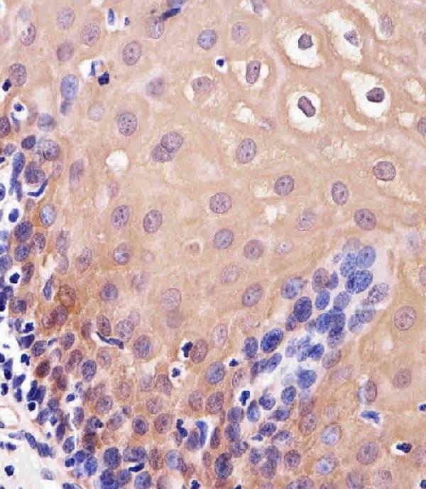
Real IHC data Immunohistochemical analysis of paraffin-embedded H. esophagus section using TXN Antibody (C-term) (Cat#A01219). A01219 was diluted at 1:25 dilution. A undiluted biotinylated goat polyvalent antibody was used as the secondary, followed by DAB staining.
Anti-TXN Antibody (C-term)
Cat # A01219

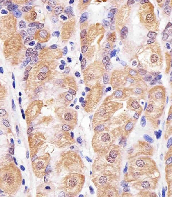
A01219 has DAB IHC images of paraffin-embedded human esophagus and stomach (A01219 image captions). M01219 is listed for human IHC and ICC/IF, but has no IHC or IF image in the payload (M01219 applications, reactivity and image inventory).

Which to pick: For tissue IHC, choose A01219: its own paraffin-section caption documents DAB staining at 1:25; the fixative is unreported (A01219 image caption). For IF/ICC, M01219 is the listed option, with a 1:50–1:200 range but no supplied IF figure (M01219 applications, dilution and image inventory). Neither SKU supports a cross-species recommendation because both list human reactivity only (A01219 and M01219 reactivity).

Each figure is that product's own IHC / IF validation image from its datasheet.

References

  1. UniProt Consortium. UniProt entry P10599 (THIO_HUMAN, Thioredoxin).
  2. Human Protein Atlas. TXN tissue IHC expression (reliability: Enhanced).
  3. Human Protein Atlas. TXN subcellular location (ICC-IF): Mainly localized to the nucleoplasm and cytosol..
  4. Human Protein Atlas. TXN antibody validation summary (3 antibodies).
  5. Identification of thioredoxin-1 as a biomarker of lung cancer and evaluation of its prognostic value based on bioinformatics analysis. Frontiers in oncology 2023 — PMC9911911.
  6. Increased level of TXNIP and nuclear translocation of TXN is associated with end stage renal disease and development of multiplex renal tumours. BMC nephrology 2024 — PMC11256699.
  7. Correlation of five secretory proteins with the nasopharyngeal carcinoma metastasis and the clinical applications. Oncotarget 2017 — PMC5438738.
  8. Thioredoxin 1 mediates TGF-β-induced epithelial-mesenchymal transition in salivary adenoid cystic carcinoma. Oncotarget 2015 — PMC4694848.
  9. PubMed PMID:3170595 — UniProt-cited evidence.
  10. PubMed PMID:2785919 — UniProt-cited evidence.
  11. PubMed PMID:1874447 — UniProt-cited evidence.