U2AF2 / Splicing factor U2AF 65 kDa subunit · IHC design guide

Design Immunohistochemistry for U2AF2

Plan paraffin-section U2AF2 IHC around its ubiquitous nuclear pattern (HPA tissue IHC). Use high-staining glandular cells or kidney tubules to assess nuclear signal, while accounting for lower staining in glial cells and myocytes (HPA tissue IHC).

Evidence assembled Oct 2026 · For research use; verify linked source records and product datasheet before use
Immunohistochemistry protocol sheet for U2AF2 (IHC for U2AF2): expected localisation Ubiquitous nuclear staining (HPA tissue IHC), antibody M03639-1, validated IHC image, and IHC protocol steps
Printable U2AF2 IHC protocol sheet — expected localisation Ubiquitous nuclear staining (HPA tissue IHC), antibody M03639-1, controls and protocol steps. Open the full U2AF2 IHC guide →

U2AF2 Immunohistochemistry Experimental Design Guide

Expected localisation, validated protocols, controls and antibodies — the at-a-glance facts below, then the full design guide.

Must know before staining
Expected localisation Ubiquitous nuclear staining (HPA tissue IHC)
Staining pattern Nuclear staining across many tissue cell types (HPA tissue IHC)
Antigen retrieval EDTA pH 8.0 HIER, heat-mediated (datasheet M03639-1)
Positive control ⓘ Adrenal gland+4 more · see all
Negative control ⓘ None in HPA (detected in all 45 tissues); use no-primary + isotype controls
Important caveats
Reasons your staining may differ from the expected pattern.
Fixation Keep fixation consistent across sections. (standard IHC practice; not target-specific)
Caveat Glial cells and myocytes may stain weakly (HPA tissue IHC)
Regulation Low tissue specificity (HPA tissue RNA)
Isoform / epitope Two isoforms; epitope coverage is unspecified (UniProt; datasheets)
Section 1

Recommended U2AF2 IHC & IF Protocols

Compare the catalog antibody’s IHC-P protocol (datasheet M03639-1) with three published U2AF2 IHC methods (PMC12924209; PMC7487667; PMC5033661).

Recommended immunohistochemistry (IHC-P) protocol parameters
SampleParaffin-embedded human gallbladder adenocarcinoma tissue; fixative not specified (datasheet M03639-1)
FixationImage fixative and duration unreported (datasheet M03639-1); verify before use.
Sectioning4–5 µm sections on charged slides (standard)
DeparaffinisationXylene, graded ethanol series to water (standard)
Antigen retrievalHeat retrieval: EDTA pH 8.0 (datasheet M03639-1); 20 min, 95–100 °C (standard)
Peroxidase block3% H2O2, 10 min, room temperature (standard)
Blocking10% goat serum (datasheet M03639-1)
Primary antibodyMouse monoclonal (clone 3G9) anti-U2AF2, 2μg/ml (datasheet M03639-1)
Primary incubationOvernight at 4 °C (datasheet M03639-1)
DetectionStreptavidin-biotin complex (SABC), DAB chromogen (datasheet M03639-1)
CounterstainHematoxylin, blue, dehydrate and mount (standard)
Expected resultU2AF2-positive staining in glandular cells of adrenal gland (HPA tissue IHC: High). HPA tissue profile: Ubiquitous nuclear expression. No signal in the no-primary control.
💡Decision noteStart with heat-mediated EDTA pH 8.0 retrieval for the catalog antibody (datasheet M03639-1). The aortic study does not specify its retrieval solution (PMC12924209).
Section 2

What Is the Expected U2AF2 Staining Pattern?

U2AF2 is a nuclear protein without a transmembrane segment (UniProt P26368: subcellular location and topology). In paraffin-section IHC, expect nuclear staining across many cell types, with high staining reported in colon glandular cells, kidney tubule cells and lymph-node germinal center cells (HPA: ubiquitous nuclear expression; High in those cells). HPA rates its tissue IHC profile Supported, with medium consistency between antibody staining and RNA expression (HPA: tissue IHC reliability).

What am I looking at on my slide?
Clear nuclear chromogen in colon glandular cells or kidney tubule cells, with discernible nuclear boundaries.This fits the reported compartment and high-staining cell populations (HPA: ubiquitous nuclear expression; High in colon glandular cells and kidney tubule cells). Compare intensity within the same run; a strong nucleus in one section is not an absolute intensity standard across preparations (general IHC practice).
Predominantly cytoplasmic or membranous signal, with little nuclear staining.This conflicts with the expected nuclear distribution (UniProt P26368: Nucleus; HPA: ubiquitous nuclear expression). Review antibody specificity and staining controls before interpreting it as U2AF2; the supplied sources do not establish a cytoplasmic IHC pattern (HPA: tissue IHC profile).
Strong signal in skeletal myocytes, smooth muscle cells or breast adipocytes, especially when expected high-staining cells are weak.HPA lists these populations as Low, not negative (HPA: Low in skeletal myocytes, smooth muscle cells and breast adipocytes). Unexpected dominance could reflect cross-reactivity or endogenous detection activity; inspect controls before assigning a cause (general IHC practice).
Chromogen appears broadly over tissue, including spaces between cells, so nuclei cannot be distinguished.Diffuse background cannot establish the reported nuclear pattern (HPA: ubiquitous nuclear expression). Check the no-primary control, blocking and detection conditions; these checks assess general IHC background and do not identify a U2AF2-specific mechanism (general IHC practice).
Little or no nuclear signal in colon glandular cells while other staining controls perform as expected.This is discordant with a reported high-staining population (HPA: High in colon glandular cells). Recheck the antibody's IHC-P instructions and run controls. A single negative section cannot establish absence of U2AF2, and HPA supplies no negative tissue reference (HPA: tissue IHC profile).
💡Expected U2AF2 appearanceCall a result positive when nuclear chromogen is clear in a reported high-staining population such as colon glandular cells or kidney tubule cells (HPA: High in those cells; ubiquitous nuclear expression); predominantly cytoplasmic, membranous or diffuse field-wide color is suspect (UniProt P26368: Nucleus; general IHC practice).
How each factor affects the staining
Compartment and tissue context (UniProt P26368: Nucleus; HPA: tissue IHC profile).U2AF2 has no transmembrane segment (UniProt P26368: topology). Score nuclear signal in identifiable cells; HPA's ubiquitous nuclear profile makes a completely unstained tissue a poor assumed negative control (HPA: ubiquitous nuclear expression; no negative tissue listed).
Relative staining levels (HPA: tissue IHC profile).HPA reports High staining in adrenal, colon, duodenal, epididymal, fallopian-tube and gallbladder glandular cells, kidney tubule cells, and lymph-node germinal center cells (HPA: High). Glial cells in hippocampus and caudate and several other listed populations are Low, not proven absent (HPA: Low).
Antibody evidence (HPA: antibody validation).HPA043562 and CAB010910 are each listed as IHC Supported; HPA041943 has no IHC status in the supplied list (HPA: antibody validation). The tissue profile is Supported with medium RNA–staining consistency, so resolve conflicting slides with controls rather than treating the profile as absolute (HPA: tissue IHC reliability).
Isoforms and antibody recognition (UniProt P26368: isoforms and domains).UniProt lists two isoforms and three RNA recognition motifs, at residues 149–231, 259–337 and 385–466 (UniProt P26368: isoforms; RRM domains). The payload does not map any listed antibody's epitope, so isoform coverage and epitope-specific interpretation remain undetermined (HPA: supplied antibody list).
Processing and modifications (UniProt P26368: processing and modified residues).The supplied record lists no signal peptide or propeptide, a chain spanning residues 2–475, and several modified residues (UniProt P26368: processing; modified residues). It supplies no evidence that these features change paraffin-section staining or antigen retrieval; use the catalog antibody's IHC-P instructions for its workflow (general IHC practice).
IF/ICC question: what should fluorescence look like? (HPA: subcellular ICC-IF).HPA supports nucleoplasmic and nuclear-speckle localization in ICC-IF and additionally lists a cytokinetic bridge (HPA: subcellular ICC-IF). That cellular detail informs interpretation, but does not establish a speckled pattern in chromogenic paraffin-section IHC (HPA: subcellular ICC-IF; tissue IHC profile).
Why is my staining missing, weak or wrong?
SituationLikely causeNext action
The expected high-staining population lacks nuclear color (HPA: High in colon glandular cells).Assay failure or weak detection is possible; HPA does not establish target-specific fixation sensitivity (HPA: tissue IHC profile; general IHC practice).Check a reported high-staining control, the run's detection controls and the catalog antibody's IHC-P instructions before interpreting the section (HPA: High in colon glandular cells; general IHC practice).
Nuclei look weak, but the whole section has a light haze (HPA: expected nuclear profile).Background may obscure true nuclear contrast; its source is not determined by the U2AF2 profile (HPA: tissue IHC profile; general IHC practice).Inspect no-primary and detection controls; review blocking, washes and chromogen development under the established IHC workflow (general IHC practice).
Signal is mainly cytoplasmic or outlines cell borders (UniProt P26368: Nucleus).The distribution disagrees with both UniProt nuclear localization and HPA nuclear tissue staining (UniProt P26368: Nucleus; HPA: ubiquitous nuclear expression).Compare a reported high-staining tissue and a no-primary control; reassess specificity before scoring this signal as U2AF2 (HPA: High in colon glandular cells; general IHC practice).
Low-listed cells stain more strongly than nearby high-listed cells (HPA: tissue IHC levels).Cross-reactivity or endogenous detection activity is possible, but this contrast alone does not prove either cause (HPA: Low and High populations; general IHC practice).Check no-primary and detection controls, then compare nuclear localization and cell identity across the same section (HPA: ubiquitous nuclear expression; general IHC practice).
Different IHC runs give conflicting nuclear intensity (HPA: tissue IHC reliability).Run variation is possible; HPA reports medium consistency between antibody staining and RNA expression, not an invariant intensity scale (HPA: tissue IHC reliability; general IHC practice).Compare matched controls and scoring criteria across runs; follow the catalog antibody's IHC-P instructions for dilution and retrieval rather than inferring settings from HPA (general IHC practice).
A nucleus lacks visible speckles in paraffin-section IHC (HPA: ICC-IF nuclear speckles).Speckles are supported in HPA ICC-IF; the supplied tissue IHC profile specifies ubiquitous nuclear expression without a speckle requirement (HPA: subcellular ICC-IF; tissue IHC profile).Judge chromogenic IHC against nuclear localization in reported high-staining cells; consult the separate IF/ICC guide when assessing fluorescence detail (HPA: tissue IHC profile; subcellular ICC-IF).

Sample controls for U2AF2 IHC & IF

🧪Run colon first and assess its glandular cells for nuclear staining; HPA scores them High (HPA: Colon, glandular cells, High; UniProt: nucleus). HPA detects U2AF2 in all 45 scored tissues, so use no-primary and isotype controls for the negative comparison; no cell type on the positive slide is established as an internal negative, though cells without signal should lack nuclear DAB (HPA: no negative tissue rows; standard IHC interpretation).
Positive control tissue: Adrenal gland (Glandular cells, HPA High)
Negative control tissue: None in HPA: U2AF2 is detected in all 45 scored tissues. Use a no-primary (secondary-only) and an isotype control instead.
ICC-IF cell lines (HPA subcellular resource): HPA ICC-IF images show U2AF2 in A-431, U-251MG, U2OS, HEK293, MCF-7, NIH 3T3, with annotated localisation: Nucleoplasm (supported), Nuclear speckles (supported) (HPA subcellular).
Technical controls: Include a no-primary, secondary-only slide and a species- and class-matched nonimmune mouse IgG isotype control; use U2AF2 knockout material as a biological specificity control if available (caption: mouse primary antibody; standard IHC controls). Block endogenous peroxidase and check endogenous biotin background in colon sections when using the caption’s biotin-based SABC/DAB detection (caption: SABC/DAB; standard IHC practice).
⚠️Feasibility: No target-specific fixation window or fixation effect is reported, and the selected M03639-1 paraffin-section caption does not state a fixative (caption: fixative unreported). The caption uses heat retrieval in EDTA at pH 8.0, but does not establish that retrieval is required (caption: EDTA retrieval). HPA ICC-IF images support nuclear localisation in cultured cells, but the supplied evidence does not show that IF or frozen sections are easier; in colon, assess glandular staining against possible endogenous peroxidase or biotin background with the stated controls (HPA: nucleoplasm and nuclear speckles; HPA: colon glandular cells; standard IHC practice).

HPA tissue IHC evidence for U2AF2

Comprehensive Human Protein Atlas IHC scoring per tissue (reliability: Supported — Medium consistency between antibody staining and RNA expression data.). Rows are taken directly from the HPA tissue chart — click any row's HPA link to view the source.

Positive expression · recommended positive controls

TissueCell typeLevelEvidenceSource
Adrenal gland Glandular cells High Protein (IHC) HPA →
Colon Glandular cells High Protein (IHC) HPA →
Duodenum Glandular cells High Protein (IHC) HPA →
Epididymis Glandular cells High Protein (IHC) HPA →
Fallopian tube Glandular cells High Protein (IHC) HPA →

Undetected expression · recommended negative controls

TissueCell typeLevelEvidenceSource
None in HPA: U2AF2 is detected in all 45 scored tissues. Use a no-primary (secondary-only) and an isotype control instead.
Section 3

Advanced U2AF2 IHC Tips

Troubleshoot U2AF2 staining in paraffin sections by checking retrieval, nuclear localisation, detection background, and compartment specific scoring.

What should I change if U2AF2 nuclear staining is weak after retrieval?
Start with heat mediated retrieval in EDTA buffer at pH 8.0 for paraffin sections (datasheet M03639-1). The catalog antibody detected U2AF2 in a paraffin section after this retrieval, followed by 2 µg/mL primary antibody overnight at 4°C (datasheet M03639-1). If nuclei remain weak, compare retrieval heating and cooling conditions on adjacent sections while keeping antibody concentration and detection constant (standard IHC practice). Check whether a known positive compartment stains before increasing retrieval intensity: U2AF2 is nuclear, and glandular cells in gallbladder show high staining in the tissue atlas (UniProt P26368 localisation; HPA: High in gallbladder glandular cells). Excessive retrieval can damage morphology and make nuclear scoring unreliable (standard IHC practice).
Could fixation explain weak or uneven U2AF2 staining?
The selected paraffin section caption does not report a fixative, so target specific fixation sensitivity is unknown (datasheet M03639-1: fixative not stated). Record the fixative and fixation duration for each specimen, then compare sections with similar processing histories before changing the antibody concentration (standard IHC practice). If fixation varies, test adjacent sections using the documented EDTA retrieval at pH 8.0 and the same 2 µg/mL primary concentration (datasheet M03639-1; standard IHC practice). Judge any improvement by nuclear signal and preserved morphology together, since damaged or poorly preserved nuclei are difficult to score (UniProt P26368 localisation; standard IHC practice). Neither the atlas staining pattern nor the protein’s topology establishes a fixation effect for this antibody (HPA: tissue IHC profile; UniProt P26368 topology).
Where should convincing U2AF2 staining appear in paraffin sections?
Expect staining predominantly in nuclei: U2AF2 is annotated as nuclear, and tissue IHC shows a ubiquitous nuclear pattern (UniProt P26368 localisation; HPA: tissue IHC profile). Subcellular imaging further supports nucleoplasm and nuclear speckles, although small speckles may be difficult to resolve with chromogenic section microscopy (HPA: subcellular localisation; standard IHC practice). Compare the DAB signal with a nuclear counterstain and score nuclear staining separately from cytoplasmic deposits (standard IHC practice). Gallbladder glandular cells provide a documented high staining reference, while smooth muscle cells are listed as low (HPA: gallbladder glandular cells High; smooth muscle cells Low). Diffuse cytoplasmic signal without corresponding nuclei warrants a background check before biological interpretation (UniProt P26368 localisation; standard IHC practice).
Can this IHC stain distinguish U2AF2 isoforms or modified epitopes?
Do not assign isoform identity from this chromogenic stain alone: the record lists 2 U2AF2 isoforms, but the supplied caption does not identify this antibody’s epitope (UniProt P26368 isoforms; datasheet M03639-1). The protein contains 3 RNA recognition motifs at residues 149–231, 259–337, and 385–466 (UniProt P26368 domains). Its annotated modified residues include phosphorylation, acetylation, and hydroxylation, but their effects on this antibody’s staining are untested here (UniProt P26368 modified residues; datasheet M03639-1). If samples differ unexpectedly, request the epitope specification and assess whether it is shared by the isoforms before making an isoform claim (standard IHC practice). Keep retrieval and detection matched across sections so processing differences do not masquerade as epitope differences (standard IHC practice).
How can IF help assess a questionable U2AF2 IHC pattern?
Use IF as a separate localisation check, with a nuclear counterstain and a marker for the cell population being compared, such as glandular epithelial cells in gallbladder (HPA: High in gallbladder glandular cells; standard IF practice). Expect nucleoplasmic signal with possible nuclear speckles (HPA: supported nucleoplasm and nuclear speckles). Choose a fluorophore channel with low tissue autofluorescence and inspect a no primary control in that channel before interpreting faint signal (standard IF practice). Because U2AF2 is nuclear and has no transmembrane segment, optimise permeabilisation for access to a nuclear epitope without assuming a membrane facing side (UniProt P26368 localisation and topology; standard IF practice). The supplied paraffin IHC caption establishes no IF fixation or permeabilisation condition for this antibody (datasheet M03639-1).
How do I separate nuclear U2AF2 signal from chromogenic background?
The documented detection used biotinylated goat anti mouse IgG, an avidin–biotin complex, and DAB after 2 µg/mL primary antibody overnight at 4°C (datasheet M03639-1). Run no primary and secondary only controls to identify detection background, and include a peroxidase block when using DAB (standard IHC practice). If staining persists without primary antibody, investigate endogenous enzyme activity and, where relevant, endogenous biotin before changing retrieval (standard IHC practice). Compare the signal with counterstained nuclei because U2AF2 has a documented nuclear pattern (UniProt P26368 localisation; HPA: ubiquitous nuclear expression). Diffuse surface deposits or staining concentrated at section edges should trigger a review of washing, drying, and detection conditions (standard IHC practice).
How should I quantify U2AF2 staining across tissue sections? ⚠ ANSWER MARKED FOR VERIFICATION
Define the cell compartment before scoring and count nuclear positive cells within that compartment, excluding damaged tissue and section edges (UniProt P26368 localisation; standard IHC practice). Report either the percentage of positive nuclei or a nuclear H-score calculated as the sum of percentages at intensity grades 0–3, giving a 0–300 scale (standard IHC practice). Normalise counts to the number of evaluable nuclei in each region, or report positive nuclei per mm² when cell density is the question (standard IHC practice). Analyse glandular and other cell populations separately because HPA reports high staining in gallbladder glandular cells and low staining in smooth muscle cells (HPA: cell specific tissue IHC levels). Keep retrieval, DAB development, counterstaining, and imaging settings consistent across compared sections (standard IHC practice).
When is an apparent U2AF2 positive IHC result likely artefactual?
Give greatest weight to signal within intact nuclei because U2AF2 is nuclear and the tissue atlas describes ubiquitous nuclear expression (UniProt P26368 localisation; HPA: tissue IHC profile). Interpret isolated cytoplasmic staining cautiously, especially when adjacent nuclei are negative, and check a no primary control (UniProt P26368 localisation; standard IHC practice). Compare cell identity as well as intensity: high staining is documented in gallbladder glandular cells, whereas smooth muscle cells are listed as low (HPA: gallbladder glandular cells High; smooth muscle cells Low). Exclude necrotic areas and section edges where morphology or reagent distribution can distort interpretation (standard IHC practice). With the documented biotin based DAB method, persistent control staining also calls for checks of endogenous peroxidase and biotin related background (datasheet M03639-1; standard IHC practice).
Boster reagents

Best U2AF2 / Splicing factor U2AF 65 kDa subunit IHC Antibodies

Three anti-U2AF2 antibodies have paraffin-section IHC images (catalog IHC captions); each also lists IF/ICC and human, mouse, and rat reactivity (catalog applications/reactivity).

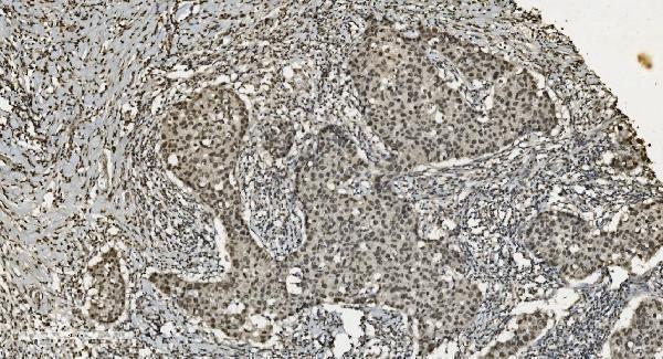
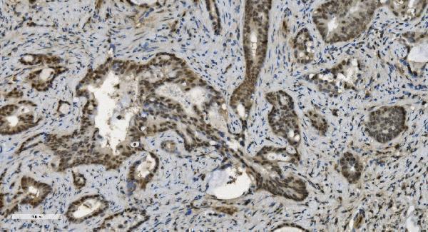
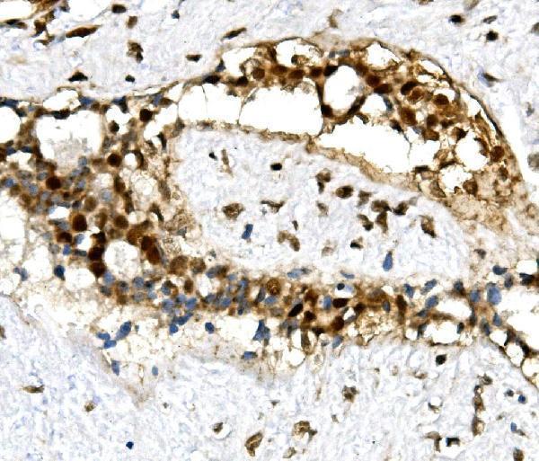
Real IHC data IHC analysis of U2AF65/U2AF2 using anti U2AF65/U2AF2 antibody (M03639-1). U2AF65/U2AF2 was detected in paraffin-embedded section of human gallbladder adenocarcinoma tissue. Heat mediated antigen retrieval was performed in EDTA buffer (pH8.0, epitope retrieval solution). The tissue section was blocked with 10% goat serum. The tissue section was then incubated with 2μg/ml mouse anti-U2AF65/U2AF2 Antibody (M03639-1) overnight at 4°C. Biotinylated goat anti-mouse IgG was used as secondary antibody and incubated for 30 minutes at 37°C. The tissue section was developed using Strepavidin-Biotin-Complex (SABC) (Catalog # SA1021) with DAB as the chromogen.
Anti-U2AF65/U2AF2 ® Antibody (monoclonal, 3G9)
Cat # M03639-1
Real IHC data IHC analysis of U2AF65/U2AF2 using anti U2AF65/U2AF2 antibody (M03639). U2AF65/U2AF2 was detected in paraffin-embedded section of human gallbladder adenocarcinoma tissue. Heat mediated antigen retrieval was performed in EDTA buffer (pH8.0, epitope retrieval solution). The tissue section was blocked with 10% goat serum. The tissue section was then incubated with 2μg/ml mouse anti-U2AF65/U2AF2 Antibody (M03639) overnight at 4°C. Biotinylated goat anti-mouse IgG was used as secondary antibody and incubated for 30 minutes at 37°C. The tissue section was developed using Strepavidin-Biotin-Complex (SABC) (Catalog # SA1021) with DAB as the chromogen.
Anti-U2AF65/U2AF2 ® Antibody (monoclonal, 10F4)
Cat # M03639
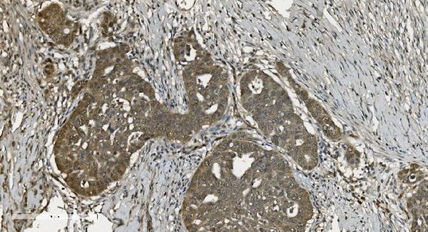
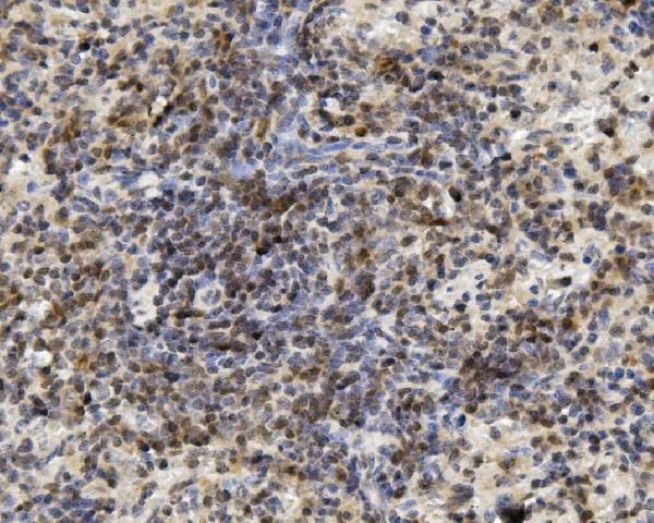
Real IHC data IHC analysis of U2AF2 using anti-U2AF2 antibody (A03639-2). U2AF2 was detected in paraffin-embedded section of human mammary cancer tissue. Heat mediated antigen retrieval was performed in EDTA buffer (pH8.0, epitope retrieval solution). The tissue section was blocked with 10% goat serum. The tissue section was then incubated with 1μg/ml rabbit anti-U2AF2 Antibody (A03639-2) overnight at 4°C. Biotinylated goat anti-rabbit IgG was used as secondary antibody and incubated for 30 minutes at 37°C. The tissue section was developed using Strepavidin-Biotin-Complex (SABC) (Catalog # SA1022) with DAB as the chromogen.
Anti-U2AF65/U2AF2 Antibody ®
Cat # A03639-2

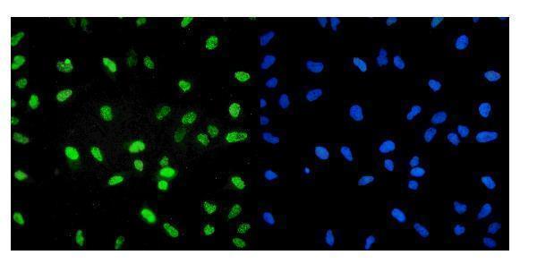
M03639-1 and M03639 each show IHC in human gallbladder adenocarcinoma paraffin sections (respective catalog IHC captions); both list IF/ICC (catalog applications). A03639-2 shows IHC in human mammary cancer and mouse and rat spleen paraffin sections (catalog IHC captions), plus IF in HeLa cells (catalog IF caption).

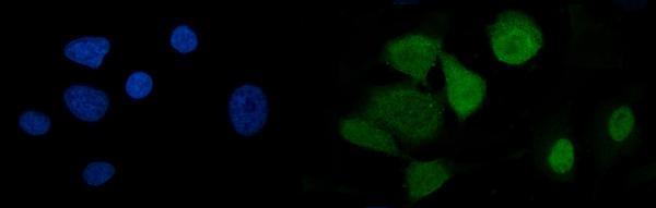
Which to pick: For tissue IHC, choose A03639-2 when cross-species image evidence matters: its own IHC captions show human mammary cancer and mouse and rat spleen paraffin sections (catalog IHC captions). For IF/ICC, A03639-2 has a HeLa cell IF image (catalog IF caption); M03639-1 and M03639 each have an A549 cell IF image and are mouse monoclonals (catalog IF captions; catalog clone/host). The IHC captions specify paraffin sections but do not report the fixative (catalog IHC captions).

Each figure is that product's own IHC / IF validation image from its datasheet.

References

  1. UniProt Consortium. UniProt entry P26368 (U2AF2_HUMAN, Splicing factor U2AF 65 kDa subunit).
  2. Human Protein Atlas. U2AF2 tissue IHC expression (reliability: Supported).
  3. Human Protein Atlas. U2AF2 subcellular location (ICC-IF): Localized to the nucleoplasm and nuclear speckles..
  4. Human Protein Atlas. U2AF2 antibody validation summary (3 antibodies).
  5. Vascular smooth muscle cell RNA-binding protein U2AF2 induces copper death by regulating C1qbp expression, delaying development of atherosclerosise. Biological research 2026 — PMC12924209.
  6. The U2AF2 /circRNA ARF1/miR-342-3p/ISL2 feedback loop regulates angiogenesis in glioma stem cells. Journal of experimental & clinical cancer research : CR 2020 — PMC7487667.
  7. CD82 suppresses CD44 alternative splicing-dependent melanoma metastasis by mediating U2AF2 ubiquitination and degradation. Oncogene 2016 — PMC5033661.
  8. OTUB2-mediated deubiquitination upregulates U2AF2 to promote colorectal cancer evasion of autophagy-ferroptosis. Cell death & disease 2026 — PMC13319764.
  9. PubMed PMID:1538748 — UniProt-cited evidence.
  10. PubMed PMID:15489334 — UniProt-cited evidence.
  11. PubMed PMID:12176931 — UniProt-cited evidence.