This website uses cookies to ensure you get the best experience on our website.


- Table of Contents
Plan paraffin IHC around nuclear staining in most cell types, with placental trophoblasts as an exception (HPA tissue IHC). The catalog antibody has a documented paraffin IHC workflow; score staining by cell population (datasheet A03816-2; standard IHC practice).

Expected localisation, validated protocols, controls and antibodies — the at-a-glance facts below, then the full design guide.
| Expected localisation | Nuclear staining in tissue; cytoplasmic shuttling is possible (HPA tissue IHC; UniProt) | |
| Staining pattern | Nuclear staining in nearly all cell types except trophoblasts (HPA tissue IHC) | |
| Antigen retrieval | Citrate pH 6 HIER, heat-mediated (datasheet A03816-2) | |
| Positive control | Adipose tissue+4 more · see all | |
| Negative control | Placenta |
| Fixation | Keep fixation consistent (standard IHC practice; not target-specific); Matched tissue-IHC evidence does not establish the fixation claim. Validate the specimen-specific method before use. (selected-SKU IHC image A03816-2) | |
| Caveat | Placental trophoblasts may lack detectable staining (HPA tissue IHC) | |
| Regulation | No expression regulator annotated (UniProt) | |
| Isoform / epitope | 2 isoforms; epitope coverage is unspecified (UniProt; datasheet A03816-2) |
Compare the catalog antibody’s citrate pH 6 IHC-P protocol (datasheet A03816-2) with published colorectal, gastric, and synovial tissue protocols (PMC6236309; PMC6235804; PMC7526534).
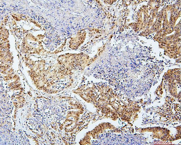
| Sample | Paraffin-embedded human colon cancer tissue; fixative not specified (datasheet A03816-2) |
| Fixation | Image fixative and duration unreported (datasheet A03816-2); verify before use. |
| Sectioning | 4–5 µm sections on charged slides (standard) |
| Deparaffinisation | Xylene, graded ethanol series to water (standard) |
| Antigen retrieval | Heat retrieval: Citrate pH 6, 20 min (datasheet A03816-2) |
| Peroxidase block | 3% H2O2, 10 min, room temperature (standard) |
| Blocking | 10% goat serum (datasheet A03816-2) |
| Primary antibody | Rabbit anti-UBA2, 0.5-1μg/ml (datasheet A03816-2) |
| Primary incubation | Overnight at 4 °C (datasheet A03816-2) |
| Detection | Streptavidin-biotin complex (SABC), DAB chromogen (datasheet A03816-2) |
| Counterstain | Hematoxylin, blue, dehydrate and mount (standard) |
| Expected result | UBA2-positive staining in adipocytes of adipose tissue (HPA tissue IHC: High). HPA tissue profile: Nuclear expression in essentially all cell types, except for placental trophoblasts. No signal in the no-primary control. |
UBA2 should stain mainly in nuclei across essentially all cell types in paraffin-section IHC; placental trophoblasts are the reported exception (HPA: tissue IHC profile). Cytoplasmic signal can occur because UBA2 shuttles between nucleus and cytoplasm, but it should be interpreted against the predominantly nuclear tissue pattern (UniProt Q9UBT2: subcellular location; HPA: tissue IHC profile). HPA rates the tissue pattern Supported, with medium consistency between antibody staining and RNA expression (HPA: tissue IHC reliability).
| Clear nuclear staining in many cell types, including adipocytes, glandular cells or hematopoietic cells. | This matches the broad nuclear pattern. HPA reports High staining in these cell types in specified tissues; their intensity does not set a threshold for every section or preparation (HPA: tissue IHC profile and listed tissue observations). |
| Predominantly cytoplasmic staining with little or no nuclear signal. | Review localisation and background controls: UBA2 can shuttle through the cytoplasm, so cytoplasmic staining alone is not proof of artefact, but a cytoplasm-only result departs from HPA's predominantly nuclear IHC pattern (UniProt Q9UBT2: subcellular location; HPA: tissue IHC profile). |
| Strong staining assigned specifically to placental trophoblasts. | Investigate cell identification and nonspecific staining. HPA reports UBA2 as Not detected in trophoblastic cells; this observation does not establish that every other placental cell should be negative (HPA: placenta tissue IHC observation). |
| Diffuse chromogen across cells and extracellular areas, obscuring nuclei. | Treat the result as uninterpretable until background is controlled. Examine a primary-antibody omission control and the detection system for nonspecific or endogenous activity; those are general chromogenic IHC checks, not reported UBA2 properties (general IHC practice). |
| No staining in a section expected to contain HPA High cells. | Check whether the relevant cells are present, then review the IHC-validated antibody, retrieval and detection controls. An absent signal alone cannot distinguish low assay sensitivity from a biological difference (HPA: listed tissue observations; general IHC practice). |
| Cell type and tissue context | HPA describes nuclear expression in essentially all cell types, with trophoblasts excepted; High examples include adipocytes, adrenal glandular cells and bone marrow hematopoietic cells (HPA: tissue IHC profile and listed tissue observations). |
| Nuclear and cytoplasmic distribution | UniProt records both compartments and nuclear–cytoplasmic shuttling; HPA tissue IHC nevertheless describes a nuclear pattern. Score the dominant compartment and assess mixed staining in its cellular context (UniProt Q9UBT2: subcellular location; HPA: tissue IHC profile). |
| Isoforms and epitope coverage | Two isoforms are listed, but the supplied records do not locate an antibody epitope or establish isoform-specific staining. Verify the IHC-validated antibody's stated epitope before making an isoform claim (UniProt Q9UBT2: isoforms; supplied HPA antibody record). |
| Protein topology and processing | UBA2 has no transmembrane segment, signal peptide or propeptide, and its annotated chain spans residues 1–640. These annotations do not by themselves predict retrieval needs or fixation sensitivity (UniProt Q9UBT2: topology and processing). |
| IF/ICC Q&A: where should UBA2 appear? | Mainly in the nucleoplasm; HPA calls that location Supported in ICC-IF. Cytoplasmic localisation is also recorded by UniProt, so assess additional signal with controls and imaging context (HPA: subcellular ICC-IF; UniProt Q9UBT2: subcellular location). |
| Evidence strength | HPA rates tissue IHC Supported, describes medium agreement with RNA data, and lists antibody HPA041436 as Supported for IHC. Treat the pattern as a reference for interpretation, not a guaranteed intensity for each specimen (HPA: tissue IHC reliability and antibody record). |
| Situation | Likely cause | Next action |
|---|---|---|
| Known HPA High cells are present, but nuclei show no chromogen. | The assay may have insufficient signal, or the expected cell population may have been misidentified; this result does not establish a UBA2-specific retrieval problem (HPA: listed tissue observations; general IHC practice). | Confirm the cell population, check a known-positive section and detection controls, then review the IHC-validated antibody's IHC conditions (HPA: listed tissue observations; general IHC practice). |
| Signal appears cytoplasmic with weak or absent nuclear staining. | UBA2 can shuttle, but a cytoplasm-dominant result conflicts with the principal HPA tissue pattern and may include background (UniProt Q9UBT2: subcellular location; HPA: tissue IHC profile). | Reassess nuclear boundaries with the counterstain; compare a known-positive tissue and an omission control before interpreting cytoplasmic signal (general IHC practice). |
| Placental trophoblasts appear strongly positive. | This differs from HPA's Not detected trophoblast observation; cell assignment or nonspecific chromogen may explain the discrepancy (HPA: placenta tissue IHC observation; general IHC practice). | Verify trophoblast identity on the section and compare primary-antibody omission and positive-tissue controls; report a reproducible discrepancy explicitly (general IHC practice). |
| Staining is diffuse or extracellular and nuclei cannot be scored. | Nonspecific antibody or detection signal, including endogenous detection activity, can obscure cellular localisation in chromogenic IHC (general IHC practice). | Inspect omission and detection-only controls; adjust blocking, washing or detection conditions according to the assay controls before scoring (general IHC practice). |
| Signal intensity varies among cell types in one section. | HPA reports High staining in selected cell populations but does not provide a universal intensity cutoff; heterogeneous staining needs cell-specific interpretation (HPA: listed tissue observations). | Score identifiable cell populations separately, record nuclear localisation and compare with appropriate controls rather than assigning one score to the whole section (HPA: tissue IHC profile; general IHC practice). |
| ICC-IF shows signal outside the nucleoplasm. | HPA's main ICC-IF location is the nucleoplasm, while UniProt also records cytoplasmic UBA2 and shuttling; imaging background remains possible (HPA: subcellular ICC-IF; UniProt Q9UBT2: subcellular location). | Check nuclear segmentation and fluorescence controls, then describe any reproducible cytoplasmic component alongside the dominant compartment (general IF practice). |


Comprehensive Human Protein Atlas IHC scoring per tissue (reliability: Supported — Medium consistency between antibody staining and RNA expression data.). Rows are taken directly from the HPA tissue chart — click any row's HPA link to view the source.
| Tissue | Cell type | Level | Evidence | Source |
|---|---|---|---|---|
| Adipose tissue | Adipocytes | High | Protein (IHC) | HPA → |
| Adrenal gland | Glandular cells | High | Protein (IHC) | HPA → |
| Appendix | Glandular cells | High | Protein (IHC) | HPA → |
| Bone marrow | Hematopoietic cells | High | Protein (IHC) | HPA → |
| Breast | Adipocytes | High | Protein (IHC) | HPA → |
| Tissue | Cell type | Level | Evidence | Source |
|---|---|---|---|---|
| Placenta | Trophoblastic cells | Not detected | Protein (IHC) | HPA → |
Troubleshoot UBA2 chromogenic IHC in paraffin sections using the catalog antibody’s tissue image, reference staining patterns, and controlled comparisons.
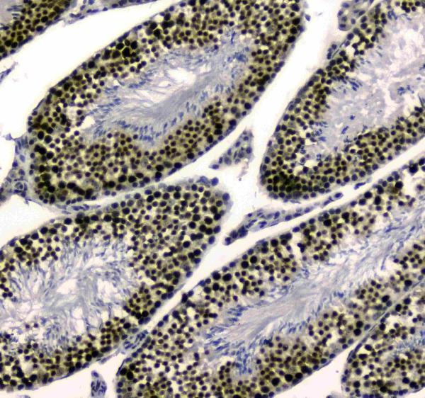
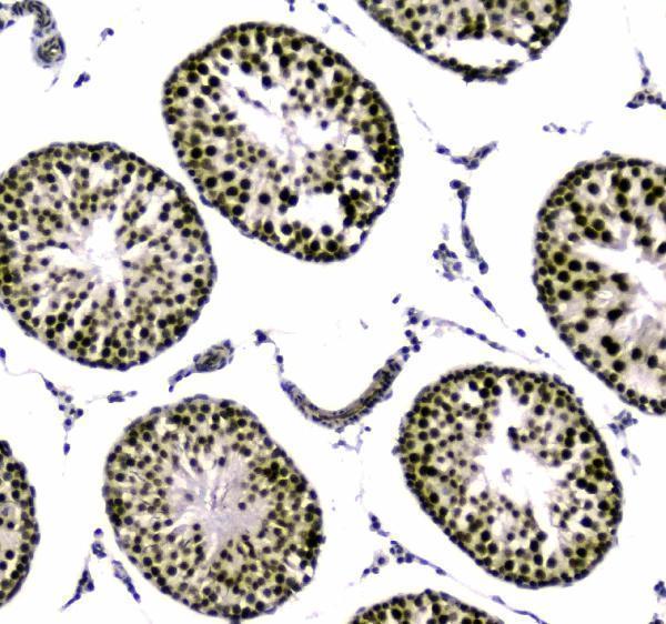
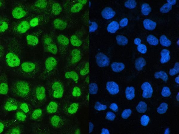
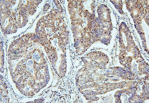
Catalog images show UBA2 IHC in paraffin sections of human cancers and mouse and rat testis, plus IF in A431 cells (catalog image captions).
A03816-2 has IHC images from human colon and mammary cancers and mouse and rat testis, plus an IF image from A431 cells (A03816-2 image captions). M03816-2 has IHC images from human intestinal cancer paraffin sections (M03816-2 image captions).
Which to pick: For tissue IHC across human, mouse and rat, choose A03816-2 because its paraffin-section images cover all three species (A03816-2 IHC captions). For human tissue IHC with a monoclonal antibody, choose M03816-2, clone 5B13; its supplied IHC images show human intestinal cancer (catalog clone; M03816-2 IHC captions). For IF/ICC, choose A03816-2, which has an A431 IF image; the fixative is unreported in both SKUs’ IHC captions (A03816-2 IF caption; A03816-2 and M03816-2 IHC captions).