UBE2I / SUMO-conjugating enzyme UBC9 · IHC design guide

Design Immunohistochemistry for UBE2I

Plan UBE2I staining in paraffin sections around the nuclear and cytoplasmic tissue pattern (HPA tissue IHC). The catalog antibody has an IHC-P working range of 0.5–1 μg/mL (datasheet A02295).

Evidence assembled Oct 2026 · For research use; verify linked source records and product datasheet before use
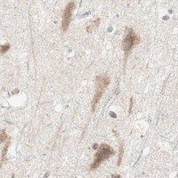
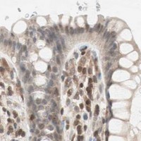
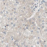
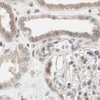
Immunohistochemistry protocol sheet for UBE2I (IHC for UBE2I): expected localisation Nuclear and cytoplasmic staining in most tissues (HPA tissue IHC), antibody A02295, validated IHC image, and IHC protocol steps
Printable UBE2I IHC protocol sheet — expected localisation Nuclear and cytoplasmic staining in most tissues (HPA tissue IHC), antibody A02295, controls and protocol steps. Open the full UBE2I IHC guide →

UBE2I Immunohistochemistry Experimental Design Guide

Expected localisation, validated protocols, controls and antibodies — the at-a-glance facts below, then the full design guide.

Must know before staining
Expected localisation Nuclear and cytoplasmic staining in most tissues (HPA tissue IHC)
Staining pattern Cells in most tissues show nuclear and cytoplasmic staining (HPA tissue IHC)
Antigen retrieval Citrate pH 6 HIER, heat-mediated (datasheet A02295)
Positive control ⓘ Bronchus+4 more · see all
Negative control ⓘ Adipose tissue+4 more · see all
Important caveats
Reasons your staining may differ from the expected pattern.
Fixation Keep fixation conditions consistent across sections (standard IHC practice; not target-specific)
Caveat Broad tissue staining can mask unstained cell types, such as adipocytes (HPA tissue IHC)
Regulation No expression-inducing stimulus reported (UniProt)
Isoform / epitope No reported isoforms; mature chain spans residues 2–158 (UniProt)
Section 1

Recommended UBE2I IHC & IF Protocols

The catalog antibody’s IHC-P protocol (datasheet A02295) is accompanied by published UBE2I IHC methods for kidney tissue (PMC8861443) and cartilage (PMC12249805).

Recommended immunohistochemistry (IHC-P) protocol parameters
SampleParaffin-embedded human appendicitis tissue; fixative not specified (datasheet A02295)
FixationImage fixative and duration unreported (datasheet A02295); verify before use.
Sectioning4–5 µm sections on charged slides (standard)
DeparaffinisationXylene, graded ethanol series to water (standard)
Antigen retrievalHeat retrieval: Citrate pH 6, 20 min (datasheet A02295)
Peroxidase block3% H2O2, 10 min, room temperature (standard)
Blocking10% goat serum (datasheet A02295)
Primary antibodyRabbit anti-UBE2I, 0.5-1μg/ml (datasheet A02295)
Primary incubationOvernight at 4 °C (datasheet A02295)
DetectionStreptavidin-biotin complex (SABC), DAB chromogen (datasheet A02295)
CounterstainHematoxylin, blue, dehydrate and mount (standard)
Expected resultUBE2I-positive staining in basal cells of bronchus (HPA tissue IHC: High). HPA tissue profile: Nuclear and cytoplasmic expression in most tissues. No signal in the no-primary control.
💡Decision noteStart with heat-mediated citrate pH 6 retrieval (datasheet A02295); optimize against the published conditions for the tissue being stained (PMC12249805).
Section 2

What Is the Expected UBE2I Staining Pattern?

UBE2I should show nuclear and cytoplasmic staining in many tissue cell types, with a mainly nuclear distribution (HPA: nuclear and cytoplasmic expression in most tissues; UniProt P63279: mainly nuclear). It is an intracellular protein with no transmembrane segment (UniProt P63279 topology). HPA rates its tissue IHC profile Approved, with medium consistency between antibody staining and RNA expression; judge the pattern by the specific cell population as well as the tissue (HPA: tissue IHC reliability).

What am I looking at on my slide?
Nuclear staining with some cytoplasmic staining in bronchial basal cells or placental trophoblasts.This fits the reported compartments and cell populations (HPA: nuclear and cytoplasmic expression; High in bronchial basal cells and placental trophoblasts). Compare nuclei and cytoplasm within the same population; a strong positive control need not have identical intensity in both compartments (UniProt P63279: mainly nuclear).
Signal outlines cell membranes or fills extracellular spaces, with little intracellular staining.Treat a predominantly surface or extracellular pattern as suspect: UBE2I has no transmembrane segment, and its reported locations are nuclear and cytoplasmic (UniProt P63279 topology and subcellular location; HPA: tissue IHC profile). Review morphology and control staining before scoring it as UBE2I.
Adipocytes or liver cholangiocytes stain as strongly as the chosen positive cells.These populations were not detected in the HPA tissue survey (HPA: adipocytes and cholangiocytes, Not detected). Consider antibody cross-reactivity or endogenous detection activity, especially if staining follows tissue edges, pigment, or damaged areas (standard IHC practice). A local exception requires independent validation.
A broad, faint wash covers nuclei, cytoplasm, and surrounding tissue.Diffuse background does not establish UBE2I localization. Excess antibody, incomplete blocking, or residual detection activity can obscure compartment boundaries (standard IHC practice). Compare a no-primary control and a known positive population; interpret only staining that rises clearly above the local background.
No convincing staining appears in a section containing bronchial basal cells.The chosen population is reported High by HPA (HPA: bronchus, basal cells, High), so a blank result calls for a technical check before a biological conclusion. Confirm that the basal cells are present, the run controls developed, and the catalog antibody is validated for IHC (standard IHC practice; HPA: IHC Approved antibodies).
💡Expected UBE2I appearanceCall the result positive when identifiable cells show nuclear with possible cytoplasmic chromogenic staining, especially High populations such as bronchial basal cells or placental trophoblasts (HPA: tissue IHC profile and High cells; UniProt P63279: mainly nuclear); isolated surface, extracellular, or uniform background color is suspect (standard IHC practice).
How each factor affects the staining
Cell population within a tissueUBE2I is broadly expressed, but HPA scores individual populations differently: bronchial basal cells are High, pancreatic endocrine cells Low, and adipocytes Not detected (HPA: tissue IHC). Select and score the specified cell population; a whole-section average can hide this variation.
Tissue IHC evidence and antibody choiceHPA calls the tissue profile Approved with medium staining–RNA consistency (HPA: tissue IHC reliability). HPA003909 and CAB009021 are IHC Approved; the supplied record does not label either IHC Enhanced (HPA: antibody validation). Keep that evidence strength in mind when judging an unexpected pattern.
Intracellular locationUBE2I is reported in nucleus, cytoplasm, and perinuclear cytoplasm, mainly nuclear, with no transmembrane segment (UniProt P63279 location and topology). These facts support an intracellular interpretation but do not establish a distinctive chromogenic perinuclear ring in every tissue section.
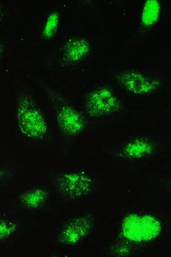
IF/ICC Q&A: Should nucleoli stain?Nucleoplasmic staining is the supported main ICC-IF location; nucleoli are an additional supported location in images from MCF-7 and U2OS cells (HPA: subcellular ICC-IF). Use that as context for an IF observation, without requiring visible nucleoli in paraffin tissue IHC (HPA: tissue IHC profile).
Negative reference populationsHPA reports Not detected staining in adipocytes, skeletal-muscle myocytes, and smooth-muscle cells, while UniProt lists expression in skeletal muscle as a tissue (HPA: tissue IHC; UniProt P63279: tissue specificity). The protein-level tissue statement does not predict staining in every resident cell type.
Why is my staining missing, weak or wrong?
SituationLikely causeNext action
The positive tissue has no visible signal.The expected positive cells may be absent from the section, or the IHC run may have failed (standard IHC practice; HPA: bronchial basal cells, High).Locate the specified cells on the counterstained section, check the run's positive control, then review the catalog antibody's IHC-P procedure and detection reagents (standard IHC practice). Avoid calling a tissue negative until these checks pass.
Every compartment has weak brown color.Diffuse background can result from excess primary antibody, inadequate blocking, or endogenous detection activity (standard IHC practice).Compare the no-primary control; review blocking, antibody dilution, washes, and chromogen development using the catalog IHC-P procedure (standard IHC practice). Score only cell-associated signal above background.
Only cell borders or extracellular material stain.That distribution conflicts with reported intracellular UBE2I localization (UniProt P63279 location and topology; HPA: tissue IHC profile).Check a second cell population with reported staining and inspect the no-primary control. If the border pattern persists only with primary antibody, seek independent antibody validation before assigning it to UBE2I (standard IHC practice).
HPA Not detected cells show strong staining.Cross-reactivity or endogenous chromogenic activity is possible; HPA reports adipocytes and cholangiocytes as Not detected (HPA: tissue IHC).Verify cell identity against morphology and counterstain, compare no-primary and positive controls, and repeat with an independently validated IHC antibody if the discrepancy matters (standard IHC practice; HPA: IHC Approved antibodies).
Nuclei are positive, but cytoplasm appears blank.A nuclear-dominant result can fit the mainly nuclear UniProt location; HPA describes nuclear and cytoplasmic expression across most tissues, not a required intensity ratio for each cell (UniProt P63279; HPA: tissue IHC profile).Check that nuclear signal is specific and cell-associated, then record the compartments actually visible. Do not force a cytoplasmic score solely to match the broad HPA summary (standard IHC scoring practice).
Tissue IHC does not show the nucleoli seen in ICC-IF.HPA supports nucleolar staining as an additional ICC-IF location, whereas its tissue IHC summary reports nuclear and cytoplasmic expression (HPA: subcellular ICC-IF and tissue IHC).Assess tissue IHC against its cell-type and compartment references. If nucleolar detail matters, evaluate it on the separate IF/ICC guide page; do not treat its absence in chromogenic sections alone as assay failure (HPA: ICC-IF and tissue IHC).

Sample controls for UBE2I IHC & IF

🧪Run placenta first: trophoblastic cells should stain (HPA: High in trophoblastic cells). Use adipose tissue as the negative comparator because adipocytes are listed as not detected (HPA: Not detected in adipocytes); on the placenta slide, non-trophoblastic cells can serve as internal background comparators only if they lack specific staining, since their negative status is unreported (HPA: placenta row).
Positive control tissue: Bronchus (Basal cells, HPA High)
Negative control tissue: Adipose tissue (HPA Not detected)
ICC-IF cell lines (HPA subcellular resource): HPA ICC-IF images show UBE2I in MCF-7, U2OS, with annotated localisation: Nucleoplasm (supported) (HPA subcellular).
Technical controls: Include a no-primary, secondary-only control, a rabbit IgG isotype control matched to the primary antibody’s clonality if known, and a verified UBE2I knockout control (caption: rabbit primary; standard IHC practice). For placenta, block endogenous peroxidase and check endogenous biotin before using the caption’s biotin-based DAB detection (caption: biotinylated secondary and SABC-DAB; standard IHC practice).
⚠️Feasibility: A target-specific fixation window is unreported, and the selected A02295 tissue-IHC caption does not state a fixative (caption: fixative not stated). The paraffin-section example uses citrate retrieval at pH 6 for 20 minutes, but this does not establish that retrieval is required under every condition (caption: citrate retrieval). HPA has ICC-IF images in MCF-7 and U2OS, but the supplied evidence does not establish that frozen sections or IF are easier; placenta’s endogenous biotin warrants a detection control (HPA: ICC-IF cell lines; standard IHC practice).

HPA tissue IHC evidence for UBE2I

Comprehensive Human Protein Atlas IHC scoring per tissue (reliability: Approved — Medium consistency between antibody staining and RNA expression data.). Rows are taken directly from the HPA tissue chart — click any row's HPA link to view the source.

Positive expression · recommended positive controls

TissueCell typeLevelEvidenceSource
Bronchus Basal cells High Protein (IHC) HPA →
Cervix Glandular cells High Protein (IHC) HPA →
Fallopian tube Non-ciliated cells High Protein (IHC) HPA →
Lymph node Non-germinal center cells High Protein (IHC) HPA →
Placenta Trophoblastic cells High Protein (IHC) HPA →

Undetected expression · recommended negative controls

TissueCell typeLevelEvidenceSource
Adipose tissue Adipocytes Not detected Protein (IHC) HPA →
Hippocampus Glial cells Not detected Protein (IHC) HPA →
Liver Cholangiocytes Not detected Protein (IHC) HPA →
Parathyroid gland Glandular cells Not detected Protein (IHC) HPA →
Seminal vesicle Glandular cells Not detected Protein (IHC) HPA →
Section 3

Advanced UBE2I IHC Tips

Troubleshoot UBE2I staining in paraffin sections by checking retrieval, cell compartment, controls and scoring against the documented IHC result (datasheet A02295; HPA tissue IHC).

How should I troubleshoot weak UBE2I staining after antigen retrieval?
Start with heat mediated citrate buffer at pH 6 for 20 minutes (datasheet A02295). Compare sections processed together, including a positive control, before changing retrieval conditions; the documented UBE2I result used 1 μg/mL primary antibody overnight at 4 °C (datasheet A02295; standard IHC practice). If staining remains weak, compare a longer citrate heating time or a different retrieval buffer on matched sections, recording both nuclear signal and tissue damage (standard IHC practice). Judge improvement against the expected nuclear and cytoplasmic pattern, since stronger staining alone does not establish specificity (HPA tissue IHC; standard IHC practice).
Can I infer which fixative preserves UBE2I staining best?
Target specific fixation sensitivity is unknown: the selected paraffin section caption does not state a fixative (datasheet A02295). Document each specimen’s actual fixative and fixation interval, then compare similarly processed sections before attributing a weak result to UBE2I biology (standard IHC practice). Keep citrate retrieval at pH 6 for 20 minutes as the documented starting condition while assessing the effect of preparation differences (datasheet A02295). Use morphology, a negative reagent control and an expected positive cell population to assess the stain; the HPA staining pattern does not establish which fixative is suitable (HPA tissue IHC; standard IHC practice).
Is cytoplasmic UBE2I staining credible when I expected nuclear staining?
Nuclear and cytoplasmic staining can both be credible in tissue: HPA describes expression in both compartments across most tissues (HPA tissue IHC). UniProt places UBE2I in the nucleus, cytoplasm and perinuclear region, with mainly nuclear localisation noted by similarity (UniProt P63279 subcellular). Score nuclear and cytoplasmic signal separately in intact cells, using a consistent counterstain and the same exposure or DAB development conditions across comparison sections (standard IHC practice). If staining appears only at torn edges or in damaged areas, repeat the section and compare with a negative reagent control before assigning biological meaning to that distribution (standard IHC practice).
Could an isoform or hidden epitope explain inconsistent staining?
The supplied record lists 0 isoforms, so an isoform specific explanation lacks support here (UniProt P63279 isoforms). UBE2I has a UBC core spanning residues 4–157, no transmembrane segment and reported modifications at residues 2, 65 and 71 (UniProt P63279 domains, topology and modified residues). The antibody epitope is not specified in the supplied caption, so these features cannot identify whether fixation or retrieval masks its binding site (datasheet A02295). Compare matched sections using the documented citrate pH 6 retrieval, and seek epitope information before making residue level claims about staining differences (datasheet A02295; standard IHC practice).
How can IF help check a surprising chromogenic UBE2I pattern?
Use IF as a separate localisation check: HPA reports supported nucleoplasmic and additional nucleolar localisation in cell images (HPA subcellular). Multiplex UBE2I with a marker for the cell type being assessed, and choose a fluorophore channel away from the specimen’s strongest autofluorescence (standard IF practice). Permeabilise cells to allow antibody access to nuclear and cytoplasmic UBE2I, which has no transmembrane segment; optimise that step for the IF specimen and antibody (UniProt P63279 topology and subcellular; standard IF practice). Compare nuclear and cytoplasmic IF signal with matched controls, then evaluate the paraffin section independently using its documented citrate pH 6 IHC retrieval (datasheet A02295; standard IF/IHC practice).
What should I check when DAB obscures UBE2I staining?
Check a section processed without primary antibody to reveal detection related staining, and inspect tissue edges and damaged regions for uneven background (standard IHC practice). The documented method used 10% goat serum, biotinylated goat anti rabbit secondary antibody and a streptavidin biotin complex with DAB (datasheet A02295). Include a peroxidase block and assess endogenous biotin where relevant to this detection chemistry; these are general controls rather than evidence about UBE2I expression (standard IHC practice). Adjust blocking, washes or DAB development one variable at a time, retaining a positive control so reduced background is not mistaken for successful staining (standard IHC practice).
How should I quantify UBE2I across paraffin sections? ⚠ ANSWER MARKED FOR VERIFICATION
Define the cell population and tissue region before scoring, and exclude folds, necrosis and poorly preserved edges (standard IHC practice). Record nuclear and cytoplasmic staining separately because both compartments are reported for UBE2I in tissue (HPA tissue IHC). For an intensity weighted summary, calculate an H score from the percentage of cells at each intensity; alternatively report percentage positive or positive cell density per mm² (standard IHC practice). Normalise cell counts to the number of eligible cells or density to analysed tissue area, and apply the same positivity threshold, counterstain assessment and DAB development criteria to every section (standard IHC practice).
How do I distinguish genuine UBE2I staining from artefact?
A plausible result includes nuclear and cytoplasmic staining in intact cells, although intensity depends on the cell population examined (HPA tissue IHC). HPA reports high staining in bronchial basal cells and placental trophoblastic cells, while adipocytes are listed as not detected; use these observations as context, not universal controls (HPA tissue IHC). Isolated edge staining, signal confined to necrosis or staining reproduced without primary antibody warrants investigation before calling cells positive (standard IHC practice). Check peroxidase related DAB signal with a negative reagent control, and reconcile unexpected compartment staining with the documented nuclear, cytoplasmic and perinuclear locations (standard IHC practice; UniProt P63279 subcellular).
Boster reagents

Best UBE2I / SUMO-conjugating enzyme UBC9 IHC Antibodies

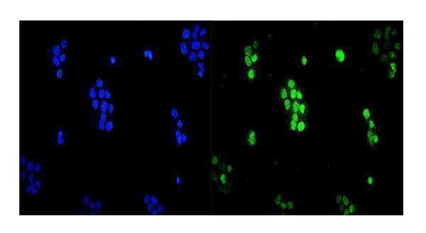
The catalog shows UBE2I IHC images from human, mouse and rat paraffin sections (IHC captions), plus IF/ICC images from U20S and A431 cells (A02295 and A02295-1 IF captions).

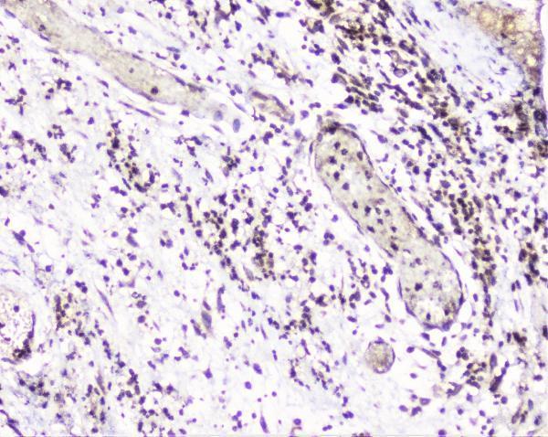
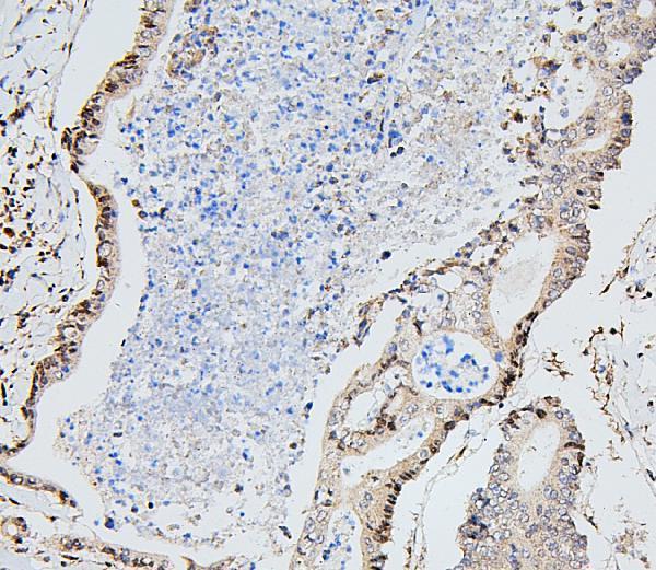
Real IHC data IHC analysis of UBE2I UBC9 using anti-UBE2I UBC9 antibody (A02295). UBE2I UBC9 was detected in paraffin-embedded section of human appendicitis tissue. Heat mediated antigen retrieval was performed in citrate buffer (pH6, epitope retrieval solution) for 20 mins. The tissue section was blocked with 10% goat serum. The tissue section was then incubated with 1μg/ml rabbit anti-UBE2I UBC9 Antibody (A02295) overnight at 4°C. Biotinylated goat anti-rabbit IgG was used as secondary antibody and incubated for 30 minutes at 37°C. The tissue section was developed using Strepavidin-Biotin-Complex (SABC)(Catalog # SA1022) with DAB as the chromogen.
Anti-UBE2I UBC9 Antibody ®
Cat # A02295
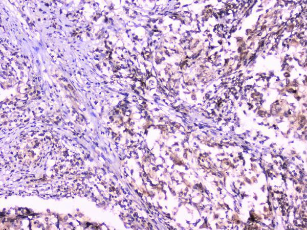
Real IHC data IHC analysis of UBE2I/UBC9 using anti-UBE2I/UBC9 antibody (A02295-1). UBE2I/UBC9 was detected in paraffin-embedded section of human colon cancer tissue. Heat mediated antigen retrieval was performed in EDTA buffer (pH8.0, epitope retrieval solution). The tissue section was blocked with 10% goat serum. The tissue section was then incubated with 1μg/ml rabbit anti-UBE2I/UBC9 Antibody (A02295-1) overnight at 4°C. Biotinylated goat anti-rabbit IgG was used as secondary antibody and incubated for 30 minutes at 37°C. The tissue section was developed using Strepavidin-Biotin-Complex (SABC) (Catalog # SA1022) with DAB as the chromogen.
Anti-UBE2I/UBC9 Antibody ®
Cat # A02295-1
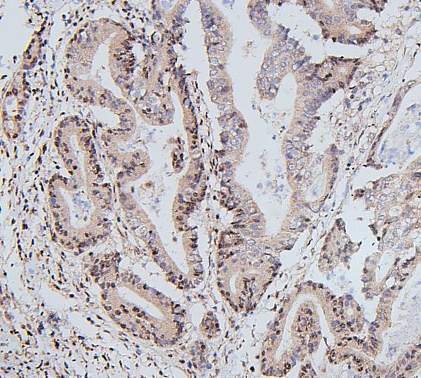
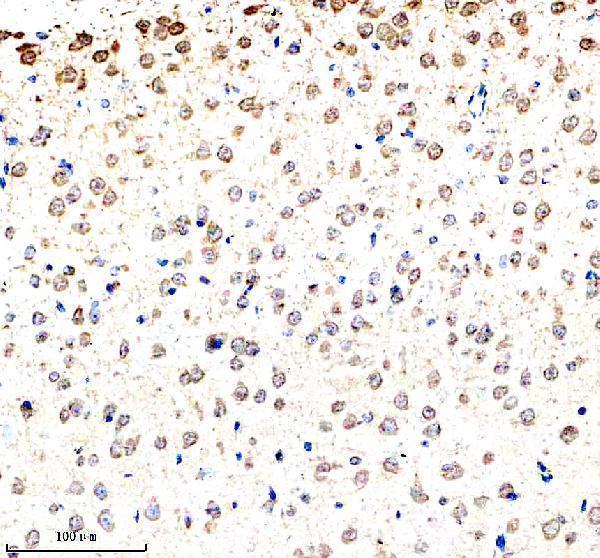
Real IHC data IHC analysis of UBE2I using anti-UBE2I antibody (M02295). UBE2I was detected in a paraffin-embedded section of mouse brain tissue. Heat mediated antigen retrieval was performed in EDTA buffer (pH 8.0, epitope retrieval solution). The tissue section was blocked with 10% goat serum. The tissue section was then incubated with 1:50 rabbit anti-UBE2I Antibody (M02295) overnight at 4°C. Peroxidase Conjugated Goat Anti-rabbit IgG was used as secondary antibody and incubated for 30 minutes at 37°C. The tissue section was developed using HRP Conjugated Rabbit IgG Super Vision Assay Kit (Catalog # SV0002) with DAB as the chromogen.
Anti-UBE2I/Ubc9 Rabbit Monoclonal Antibody
Cat # M02295

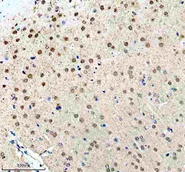
A02295 shows IHC in human appendicitis and lung cancer and mouse and rat brain, plus IF/ICC in U20S cells (A02295 image captions); A02295-1 shows IHC in human colon cancer, mammary cancer and tonsil, plus IF/ICC in A431 cells (A02295-1 image captions). M02295 shows IHC in mouse brain, rat brain and rat hippocampus (M02295 IHC captions).

Which to pick: For tissue IHC, choose A02295 for citrate retrieval at pH 6 for 20 minutes (A02295 IHC captions), A02295-1 for EDTA retrieval at pH 8.0 (A02295-1 IHC captions), or monoclonal M02295 for mouse or rat brain sections with EDTA retrieval at pH 8.0 (catalog: clone EDF-21; M02295 IHC captions). For IF/ICC, A02295 and A02295-1 each have a cell image; M02295 lists IF/ICC but has no IF image in the payload (catalog applications and IF captions). For work across species, A02295 has human, mouse and rat IHC images (A02295 IHC captions); the fixative is unreported in the paraffin-section captions for all three SKUs (IHC captions).

Each figure is that product's own IHC / IF validation image from its datasheet.

References

  1. UniProt Consortium. UniProt entry P63279 (UBC9_HUMAN, SUMO-conjugating enzyme UBC9).
  2. Human Protein Atlas. UBE2I tissue IHC expression (reliability: Approved).
  3. Human Protein Atlas. UBE2I subcellular location (ICC-IF): Mainly localized to the nucleoplasm. In addition localized to the nucleoli..
  4. Human Protein Atlas. UBE2I antibody validation summary (3 antibodies).
  5. Identification of UBE2I as a Novel Biomarker in ccRCC Based on a Large-Scale CRISPR-Cas9 Screening Database and Immunohistochemistry. Frontiers in molecular biosciences 2022 — PMC8861443.
  6. UBC9-Mediated SUMO Pathway Drives Prohibitin-1 Nuclear Accumulation and PITX1 Repression in Primary Osteoarthritis. International journal of molecular sciences 2025 — PMC12249805.
  7. Targeting UBC9-mediated protein hyper-SUMOylation in cystic cholangiocytes halts polycystic liver disease in experimental models. Journal of hepatology 2021 — PMC8157180.
  8. Increased PD-L1 Restricts Liver Injury in Nonalcoholic Fatty Liver Disease. Oxidative medicine and cellular longevity 2022 — PMC9126662.
  9. PubMed PMID:8668529 — UniProt-cited evidence.
  10. PubMed PMID:9067428 — UniProt-cited evidence.
  11. PubMed PMID:8565643 — UniProt-cited evidence.