UBE3A / Ubiquitin-protein ligase E3A · IHC design guide

Design Immunohistochemistry for UBE3A

Plan chromogenic paraffin IHC for UBE3A using catalog antibody A00582 at 0.5–1 μg/mL (datasheet: A00582). Score cytoplasmic and nuclear staining separately, using the tissue profile to select positive and low-signal controls (HPA tissue IHC).

Evidence assembled Oct 2026 · For research use; verify linked source records and product datasheet before use
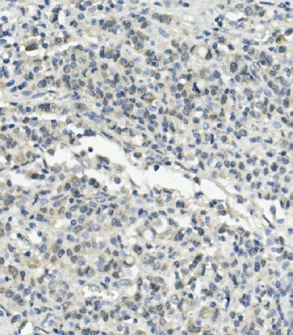
Immunohistochemistry protocol sheet for UBE3A (IHC for UBE3A): expected localisation Mainly cytoplasmic, with nuclear staining (HPA tissue IHC), antibody A00582, validated IHC image, and IHC protocol steps
Printable UBE3A IHC protocol sheet — expected localisation Mainly cytoplasmic, with nuclear staining (HPA tissue IHC), antibody A00582, controls and protocol steps. Open the full UBE3A IHC guide →

UBE3A Immunohistochemistry Experimental Design Guide

Expected localisation, validated protocols, controls and antibodies — the at-a-glance facts below, then the full design guide.

Must know before staining
Expected localisation Mainly cytoplasmic, with nuclear staining (HPA tissue IHC)
Staining pattern Glandular, neuronal and glial cells: cytoplasmic and nuclear signal (HPA tissue IHC)
Antigen retrieval EDTA pH 8.0 HIER, heat-mediated (datasheet A00582)
Positive control ⓘ Adrenal gland+4 more · see all
Negative control ⓘ Adipose tissue
Important caveats
Reasons your staining may differ from the expected pattern.
Fixation Keep fixation consistent across sections. (standard IHC practice; not target-specific)
Caveat Adipocytes may lack signal despite broad tissue staining (HPA tissue IHC)
Regulation Expression regulation unreported (UniProt)
Isoform / epitope 3 isoforms; epitope coverage is unreported (UniProt)
Section 1

Recommended UBE3A IHC & IF Protocols

The catalog antibody’s IHC-P protocol is followed by published UBE3A staining conditions for brain sections and human cortex (PMC6532248; PMC5758499; PMC11169893; PMC6194692).

Recommended immunohistochemistry (IHC-P) protocol parameters
SampleParaffin-embedded human gastric cancer tissue; fixative not specified (datasheet A00582)
FixationImage fixative and duration unreported (datasheet A00582); verify before use.
Sectioning4–5 µm sections on charged slides (standard)
DeparaffinisationXylene, graded ethanol series to water (standard)
Antigen retrievalHeat retrieval: EDTA pH 8.0 (datasheet A00582); 20 min, 95–100 °C (standard)
Peroxidase block3% H2O2, 10 min, room temperature (standard)
Blocking10% goat serum (datasheet A00582)
Primary antibodyRabbit anti-UBE3A, 0.5-1μg/ml (datasheet A00582)
Primary incubationOvernight at 4 °C (datasheet A00582)
DetectionStreptavidin-biotin complex (SABC), DAB chromogen (datasheet A00582)
CounterstainHematoxylin, blue, dehydrate and mount (standard)
Expected resultUBE3A-positive staining in glandular cells of adrenal gland (HPA tissue IHC: High). HPA tissue profile: Mainly cytoplasmic but also nuclear expression in all tissues. No signal in the no-primary control.
💡Decision noteStart with heat-mediated EDTA pH 8.0 retrieval for the catalog antibody (datasheet: A00582). The published conditions below describe separate tissue preparations and antibodies.
Section 2

What Is the Expected UBE3A Staining Pattern?

UBE3A should show mainly cytoplasmic staining with some nuclear staining across tissues (HPA tissue IHC); UniProt also places it in the cytoplasm and nucleus, with no transmembrane segment (UniProt Q05086). Expect readily visible staining in HPA-high glandular, respiratory epithelial, neuronal, granular-layer or glial cells, depending on the section (HPA tissue IHC). HPA rates its tissue staining Approved, with medium RNA–protein consistency and external verification pending (HPA tissue IHC).

What am I looking at on my slide?
Cytoplasmic staining with a nuclear component in the expected cells.This matches the reported tissue pattern (HPA tissue IHC) and UniProt localization (UniProt Q05086). Compare cells within the same section before scoring intensity: HPA reports high staining in selected cell types, while its overall tissue profile is broad (HPA tissue IHC).
Staining is confined to membranes or extracellular material, with little cellular signal.A membrane-only or extracellular pattern does not match cytoplasmic and nuclear UBE3A (HPA tissue IHC; UniProt Q05086). Treat it as possible nonspecific staining or a detection artefact; confirm the cell boundaries and repeat with appropriate controls (standard IHC practice).
Adipocytes stain strongly while expected positive cells show little signal.HPA reports UBE3A as not detected in adipocytes (HPA tissue IHC). Consider antibody cross-reactivity or endogenous chromogenic detection activity, then compare a reagent control and an HPA-high cell population processed in the same run (standard IHC practice; HPA tissue IHC).
Color is spread evenly over cells, stroma and blank areas.Diffuse deposit without a cell-shaped cytoplasmic or nuclear pattern is difficult to score as UBE3A (HPA tissue IHC). Check blocking, washes, detection reagents and section handling with suitable controls (standard IHC practice).
No cellular signal appears in a section containing HPA-high cells.An absent result in, for example, adrenal glandular cells or caudate neuronal cells conflicts with HPA's high staining reports (HPA tissue IHC). First check that the expected cells are present, then review antibody suitability and the run's positive and detection controls (standard IHC practice).
💡Expected UBE3A appearanceCall a result positive when expected cells show chiefly cytoplasmic color with possible nuclear color and readily visible staining in an HPA-high population; isolated membrane, extracellular or uniform background color is suspect (HPA tissue IHC; standard IHC practice).
How each factor affects the staining
Tissue and cell choiceHPA reports high staining in adrenal, appendix, breast and cervical glandular cells; bronchial respiratory epithelial cells; caudate neurons; cerebellar granular-layer cells; and cortical glia (HPA tissue IHC). Adipocytes are not detected and marrow hematopoietic cells are low in the supplied profile (HPA tissue IHC). Select and score by cell type, not tissue name alone (standard IHC practice).
Antibody evidenceHPA lists three rabbit polyclonals with Approved IHC status: HPA039410, HPA040380 and CAB009723 (HPA antibodies). This is an approval record, not an Enhanced IHC claim; the tissue profile has medium consistency with RNA and awaits external verification (HPA antibodies; HPA tissue IHC). Interpret marginal staining with those limits in view.
Isoforms and epitope coverageUniProt lists three UBE3A isoforms and a HECT domain at residues 776–875 (UniProt Q05086). The supplied records do not locate any antibody epitope or establish equal detection of all isoforms (HPA antibodies; UniProt Q05086). If comparing variants, obtain epitope information before attributing a staining difference to expression.
Processing and compartmentUniProt reports no signal peptide, propeptide or transmembrane segment, and a single annotated chain spanning residues 1–875 (UniProt Q05086). A secreted or membrane-only pattern is therefore unsupported by this record (UniProt Q05086). These annotations do not establish how fixation or antigen retrieval affects staining.
IF/ICC Q: where should signal appear?A: HPA's ICC-IF summary places UBE3A mainly in the nucleoplasm, additionally in the cytosol; HPA039410 has Approved ICC status (HPA subcellular; HPA antibodies). This is an ICC-IF observation, while tissue IHC is described as mainly cytoplasmic with nuclear staining (HPA tissue IHC). Interpret each application against its own reference pattern.
Why is my staining missing, weak or wrong?
SituationLikely causeNext action
No staining in expected positive cells.The selected cell population may be absent, or the antibody or detection run may have failed (HPA tissue IHC; standard IHC practice).Locate HPA-high cells in the section; check a positive control, reagent setup and detection sequence before calling the tissue negative (HPA tissue IHC; standard IHC practice).
Nuclear staining appears without much cytoplasmic color.Nuclear UBE3A is supported, but tissue IHC is described as mainly cytoplasmic (HPA tissue IHC; UniProt Q05086).Assess the specific cell type and compare another section or control; avoid rejecting genuine nuclear signal solely because the cytoplasm is faint (HPA tissue IHC; standard IHC practice).
Strong color appears in adipocytes.HPA lists adipocytes as not detected; cross-reactivity or endogenous detection activity is possible (HPA tissue IHC; standard IHC practice).Review a detection-only control and compare HPA-high cells in the same run before assigning adipocyte positivity (HPA tissue IHC; standard IHC practice).
Color coats the section without clear cellular boundaries.Diffuse background can reflect nonspecific reagent binding, insufficient washing or endogenous activity (standard IHC practice).Inspect controls, blocking and washes, then score only compartment-resolved staining in identifiable cells (standard IHC practice; HPA tissue IHC).
An apparent rim surrounds cells or extracellular spaces.That distribution conflicts with the reported intracellular localization and absence of a transmembrane segment (HPA tissue IHC; UniProt Q05086).Check morphology, counterstain and reagent controls; seek reproducible cytoplasmic or nuclear staining before interpreting UBE3A (standard IHC practice; HPA tissue IHC).
A different fixation or retrieval condition changes the result.Target-specific fixation effects are not established by the supplied assay evidence. Verify with a matched IHC source before attributing a result to fixation.Use matched positive and negative controls when comparing conditions, and report the observed change without attributing it to a proven UBE3A epitope effect (standard IHC practice).

Sample controls for UBE3A IHC & IF

🧪Run breast first: its glandular cells should stain for UBE3A (HPA: High in breast glandular cells). Use adipose tissue as the negative tissue (HPA: Not detected in adipocytes); adipocytes within a breast section are candidate internal negative cells and should show no specific chromogen, subject to confirmation on the slide (HPA: Not detected in adipose-tissue adipocytes).
Positive control tissue: Adrenal gland (Glandular cells, HPA High)
Negative control tissue: Adipose tissue (HPA Not detected)
ICC-IF cell lines (HPA subcellular resource): HPA ICC-IF images show UBE3A in A-431, U-251MG, U2OS, with annotated localisation: Nucleoplasm (approved) (HPA subcellular).
Technical controls: Include a no-primary, secondary-only control and a rabbit IgG control matched to the primary antibody’s clonality (A00582 caption: rabbit primary antibody; standard IHC practice). Confirm specificity with a UBE3A knockout sample or a validated peptide block; for SABC/DAB detection, quench endogenous peroxidase and check endogenous biotin background (A00582 caption: SABC/DAB; standard IHC practice).
⚠️Feasibility: The A00582 paraffin-section caption does not report a fixative, and the supplied evidence gives no UBE3A-specific fixation window or fixation effect (A00582 caption). The example uses heat-mediated EDTA retrieval at pH 8.0, but whether breast staining depends on that retrieval step is unreported; the supplied evidence also does not establish whether frozen sections or IF/ICC are easier (A00582 caption). Breast stromal collagen can autofluoresce in IF, so assess background with an unstained section (standard IF practice).

HPA tissue IHC evidence for UBE3A

Comprehensive Human Protein Atlas IHC scoring per tissue (reliability: Approved — Medium consistency between antibody staining and RNA expression data. Pending external verification.). Rows are taken directly from the HPA tissue chart — click any row's HPA link to view the source.

Positive expression · recommended positive controls

TissueCell typeLevelEvidenceSource
Adrenal gland Glandular cells High Protein (IHC) HPA →
Appendix Glandular cells High Protein (IHC) HPA →
Breast Glandular cells High Protein (IHC) HPA →
Bronchus Respiratory epithelial cells High Protein (IHC) HPA →
Caudate Neuronal cells High Protein (IHC) HPA →

Undetected expression · recommended negative controls

TissueCell typeLevelEvidenceSource
Adipose tissue Adipocytes Not detected Protein (IHC) HPA →
Section 3

Advanced UBE3A IHC Tips

Troubleshoot UBE3A staining in paraffin sections by checking retrieval, compartment, controls and scoring before interpreting differences between specimens.

What retrieval should I start with when UBE3A staining is weak in paraffin sections?
Start with heat-mediated retrieval in EDTA pH 8.0 for paraffin sections (datasheet A00582). The selected gastric cancer image used this retrieval before incubation with 1 μg/ml primary antibody overnight at 4°C, so keep those conditions consistent while assessing retrieval (A00582 tissue-IHC caption). Compare a retrieved section with an otherwise matched section that omits retrieval, and check whether stronger staining retains the expected cytoplasmic and nuclear pattern (HPA: tissue IHC profile; UniProt Q05086: cytoplasm and nucleus). If staining remains weak, adjust heating duration on matched sections while monitoring tissue integrity; record the actual time and temperature because the caption does not specify them (A00582 tissue-IHC caption).
Can I infer a suitable fixative from the published UBE3A tissue staining pattern?
No target-specific fixation sensitivity is established by the supplied evidence, and the selected paraffin-section caption does not state a fixative (A00582 tissue-IHC caption). Record the fixative and fixation duration for every specimen, then compare sections processed with the same retrieval and detection conditions before attributing staining differences to UBE3A abundance. Use EDTA pH 8.0 heat-mediated retrieval as the documented starting condition for this antibody (datasheet A00582). Include a consistently processed reference section in each run; a change in signal after a fixation change establishes a workflow effect, but does not by itself identify the molecular cause.
Should UBE3A staining be nuclear, cytoplasmic, or both?
Assess both cytoplasmic and nuclear staining in paraffin sections: UBE3A is annotated in both compartments (UniProt Q05086: subcellular location), and tissue IHC is mainly cytoplasmic with some nuclear expression (HPA: tissue IHC profile). Score the compartments separately because a shift between them can be obscured by a single whole-cell intensity score. Check cell boundaries and the counterstain before calling diffuse DAB deposition nuclear; the counterstain should identify intact nuclei in the scored cells. Strong signal confined to an unexpected structure should trigger review of morphology and controls, since UBE3A has no annotated transmembrane segment (UniProt Q05086: topology).
Could epitope position or UBE3A isoforms explain inconsistent staining?
Map the antibody immunogen against UBE3A isoforms I, II and III before treating a staining difference as altered total protein (UniProt Q05086: isoforms). The supplied caption does not locate the A00582 epitope, so its coverage of individual isoforms cannot be established from that image (A00582 tissue-IHC caption). UBE3A contains a HECT domain at residues 776–875 and annotated modified residues at 218 and 659; their relevance depends on the actual epitope (UniProt Q05086: domains and modified residues). Compare the immunogen sequence with isoform sequences and document what is shared before interpreting compartment-specific staining as an isoform-specific result.
How should I check UBE3A localisation in a multiplex IF experiment?
Pair UBE3A with a marker identifying the cell population being assessed, and inspect nuclear and cytosolic channels separately (HPA: approved nucleoplasm and additional cytosol localisation in ICC/IF). Choose a fluorophore in a channel with low tissue autofluorescence after examining an unstained specimen, and include single-colour controls to assess bleed-through. UBE3A has no annotated transmembrane segment, so use controlled permeabilisation to access intracellular epitopes; optimise its strength against morphology and the marker signal (UniProt Q05086: topology and subcellular location). Treat IF localisation as supporting context for the paraffin-section result, where the tissue IHC profile is mainly cytoplasmic with some nuclear expression (HPA: tissue IHC profile).
How can I reduce diffuse DAB background without losing UBE3A signal?
First compare a no-primary section with the stained section to locate background arising from the secondary antibody or chromogenic detection. The selected image used 10% goat serum blocking, 1 μg/ml primary antibody overnight at 4°C, and a biotinylated secondary followed by SABC and DAB (A00582 tissue-IHC caption). For a peroxidase-based workflow, check the effectiveness of the endogenous peroxidase block and rinse between steps; these are general IHC controls, not evidence of a UBE3A-specific problem. If background persists, titrate primary concentration around the documented condition while retaining a reference section and checking that cytoplasmic and nuclear staining remains interpretable (HPA: tissue IHC profile).
What should I score when comparing UBE3A across paraffin sections? ⚠ ANSWER MARKED FOR VERIFICATION
Define the cell population and tissue area before scoring, then record nuclear and cytoplasmic UBE3A separately because both compartments are reported (HPA: tissue IHC profile; UniProt Q05086: subcellular location). Use an H-score based on intensity and percentage of positive cells, or report percentage positive and positive-cell density per mm² when those measures fit the question. Normalise cell counts to viable tissue area or to the number of eligible cells, and exclude folds, necrosis and section edges using the same rules throughout the series. Hold retrieval, staining and image-analysis thresholds constant; the selected antibody image used EDTA pH 8.0 retrieval (datasheet A00582).
How do I distinguish credible UBE3A staining from artefact?
Look for staining in intact cells with a plausible cytoplasmic or nuclear distribution, then compare with the no-primary section and a consistently processed reference (HPA: tissue IHC profile; UniProt Q05086: subcellular location). Treat edge-restricted colour, necrotic deposits and signal in the wrong compartment as reasons to inspect morphology and detection controls before calling a biological change. Check endogenous peroxidase when DAB appears without primary antibody; the selected image used SABC with DAB detection (A00582 tissue-IHC caption). HPA reports Approved tissue staining with medium consistency against RNA data and pending external verification, so use independent controls before making a strong claim from staining alone (HPA: tissue IHC reliability).
Boster reagents

Best UBE3A / Ubiquitin-protein ligase E3A IHC Antibodies

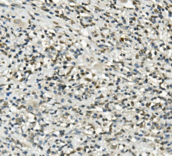
A00582 has real IHC images from human paraffin sections of gastric cancer, rectal cancer and brain (catalog image captions). No IF images are supplied (catalog IF images).

Real IHC data IHC analysis of UBE3A using anti-UBE3A antibody (A00582). UBE3A was detected in paraffin-embedded section of human gastric cancer tissue. Heat mediated antigen retrieval was performed in EDTA buffer (pH8.0, epitope retrieval solution). The tissue section was blocked with 10% goat serum. The tissue section was then incubated with 1μg/ml rabbit anti-UBE3A Antibody (A00582) overnight at 4°C. Biotinylated goat anti-rabbit IgG was used as secondary antibody and incubated for 30 minutes at 37°C. The tissue section was developed using Strepavidin-Biotin-Complex (SABC) (Catalog # SA1022) with DAB as the chromogen.
Anti-UBE3A ® Antibody
Cat # A00582

A00582 is listed for human IHC and illustrated in human gastric and rectal cancer paraffin sections (catalog applications/reactivity; catalog image captions). Human brain paraffin-section images are also supplied (catalog image captions).

Which to pick: Choose A00582 for human paraffin-section IHC; its stated dilution is 0.5–1 μg/ml, and its image captions do not report the fixative (datasheet: IHC dilution; catalog image captions). No IF/ICC recommendation is supported because A00582 has no listed IF/ICC application or IF image (catalog applications; catalog IF images). No cross-species IHC option is supported: A00582 lists Human reactivity only, and its clonality is unreported (catalog reactivity; catalog clone).

Each figure is that product's own IHC / IF validation image from its datasheet.

References

  1. UniProt Consortium. UniProt entry Q05086 (UBE3A_HUMAN, Ubiquitin-protein ligase E3A).
  2. Human Protein Atlas. UBE3A tissue IHC expression (reliability: Approved).
  3. Human Protein Atlas. UBE3A subcellular location (ICC-IF): Mainly localized to the nucleoplasm. In addition localized to the cytosol..
  4. Human Protein Atlas. UBE3A antibody validation summary (3 antibodies).
  5. Delayed loss of UBE3A reduces the expression of Angelman syndrome-associated phenotypes. Molecular autism 2019 — PMC6532248.
  6. Enhanced Transmission at the Calyx of Held Synapse in a Mouse Model for Angelman Syndrome. Frontiers in cellular neuroscience 2017 — PMC5758499.
  7. Regional and cellular organization of the autism-associated protein UBE3A/E6AP and its antisense transcript in the brain of the developing rhesus monkey. Frontiers in neuroanatomy 2024 — PMC11169893.
  8. Subcellular organization of UBE3A in human cerebral cortex. Molecular autism 2018 — PMC6194692.
  9. PubMed PMID:9143503 — UniProt-cited evidence.
  10. PubMed PMID:8988171 — UniProt-cited evidence.
  11. PubMed PMID:8988172 — UniProt-cited evidence.