This website uses cookies to ensure you get the best experience on our website.
- Table of Contents
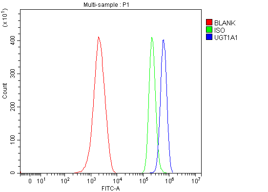
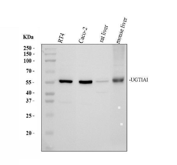
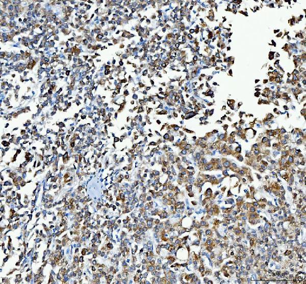
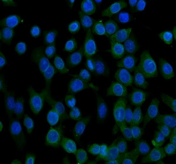
Facts about UDP-glucuronosyltransferase 1-1.
.
Rat | |
---|---|
Gene Name: | Ugt1a1 |
Uniprot: | Q64550 |
Entrez: | 24861 |
Belongs to: |
---|
UDP-glycosyltransferase family |
bilirubin UDP-glucuronosyltransferase 1-1; bilirubin UDP-glucuronosyltransferase isozyme 1; Bilirubin-specific UDPGT isozyme 1; GNT1; GNT1EC 2.4.1.17; HUG-BR1; UDP glucuronosyltransferase 1 family, polypeptide A1; UDP glycosyltransferase 1 family, polypeptide A1; UDP-glucuronosyltransferase 1-1; UDP-glucuronosyltransferase 1-A; UDP-glucuronosyltransferase 1A1; UDPGT 1-1; UDPGT; UG-BR1; UGT1; UGT1*1; UGT1.1; UGT1-01; UGT1A; UGT-1A; UGT1A1; UGT1AUDPGT 1-1; UGT1BILIQTL1
Mass (kDA):
59.663 kDA
Rat | |
---|---|
Location: | 9q35 |
Sequence: | 9; |
The antibody used by Boster Bio has been validated on multiple platforms and with both known positive and negative samples to ensure specificity and high affinity. The company rewards scientists for the first review of each of its products with product credits. This is one of the most important factors to look for when selecting an antibody. The Boster Bio website also offers a community forum where scientists from all over the world can share their ideas, suggestions, and experiences.
The reversible inhibition mechanism of kurarinone was assessed using the parallel artificial membrane permeation assay (PAM-A), a rapid in vitro assay for passive biomembrane permeability. This method was first developed to predict the passive permeability of a drug in the gastrointestinal tract. However, it was not clear whether kurarinone would inhibit CYP1A2 or UGTs in humans.
The recombinant human supersomes were used in the study. The compound was mixed with recombinant CYPs (15 nM) and incubated for 30 min. The incubation period is described in Section 2.3. In addition to the inhibition of CYP1A2, kurarinone inhibited CYP2C9 and UGT1A2.
CYP2D6 and CYP1A2 play a significant role in the biotransformation of kurarinone. Other enzymes also metabolize kurarinone with limited ability. However, when kurarinone inhibits these enzymes, it amplifies the concentration of the drug in the blood and increases the risk of adverse effects. Further research is necessary to confirm the effectiveness of this drug in inhibiting the biotransformation of phenacetin.
Inhibition of CYP1A2 is a significant pharmacological outcome of the development of new therapeutic agents for hepatitis C. Cyclosporine inhibitors inhibit CYP1A2, a metabolic enzyme that converts procarcinogens into cancer. Inhibitors inhibit CYP1A2 by more than 50%, and some of these have IC50 values of 20 nM, which would serve as starting points for lead optimization. Virtual screening has the added benefit of identifying inhibitors and determining their effects.
A study of kurarinone revealed that a prenylated flavone isolated from Sophora flavescens was able to inhibit CYP1A2 by a simple UHPLC-MS/MS method. The samples were pretreated using acetonitrile-mediated precipitation, and chromatographic separation was achieved using a Waters ACQUITY HSS T3 column.
Noncompetitive inhibition of enzymes is a process that causes the substrate to bind less efficiently. The inhibitor binds to a site outside the active site, changing its three-dimensional structure. The enzyme still binds to substrate, but it no longer has the optimal arrangement to stabilize the transition state. The inhibition process also reduces the enzyme's Vmax, so it is impossible to increase the concentration of the substrate to overcome the decreased rate of reaction.
The main difference between competitive inhibition and noncompetitive inhibition is the mechanism of inhibition. In competitive inhibition, a substrate binds to the enzyme's active site while the inhibitor binds to an allosteric site. This means that the enzyme cannot catalyze the reaction with the inhibitor. Noncompetitive inhibition, however, is reversible, meaning that the enzyme will function normally after the inhibitor has dissociated.
Tricyclic secondary alcohol 26b is the most selective inhibitor of UGT1A1 in Boster Bio. It inhibits 14 other UGT isoforms, including those from subfamily 1A. Furthermore, it is highly selective and possesses true selectivity of over 1000. This makes it an effective inhibitor of UGT1A1.
A typical example of a mixed inhibitor is product inhibitor P. It binds two forms of enzyme. This is referred to as a mixed inhibitor. The theory behind these inhibitors can be applied to any situation where inhibition and enzyme activation are involved. With mixed inhibitors, one can draw a family of intersecting lines. Inhibitor P can act as a mixed inhibitor by binding to both forms of the enzyme.
In general, the UGT1A1 gene contains several polymorphisms. Fortunately, the UGT1A1 gene can be detected using a fluorescence quantitative PCR method. Fluorescence PCR is a powerful technique because it can detect UGT1A1 polymorphisms using as little as one ng of genomic DNA. However, it is limited in its site specificity, low sensitivity, and high instrument cost.
For example, a recent study found a significant association between the UGT1A1*28 genotype and the risk of lung cancer. Further studies are needed to determine whether bilirubin fractionation is sufficient. Other research is needed to determine the optimal dosage and duration of anticancer medications. While the UGT1A1 gene is a valuable tool for assessing anticancer drugs, there is no universal screening test that will guarantee efficacy and safety.
As a cellular enzyme, UGT1A1 is essential for metabolic clearance and detoxification of bilirubin. Several therapeutic drugs are metabolized in the liver by this enzyme. However, a common mutation in the gene promoter reduces the expression level and renders it more susceptible to drug interactions. This may explain why people with Gilbert's polymorphism tend to develop cancer of the uterus and colon.
The UGT1A1 gene is important for the metabolism of pro-tease inhibitors. Certain UGT1A1 polymorphisms may affect the pharmacokinetics of pro-tease inhibitors, and may even cause severe toxicity. This toxicity must be studied extensively in African populations. When the UGT1A1 gene is regulated through drug metabolism, it can reduce or increase the risk of disease.
A source of anti-UGT1A antibody can be identified by analyzing mouse liver sections stained with DiI. HLSC-injected mice had diffuse and localized expression, and DiI-positivity was co-localized with UGT1A1 immunoreactivity. These cells lacked the proliferation marker PCNA and were not detected in mouse serum 15 days after cell injection. This suggests that the antibody may originate from different sources.
Human UGT1A1 protein is primarily metabolized in the liver and is expressed in several human organs. It is involved in the metabolism of several drugs and other important endogenous substances, including raltegravir. It has been implicated in the inactivation of anticancer drugs, including SN-38. Its level is related to a patient's response to the anticancer drug.
The serum of the mice with the UGT1A1 marker was obtained at indicated time points. The serum, liver, brain, and blood of Day 8 wt mice and HLSC-treated mice were fixed in formalin and embedded in paraffin for subsequent analysis. The UGT1A1 protein was analysed with an anti-rabbit secondary antibody. A separate test, using an anti-PCNA antibody, detected the presence of proliferating cells. The labelled cells were tracked by means of a Zeiss microscope and Apotome software.
The quantitative method of measuring UGT1A1 protein levels in human liver microsomes was validated using an external standard addition. This method is based on the addition of known amounts of peptide standards to the sample matrices. The sample matrices used for the test were spiked with three series of label-free peptide 1 and 2. The measured UGT1A1 protein content was plotted against the concentration of the standard working solution. The accuracy of this method can be calculated through an equation.
A recent study validated the UGT1A1 marker. Its genotype is associated with severe neutropenia in a patient population treated with irinotecan. Compared to other genetic markers, this marker is the strongest predictor of severe neutropenia. This study showed that UGT1A1 was highly associated with a higher risk of severe neutropenia. However, the findings of this study have limitations.
PCR was performed with primers designed to target the UGT1A1 gene and nearby intronic sequences. The sample was analyzed using a PCR amplification reaction in a volume of 30 mL. The PCR reaction mixture contained 0.5 mM of each dNTP (Fermentas) and 50 to 100 ng of genomic DNA. In addition, the PCR reaction mixture contained 1.4 mM MgCl2 and one unit of DNA polymerase (ABI).
The coding region of UGT1A1 contains four TA repeats (TA repeats) and four exonic regions. The TA repeats in the UGT1A1 gene promoter are responsible for the glucuronidation of numerous compounds. Glucuronidation occurs when a substrate undergoes biotransformation into a metabolite. There are four families of UGT enzymes. The UGT1A1 gene complex is encoded by the UGT1A gene on chromosome 2q37. The gene complex contains 13 variable exons and four common exons, with nine viable first exons.
As UGT1A1 gene is involved in the metabolism of various drugs, UGT1A1 molecular genetic testing is important to individualize treatment. Consequently, individuals with reduced UGT1A1 function are at risk of severe adverse drug reactions. Moreover, UGT1A1 is used in treating metastatic triple-negative breast cancer. The UGT1A1 gene is highly conserved, making it one of the most valuable tumor markers.
PMID: 7603447 by Coffman B.L., et al. Cloning and stable expression of a cDNA encoding a rat liver UDP- glucuronosyltransferase (UDP-glucuronosyltransferase 1.1) that catalyzes the glucuronidation of opioids and bilirubin.
PMID: 7608130 by Emi Y., et al. Drug-responsive and tissue-specific alternative expression of multiple first exons in rat UDP-glucuronosyltransferase family 1 (UGT1) gene complex.