This website uses cookies to ensure you get the best experience on our website.
- Table of Contents
Facts about Regulator of nonsense transcripts 1.
In EJC-dependent NMD, the SURF complex associates with the exon junction complex (EJC) (found 50-55 or more nucleotides downstream from the termination codon) via UPF2 and allows the formation of an UPF1-UPF2-UPF3 surveillance complex which is believed to activate NMD. Phosphorylated UPF1 is recognized by EST1B/SMG5, SMG6 and SMG7 which are thought to supply a link to the mRNA degradation machinery involving exonucleolytic and endonucleolytic pathways, and to function as adapters to protein phosphatase 2A (PP2A), thus triggering UPF1 dephosphorylation and allowing the recycling of NMD factors.
Human | |
---|---|
Gene Name: | UPF1 |
Uniprot: | Q92900 |
Entrez: | 5976 |
Belongs to: |
---|
DNA2/NAM7 helicase family |
ATP-dependent helicase RENT1; EC 3.6.1; EC 3.6.4.-; FLJ43809; HUPF1; KIAA0221FLJ46894; Nonsense mRNA reducing factor 1; NORF1; NORF1delta helicase; pNORF1; regulator of nonsense transcripts 1; RENT1; RENT1UP Frameshift 1; UPF1 regulator of nonsense transcripts homolog (yeast); UPF1; up-frameshift mutation 1 homolog; Up-frameshift suppressor 1 homolog; yeast Upf1p homolog
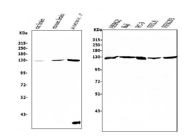
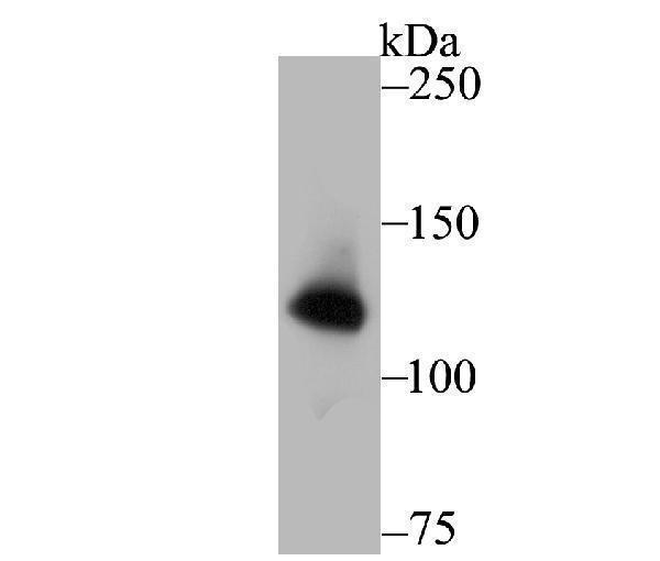
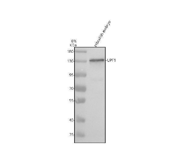
Mass (kDA):
124.345 kDA
Human | |
---|---|
Location: | 19p13.11 |
Sequence: | 19; NC_000019.10 (18831405..18868230) |
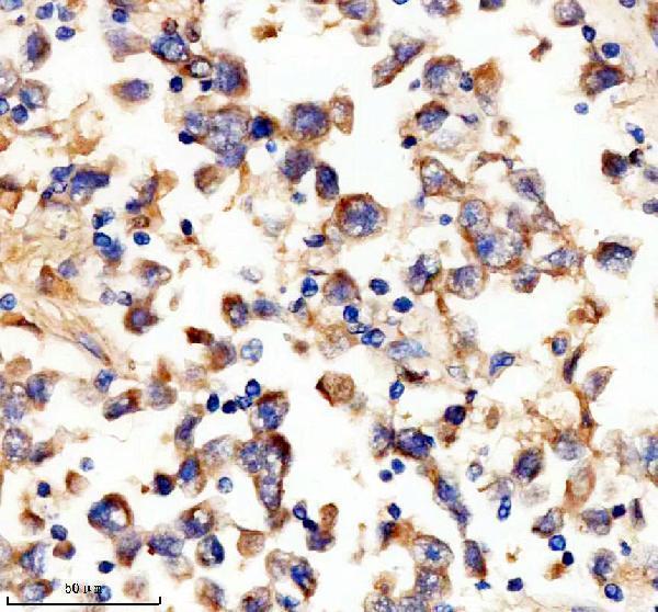
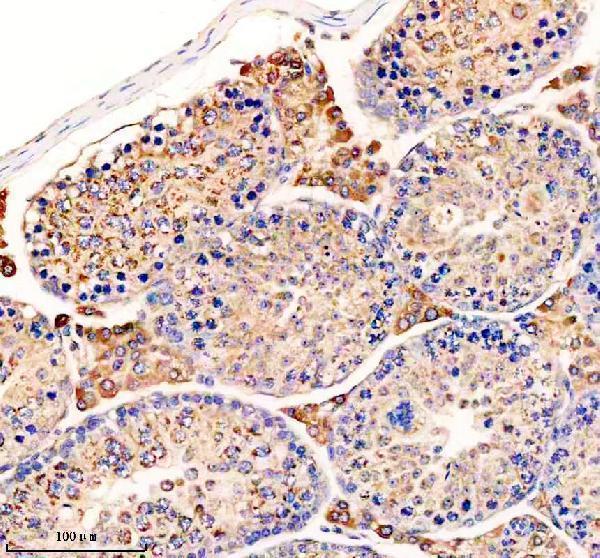
Ubiquitous.
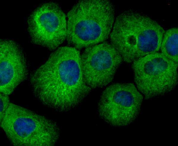
Cytoplasm. Cytoplasm, P-body. Nucleus. Hyperphosphorylated form is targeted to the P-body, while unphosphorylated protein is distributed throughout the cytoplasm. Localized in the chromatoid bodies of round spermatids (By similarity).
The UPF1 marker is one of many proteins present in the human genome. Researchers are becoming more interested in the role it plays in the process of depolymerization of mRNA. It activates NMD and inhibits replication-dependent histone mRNA degradation. This article will examine the most significant functions and potential applications of UPF1 across a variety of areas. Boster Bio: The Best Uses For The UPF1 Marker
The conclusion of the S phase of a cell's cycle is when histone mRNAs are degraded. This process is controlled by multiple steps. Translation and degradation of replication-dependent histone mRNAs are coordinated, but the molecular mechanisms underlying this process remain elusive. One such process is stem loop-mediated remodeling of stem loop-binding protein (TPF) by UPF1 when it is transitioning from an active translation state to a degradation-active state. UPF1 regulates the degradation of mRNAs that have 3'-UTRs. It is also dependent on TDRD6, which unwinds DNA double-stranded, facilitating its destruction.
The SURF complex is linked to the exon junction complex that is located 50-55 nucleotides downstream of the codon that terminates. When UPF1 and UPF2 create the UPF1 -UPF2-UPF3 surveillance complex, it activates NMD. During NMD, UPF1 interacts with EST1B/SMG1B/SP2A, a phosphorylation-dependent protein kinase complex that binds PP2A.
Three steps regulate the metabolism of histone mRNA. They are transcription, degradation, or translation. These three steps are responsible for the proper accumulation of the canonical histone proteins during the S phase. The SLBP is not the only one. The third step is the removal of excess histone protein. Both of these steps require activation cyclin A–CDK1 to be broken down.
Replication-dependent histone genes are expressed in chromatin and the Cajal body is a nucleus domain containing U7 snRNP. Cajal bodies are processing factors and are crucial in maturing snRNA. In vertebrates, Cajal bodies serve as locations for maturation of snRNAs and assembly.
Despite the importance of histones, histone genes accumulate before the cell is able to degrade them. A protein complex containing RAD53, a checkpoint kinase RAD53 which targets excessive histone proteins for degrading in yeast. Further research must be conducted before the same system can be found in metazoans. Similar systems are likely to be found in all eukaryotes.
The S phase of the cell cycle is where the gene for the histone protein, PC4, is needed. The increase in DNA replication is achieved through processing of the 3'-end that requires canonical histones that are the core. This gene set is extremely active in human, frog and animal mRNAs. Further, UPF1 regulates the expression of histone mRNA in cells.
Replication-dependent histone mRNAs are polyadenylated mRNAs, and are regulated primarily at the transcriptional level. However, the stem-loop mechanism was maintained in all metazoa. Ultimately, it has evolved to function post-transcriptionally, where it enables regulation at the post-transcriptional level.
SLBP1 can block the transcription of histone mRNA the early stages of development. The newly synthesized histone protein supplies histones until the zygotic process begins. This protein stops the growth and accumulation of histone mRNAs in early development. The nurse cells also translate the histone mRNA of the oocyte.
Autophagy is one of the main features of autophagy, a process that produces free amino acids in cells in response to starvation or metabolic stress. Interestingly, the regulation of NMD affects amino acids the most. NMD inhibition and hyperactivation decreased the levels of amino acids. Likewise, it activated NMD increasing amino acid concentrations. Therefore, autophagy may be an effective method to regulate levels of protein and to promote protein metabolism.
The RNA viruses rely on MRNA processing and mRNP remodelling for translation. Their genomic RNA contains stop codons either in the 3'UTR or in the middle. NMD activation can be activated by moving a considerable distance away from the stop codon. Many studies have suggested the molecular basis for NMD activation. This is what encodes the RNA virus that encodes the viral protein. What molecular mechanism triggers the NMD pathway to stimulate the protein?
To combat infection and maintain healthy immune balance, a person must have a strong immune response. In the absence of this, it could turn chronic or even cause death. This is why it is so vital to find and keep a healthy balance between the homeostasis and immune response. Recent research has proven that NMD plays a crucial role in the regulation of the immune response. Cytokines are signals molecules that can be induced in response of inflammation, trigger cells generate an inflammatory response and trigger a cascade of immune responses.
Another way to stop NMD. It occurs when the PTC code sequence is not recognized by the ribosome, or NMD regulators are unable to detect it. Despite the fact that the PTC is an essential requirement for NMD and translation readthrough at the PTC reduces the chance of activation. Furthermore, reading through of translations in the PTC region is able to eliminate complexes of the exon junction downstream of the PTC61.
NMD is believed to target a significant portion of functional and physiologically active wild type mRNAs. The authors of the study have demonstrated that NMD can regulate the expression levels of biologically active genes. However, this is just a simple snapshot of how the pathway operates. There are many other instances of NMD-mediated gene regulation. It is important to recognize that NMD is a key tool to regulate gene expression.
The mechanism of NMD activation is not well comprehended. Complex interactions cause changes in gene expression that aim to reduce stress, restore homeostasis and start the process of apoptosis. Different stress conditions can inhibit NMD through various mechanisms. One of the mechanisms that lead to inhibition of translation is phosphorylation of eIF2a which inhibits the initiation of several stress pathways. The increased regulation of NMD then upregulates the transcripts encoding factors and results in stronger stress responses.
The mechanism by which NMD regulates viral replication has not been identified, it is believed to act as an obstacle between host cells and viruses. A genome-wide RNAi analysis has revealed many components of NMD. These components are responsible to higher levels of viral proteins and greater viral infection if they are not controlled. Some viruses have found ways to avoid NMD-mediated destruction. The Rous sarcoma viruses for instance, have an element in the 3' UTR that prevents the virus from functioning.
Upf1 is a crucial regulator of the nonsense-mediated decay (NDD) pathway which blocks access to DNA and stimulates tumorigenesis. This pathway is activated during replication stress. Boster Bio's UPF1 inhibitors block NMD function and improve SMD efficiency. Both mechanisms are essential for the proper degradative process of histone mRNA.
Researchers have discovered a brand new inhibitor that blocks the nonsense-mediated pathway of decay of mRNA. The compound blocks mRNA decay by targeting UPF1 protein as well as the Poly(a),-binding protein that regulates mRNA destruction. The compound is also effective against BRCA1 mRNA , which is responsible for cancer. These inhibitors are now being studied for their potential to fight cancer.
In addition to blocking UPF1, this medication also targets the NMD factor gene STAT3. This transcription factor regulates gene expression and is associated with tumorigenesis. Mutations in the UPF1 gene can cause NMD to be affected and cause an increase in the expression of tumor-related genes. This could explain why tumors that have functional NMD are resistant to UPF1 inhibitors.
UPF1 inhibitors have been shown to be ineffective against cancerous cells. This suggests that they are not suitable for clinical trials. However there is evidence to suggest that tumor suppressor genes are more prone to NMD. These genes could be expressed in large numbers in non-malignant cells due to the fact that they are usually over-expressed. In addition cancer cells with MSI contain high levels UPF1 and SMG1 which enhance their NMD-mediated degradation.
PMID: 8855285 by Perlick H.A., et al. Mammalian orthologues of a yeast regulator of nonsense transcript stability.
PMID: 9064659 by Applequist S.E., et al. Cloning and characterization of HUPF1, a human homolog of the Saccharomyces cerevisiae nonsense mRNA-reducing UPF1 protein.