This website uses cookies to ensure you get the best experience on our website.
- Table of Contents
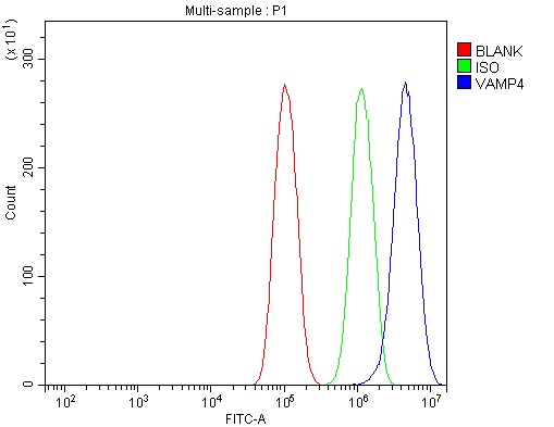
Facts about Vesicle-associated membrane protein 4.
.
Human | |
---|---|
Gene Name: | VAMP4 |
Uniprot: | O75379 |
Entrez: | 8674 |
Belongs to: |
---|
synaptobrevin family |
VAMP24; VAMP-4; vesicle-associated membrane protein 4
Mass (kDA):
16.397 kDA
Human | |
---|---|
Location: | 1q24.3 |
Sequence: | 1; NC_000001.11 (171700160..171742844, complement) |
Golgi apparatus, trans-Golgi network membrane; Single-pass type IV membrane protein. Associated with trans Golgi network (TGN) and newly formed immature secretory granules (ISG). Not found on the mature secretory organelles.
The VAMP4 Marker is a high-affinity primary antibody developed by Boster Bio. It can be used to detect toxins in vivo. This article provides an overview of the VAMP4 Marker and its value. Read on to learn more. Also, discover more information about Boster's high-affinity primary antibodies. It is available to scientists worldwide. This article summarizes the best uses of Boster bio's VAMP4 marker.
The VAMP4 marker was chosen for its specificity. This protein is present in a subset of synaptic vesicles, which are involved in the docking and fusion process. This subset is also known as synaptobrevins, and it may play an important role in trans-Golgi network-to-endosome transport. To determine its specificity, the high-affinity primary antibodies from Boster use the VAMP4 marker.
The high-affinity primary antibodies from Boster's Biolabs utilize the VAMP4 marker to visualize the vesicular domain of the membrane. Antibodies against the VAMP4 marker have been shown to colocalize with a range of proteins, including syntaxin and rbet1. This suggests that they recognize the same protein as other antibodies against different components of the membrane.
The antibodies can be used to detect proteins that are expressed in various types of tissues. The VAMP4 marker is also found in the cis-Golgi, the TGN, and recycling endosomes. When boster scientists use anti-VAMP4 antibodies, they can submit their findings for product credits. The products of Boster are applicable to scientists from all countries.
The anti-VAMP4 antibody was produced by a process known as affinity chromatography. In this process, the anti-VAMP4 antibody is bound to a protein A-Sepharose bead sorbent. Then, the beads are eluted in 0.1 M glycine (pH 2.8) buffer. The eluates containing the bound antibodies are neutralized with 0.02% sodium azide.
To optimize antibody performance, boster's high-affinity primary antibodies are conjugated to a secondary antibody to reduce unspecific binding. In addition, the antibodies have to be bought fresh. Always follow the manufacturer's recommendations for antibody storage and use. The antibodies should not be stored in cold storage as they can become contaminated with Sodium Azide. However, Boster's high-affinity primary antibodies are suitable for all research environments.
Since VAMP4 is present in large protein complexes, it is likely that it is the functional homologue of the synaptic 7S vesicle docking-fusion complex. The high-affinity primary antibodies employ the VAMP4 marker to detect this protein. The markers are particularly useful in cell biology, since they can accurately predict the location of vesicle-trafficking proteins.
To confirm that anti-VAMP4 is specifically specific for a particular membrane protein, crude membranes from rat brain were first separated through a 11% Triton X-100 glycerol velocity gradient. These sequential fractions were then resolved by SDS-PAGE. The samples were stained with Coomassie blue. In a subsequent experiment, anti-VAMP4 antibodies were co-isolated with GST-syntaxin-6.
A variety of different methods are available to detect botulinum neurotoxins, including immunoassays, molecular diagnostics, and enzyme-linked immunosorbent assays (ELISAs). These approaches may replace the use of MBA and address the three Rs of a toxicity test. However, several of these methods have limited sensitivity, and cannot distinguish fully active BoNT from protein toxin.
To measure the amount of bound antibody to the target protein, cells were incubated in the presence of the peptide antigen. NanoLuc-VAMP2 cells and Engineered cells were incubated for 60 h in the presence of BoNT/B to assess their sensitivity. Immunoblotting was performed to measure the level of protein synthesis. The cleaved VAMP2 antibody had a sensitivity of at least 40 fM, which was comparable to the mouse LD50.
Immunoassays were performed in both mice and humans. One of the advantages of immunoassays is their sensitivity. An ELISA allows multiple samples to be analyzed at the same time. While early ELISAs lacked sensitivity, newer methods with signal amplification methods have improved the sensitivity of the test. Inactivated toxin, as well as certain matrix components, may affect the antibodies or result in false-positive results.
Currently, there are two major methods to detect cyanotoxins in freshwater. These two methods are used to detect toxins in a wide variety of aquatic environments. The most common of these methods is ELISA, which requires no special training or expensive equipment. However, repeat analysis is recommended if the first screening kit detects the toxin. If this approach does not yield a positive result, repeat chromatographic analysis is necessary.
BoNTs toxinotypes cleave distinct intracellular proteins. They are known as SNARE (soluble N-ethylmaleimide-sensitive attachment receptor) and VAMP (vesicle associated membrane protein). Both types of toxins interact with the SNAP-25 molecule and bind gold nanoparticles. Soluble BoNT/A cleaves this molecule, gold nanoparticles attach to the probe particles.
In addition to identifying the VAMP4 molecule, reengineering of the VAMP molecule will allow detecting all steps of BoNT/B intoxication. Furthermore, this approach will provide a user-friendly enzyme-linked immunosorbent assay, which meets the 3Rs objectives. A sensitive reporter cell line may be suitable for monitoring the activity of toxins during vaccine manufacture.
The most sensitive method for detecting BoNT toxicity in vitro is the mass spectrometry approach, which requires both full and interlaboratory validation. Cell-based assays based on neuronal cells are highly sensitive, but they are not robust enough due to high batch-to-toxin variability. Therefore, the development of immunoassays using neuronal cells in vitro will continue.
While cell culture assays have been developed to identify the potency of BoNT in humans, these methods can be affected by non-specific effects. Adjuvants and other additives in the matrices may influence the results, making the in vivo model the gold standard for this contaminant. In vivo testing, the use of this marker is routine and does not require animal sacrifice.
The VAMP4 gene encodes a protein similar to mannose-6-phosphate receptors. This receptor packages in the TGN where it buds in CCV. It fuses with an acceptor endosome to recycle back to the TGN. However, this protein is poorly localized in the cytoplasm. Therefore, it may be difficult to detect the VAMP4 gene in human cells. In this study, we used a VAMP4 marker in the human cell line HS-7.
We found that both the VAMP3 and -A markers were present in human brain and cell culture. We performed PCR using the VAMP-1A primer, 5'-GAATCGATTCCTGCCCGCGTCTGTAAT-3'. The results showed that VAMP-4 mRNA was present in the cytosol but not in the late endosome. This indicates that this marker is essential for the accumulation and fusion of receptor-mediated cargo.
A VAMP-2 marker showed similar punctate staining and fluorescein uptake. We also observed the presence of VAMP-2 in the secretory granules of endothelial cells. We did not observe VAMP-1A in endothelial cells or the mitochondrial marker Tom20. In contrast, VAMP-1B was found at the perinuclear concentration.
Legionella dotA is inefficient in intracellular growth. In addition, mutant Legionella strains do not grow within the cell. Moreover, the mutant is non-rate-limiting for phagocytosis. Further studies are required to establish if VAMP4 is essential for a particular pathogen. So, the Value of a VAMP4 Marker in Human Cells
PMID: 9553086 by Advani R.J., et al. Seven novel mammalian SNARE proteins localize to distinct membrane compartments.
PMID: 28626000 by Zhang X., et al. BAIAP3, a C2 domain-containing Munc13 protein, controls the fate of dense-core vesicles in neuroendocrine cells.