This website uses cookies to ensure you get the best experience on our website.
- Table of Contents
16 Q&As
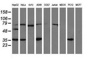
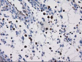
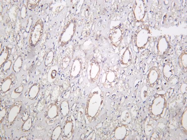
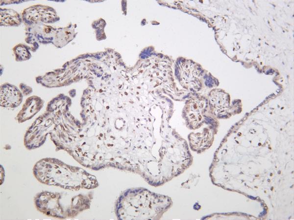
Facts about Vasodilator-stimulated phosphoprotein.
It protects the barbed end of growing actin filaments against capping and increases the rate of actin polymerization in the presence of capping protein. VASP stimulates actin filament elongation by promoting the transfer of profilin- bound actin monomers on the barbed end of growing actin filaments.
Human | |
---|---|
Gene Name: | VASP |
Uniprot: | P50552 |
Entrez: | 7408 |
Belongs to: |
---|
Ena/VASP family |
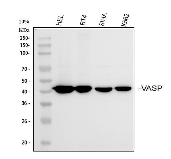
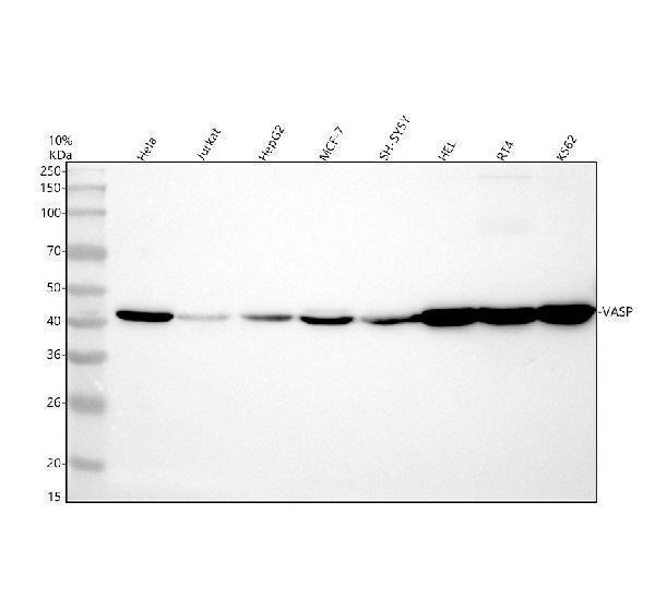
vasodilator-stimulated phosphoprotein; VASP
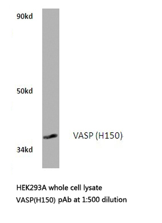
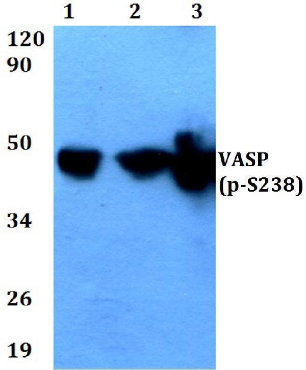
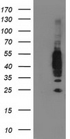
Mass (kDA):
39.83 kDA
Human | |
---|---|
Location: | 19q13.32 |
Sequence: | 19; NC_000019.10 (45507459..45526989) |
Highly expressed in platelets.
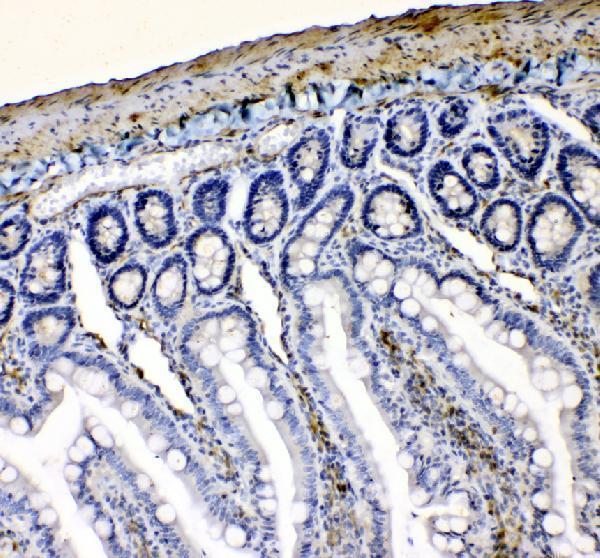
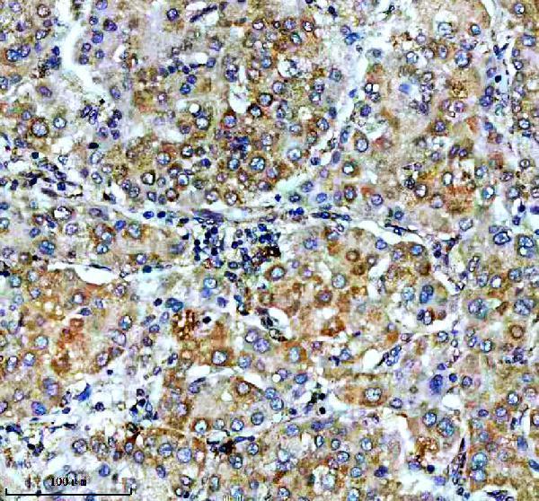
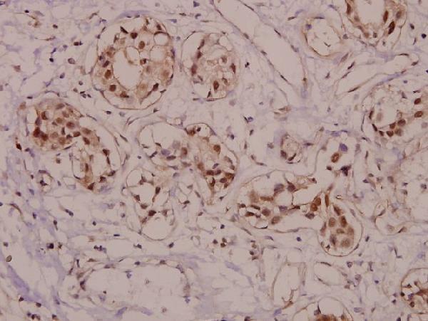
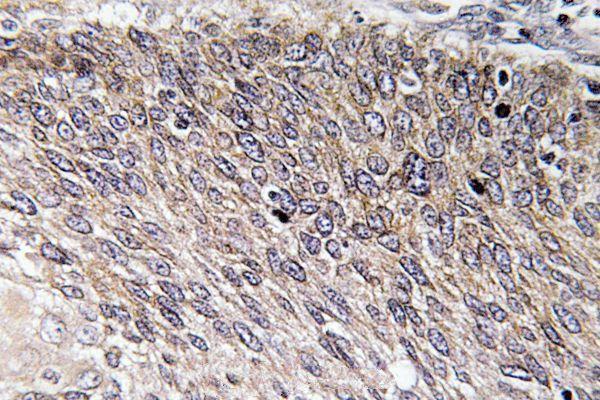
Cytoplasm. Cytoplasm, cytoskeleton. Cell junction, focal adhesion. Cell junction, tight junction. Cell projection, lamellipodium membrane. Cell projection, filopodium membrane. Targeted to stress fibers and focal adhesions through interaction with a number of proteins including MRL family members. Localizes to the plasma membrane in protruding lamellipodia and filopodial tips. Stimulation by thrombin or PMA, also translocates VASP to focal adhesions. Localized along the sides of actin filaments throughout the peripheral cytoplasm under basal conditions. In pre-apoptotic cells, colocalizes with
There are many questions that you can ask yourself when conducting experiments using Boster Bio. These guides and tips can assist you in optimizing your experiments, and improve the results. Troubleshooting is a problem that every researcher has to deal with. Although protocols and controls can be used to eliminate a lot of errors, it's vital to be able detect and fix any issues that may arise.
Phosphorylation of VASP by cGPK and cAPK alters its mobility and function in vivo. Monoclonal antibodies for cGPK and cAPK recognize VASP that is phosphorylated, an indicator that can be used to define VASP physiological function. In the current study, we found one site on VASP, Ser157, that is preferentially phosphorylated by cAPK and cGPK. The phosphorylation of this particular site alters VASP's electrophoretic mobility which results in an apparent shift in molecular mass.
The phosphorylation of VASP's serine 157 by cAPK and cGPK are similar in terms of time and intensity. When activated with cGPK, there is the highest levels of phosphorylation for VASP serine 239. However only 50% of the phosphorylation is caused by cGPK. However, the monoclonal VASP antibody 16C2 detects VASP phosphorylation in human platelets.
Phosphorylation of VASP through CGPK and cAPK is controlled differently in human platelets. In a previous study we discovered that cGPK regulates VASP and cAPK inhibits platelet inhibition. We discovered that cGPK is a phosphorylator of VASP at serine 239, which is in line with effects of CGPK on the phosphorylation of platelets.
Utilizing the commercial peptide synthesizer we generated phosphopeptides that bind the serine 239 phosphorylation site of VASP. In the process of peptide synthesis we added phosphohoserine with Fmoc-Ser(PO(OBzl)OH. We then bonded these phosphopeptides to keyhole limpet hemocyanin using a second antibody and electrophoretic gel chromatography (ECL).
We first examined a rat platelet with an antibody to VASP against Ser239 and a 10- mm PGE1 solution. To find out if cGPK inhibits cAPK's activity, we then looked at the platelet samples of the rats. The platelet sample was infused with a monoclonal VASP antibody to Ser239 as well as incubated with 100 millimeters of PGE1 as well as a 5 SNP solution of 5 percent.
These findings also suggest that cGPK and/or cAPK could have a dual effect and result in the generation of a 16C2-recognised signal for VASP. This is the first study that has shown that both signal-generating proteins are essential to regulate VASP. This study has some limitations.
The activation of cAPK through M4 VASP antibody results in an phosphorylation process of serine 239 of the VASP protein in cell extracts and intact human platelets. This phosphorylation is a distinct signal triggered by activation of cGPK. The phosphorylation of VASP serines 157 and 239 is a brief and reversible response to activation. The reaction is asymmetric, however it requires two distinct antagonists either cGPK inhibitors or substrates.
cGPK was synthesized from a cAPK subunit. It phosphorylated His6VASP to 30 degrees Celsius in 10 mm HEPES buffer. The phosphorylation-induced mobility shift of VASP was detected by 16C2 antibody. These studies indicate that 16C2 is a powerful tool to evaluate VASP function in intact cells.
A VASP shift is caused by the phosphorylation of VASP by cAMP dependent protein kinases. The VASP signal is detected by a monoclonal antibody recognizing the phosphorylation of serine 239 in VASP in human platelets. The 16C2 antibody has the highest sensitive for measuring the activation of cGMP dependent kinases within intact cells.
VASP signals that have altered phosphorylation sites result from serine 239-phosphorylation by cAPK. Both wild-type and mutant VASP exhibit altered phosphorylation sites. This makes it possible to identify VASP in various contexts. The studies have also shown that VASP is able to activate cGPK and activate cAPK and that the phosphorylation sites trigger an expression of VASP in cells.
In addition to the effects of the cAMP-dependent protein kinases VASP also functions as a substrate for profilin. Both proteins are found in the cell cortex in highly dynamic areas. Furthermore, both proteins are functionally linked and may play a role in the formation of F-actin that is spatially restricted. Profilin plays a new role in modulating the cell's response to the formation of actin filaments, as shown by this study.
Ena/VASP proteins are responsible for the regulation of actin-based mobility and growth cone guidance and are well-known regulators. They regulate the actin cytoskeleton and influence tensile strength as well as contractility. Mice with low levels of VASP activity have more thick actin stress fibers, and larger focal adhesions. Microspike rigidity is regulated by VASP activity. They are involved in numerous physiological processes inside cells.
To study the significance of cAPK in the regulation of expression of VASP, researchers employed the SU(4) spectrum to determine its decay rate. The authors also determined VASP's phosphorylation sites using old mesons. The mutations don't alter the overall properties of the VASP protein. The gene is essential for controlling cell growth.
The ActA protein of Listeria monocytogenes triggers cytoskeleton reorganization. Two profilin-binding proteins are involved in this interaction. This interaction is caused by the EVh2 domain motif. It is present in two major Eukaryotic ActA analogs. These two proteins may play an interchangeable role in activating platelets.
In this study, we used an VASP mutant to demonstrate that phosphorylation of cGPK results in a 16-C2 recognized signal. This mutation alters the electrophoretic mobility , and the phosphorylation location at Ser157. Mutations in this site may cause a shift in molecular mass.
Phosphorylation of VASP by cGPK and cAPK produces a signal that is recognized as 16C2 for VASP. Both cGPK as well as the cAPK are responsible for VASP's phosphorylation. We utilized a monoclonal antigen (M4) for VASP phosphorylation identification, and a monoclonal anti-VASP antibodies (16C2) for VASP phosphorylation identification.
The monoclonal antibody 16C2 initially created to detect phosphorylation in VASP at serine 239 which represents the best characterized the cGMP-dependent proteinkinase phosphorylation location. It also recognized mutant VASP constructs with the phosphorylation site mutated. Therefore, cGPK-induced VASP phosphorylation is a reliable indicator of cGPK activation in intact cells.
The plasma level of cytokine is determined by VASP's site of phosphorylation. If cGPK is blocked VASP phosphorylation occurs, and this is the first step towards creating a functional inhibitor of proteins. VASP phosphorylation, added to cGPK, is the most frequent biomarker of cGPK.
The activation of the cGPK gene has been associated with phosphorylation of VASP in serine 157 and serine 239 via the kinase PI3Kase. The effects of cGPK on VASP phosphorylation was similar. The highest level of phosphorylation occurs in serine 239 whereas intact platelets have only have about half the activity of the CGPK. Tests with human plates lacking cGPK demonstrated that VASP phosphorylation results in a 16C2-recognized signal.
The phosphorylation of VASP in serine 157 permits us to look for substances that can affect the function of the VASP. These compounds can be used for treating cardiovascular diseases associated with vascular damage. The various embodiments of the invention are discussed below. These examples are intended to clarify the invention but do not restrict the invention. Contact us with any questions or wish to know more about our invention.
A new monoclonal antibody known as 16C2 is being developed to detect phosphorylated ASP in biological material. This antibody is ideally suited for human biological material. The antibody itself has a particular binding partner, which allows us to detect VASP that has been phosphorylated. This method is based on a method called Kiihler and Milstein.
PMID: 7828592 by Haffner C., et al. Molecular cloning, structural analysis and functional expression of the proline-rich focal adhesion and microfilament-associated protein VASP.
PMID: 10087267 by Laurent V., et al. Role of proteins of the Ena/VASP family in actin-based motility of Listeria monocytogenes.