VGLL2 / Transcription cofactor vestigial-like protein 2 · Western blot design guide

Design a Western Blot for VGLL2

Source-linked VGLL2 Western blot protocol options, expected-band and isoform facts, troubleshooting for weak or shifted signal, and recommended anti-VGLL2 WB antibodies. Everything you need to plan the experiment before you commit precious samples.

Evidence assembled October 2026 · For research use; verify linked source records and product datasheet before use
Western blot protocol sheet for VGLL2: expected band ~33.4 kDa, hero antibody A11724, catalog values and labelled standard workflow; separate PMC comparisons on the guide
Printable VGLL2 Western blot protocol sheet — expected band ~33.4 kDa, antibody A11724, controls and PMC citations. Open the full VGLL2 WB guide →

VGLL2 Western Blot Experimental Design Guide

Expected bands, source-linked protocol options, controls and antibodies — the at-a-glance facts below, then the full design guide.

Must know before running
Expected band ~33.4 kDa
Gel 12–15% (standard starting point)
Negative control ⓘ Suggested KO / knockdown lysate
Important caveats
Reasons your observed band may differ from the expected size.
PTM —
Caveat —
Gene-set association MSigDB C7 membership
Isoform 2 isoform(s)
Section 1

Source-Linked VGLL2 Western Blot Protocol Options

The A11724 protocol combines labelled catalog values with standard starting conditions. Published comparisons retain their own sample, reagent and detection scope.

Recommended Western blot protocol parameters
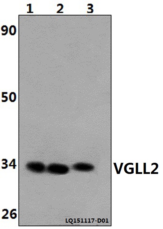
Sample / lysateThe Heart of Rat(40ug), The Heart of Mouse(40ug), Hela (40ug) (catalog A11724)
Gel %12–15% (standard starting point)
Load20–30 µg total protein per lane; optimize for abundance (standard starting point)
TransferStandard semi-dry transfer; verify efficiency (standard starting point)
Membrane0.45 µm PVDF (standard starting point)
Blocking5% milk or 5% BSA in TBST (standard starting point)
Primary antibodyA11724; use the WB datasheet starting dilution (standard starting point)
Primary incubationOvernight at 4 °C (standard starting point)
Secondary antibodySpecies-matched HRP conjugate at validated dilution (standard starting point)
Secondary incubation1 h at room temperature (standard starting point)
Wash3 × 5 min in TBST (standard starting point)
DetectionECL; bracket exposures to avoid saturation (standard starting point)
Section 2

What Is the Expected VGLL2 Western Blot Band Size?

VGLL2 is predicted at 33.4 kDa; its isoforms could affect migration, but no empirical band size or distinct isoform bands are established.

What am I looking at on my blot?
Band near 33.4 kDaConsistent with predicted VGLL2 size; confirm identity with controls
Two bands at different positionsCould reflect isoforms 1 and 2; their migration difference is unestablished
Weak band in whole-cell lysateNuclear VGLL2 may be diluted in the sample
Band enriched in nuclear fractionConsistent with VGLL2 nuclear localization
💡Expected VGLL2 appearanceVGLL2 has a predicted mass of 33.4 kDa, but no empirical band size or resolvable migration pattern for its two isoforms is supplied; confirm band identity with appropriate controls.
How each factor affects band size
Predicted VGLL2 massProvides a 33.4 kDa reference, not a validated apparent band size
Alternative splicingCould alter apparent size, but no migration difference is established
Isoform 1Its size relative to isoform 2 is unspecified
Isoform 2Its size relative to isoform 1 is unspecified
Why is my band missing or off?
SituationLikely causeNext action
No band in lysateNuclear VGLL2 may be poorly representedCheck a nuclear fraction and verify fraction recovery
Band higher than expectedBand identity or migration difference is unestablishedConfirm with VGLL2 knockdown or an independent antibody
Band lower than expectedBand identity or migration difference is unestablishedConfirm with VGLL2 knockdown or an independent antibody
Multiple bandsIsoforms 1 and 2 are annotated, but distinct bands are unprovenCompare isoform-specific expression and VGLL2 knockdown
Weak or no signalNuclear VGLL2 may be diluted in whole-cell lysateEnrich the nuclear fraction and check sample loading

Sample controls for VGLL2 Western blot

🧪For positive controls for VGLL2 in Western blot, you can use no HPA-supported tissue or cell line from the supplied evidence.
Positive control: No high/medium HPA tissue identified
Negative control: Suggested KO / knockdown lysate
Loading controls: Run GAPDH, β-actin, and a total-protein stain alongside the samples.
⚠️Feasibility: VGLL2 is nuclear, but the supplied HPA record has no expression data to guide control selection.

HPA tissue expression evidence for VGLL2

Comprehensive Human Protein Atlas IHC scoring per tissue. Rows are taken directly from the HPA tissue chart — click any row's HPA link to view the source.

Higher expression tissues · candidate positive controls from IHC

TissueCell typeLevelEvidenceSource
No high/medium HPA tissues identified in the supplied evidence.

Lower expression tissues · IHC evidence, not confirmed WB-negative controls

TissueCell typeLevelEvidenceSource
No lower-expression tissue rows available in the supplied evidence.
Section 3

Advanced VGLL2 Western Blot Tips

Deeper troubleshooting and optimisation questions for VGLL2, answered from its protein features.

How should VGLL2 band migration be interpreted?
Band shift · Use the separately labelled calculated mass and catalog-observed evidence above. A sequence annotation does not establish an observed migration shift. Verify target identity with orthogonal controls.
Could VGLL2 isoforms produce different bands?
Isoforms · Yes. UniProt lists two isoforms; isoform 2 lacks residues 131–304 in canonical UniProt numbering. It may therefore run below isoform 1, but these features do not establish where either band appears on a blot.
Could modification explain a shifted VGLL2 band?
PTM · The supplied UniProt features list no modified residues or glycosylation sites. They provide no specific modification to test as an explanation. Even a listed modification would not, by itself, establish a visible band shift.
Does this guide establish induction of VGLL2?
Induction · No general induction response is established by this guide. A pathway or gene-set association is not evidence of induction in a particular specimen. Verify the relevant treatment and control in a target-specific experiment.
What transfer method to use for VGLL2 Western blot?
Transfer · The canonical predicted mass is 33.4 kDa, while isoform 2 lacks residues 131–304. Choose transfer conditions that retain proteins in this size range, and check transfer of both bands if testing for both isoforms. The supplied features do not specify a transfer method.
How should blocking be optimized?
Blocking · Standard workflow guidance: follow the A11724 datasheet where specified. Otherwise compare 5% milk or 5% BSA in TBST; for a phospho-specific assay start with BSA. Optimize background and specific signal with matched controls.
What should guide VGLL2 quantitation?
Quantitation · Decide whether to quantify isoform 1, isoform 2, or both before selecting bands. Isoform 2 lacks canonical UniProt residues 131–304, so verify that the antibody recognizes both if combining their signals. VGLL2 is annotated as nuclear, which can guide sample preparation.
What size band is expected for VGLL2?
Interpretation · The canonical VGLL2 sequence is 317 amino acids with a predicted mass of 33.4 kDa. No observed band position is supplied, so use 33.4 kDa as a reference rather than an expected apparent mass.

Check the antibody's recognized sequence against the isoform difference. An epitope within canonical UniProt residues 131–304 is absent from isoform 2; an antibody targeting that region would not detect isoform 2.

Compare bands with the 33.4 kDa canonical prediction and consider the shorter isoform 2, which lacks canonical UniProt residues 131–304. Check whether the antibody can recognize that isoform. Without an observed reference band, band position alone cannot identify either isoform.
Boster reagents

VGLL2 Western Blot Antibodies

Catalog antibodies with Western blot application and product-specific WB images. Evaluate suitability with the reported sample, controls and experimental conditions.

Real WB data Western blot (WB) analysis of VGLL2 polyclonal antibody at 1:500 dilution Lane1:The Heart tissue lysate of Rat(40ug) Lane2:The Heart tissue lysate of Mouse(40ug) Lane3:Hela whole cell lysate(40ug)
Anti-VGLL2 Antibody
Cat # A11724

the supplier A11724 is a polyclonal anti-VGLL2 antibody listed for human, mouse, and rat. Its WB image shows rat and mouse heart lysates and HeLa whole cell lysate, each at 40 µg, using a 1:500 antibody dilution. Evidence shown is limited to those samples and conditions.

Which to pick: A11724 is the only listed VGLL2 antibody. Its WB image provides a starting reference for rat and mouse heart tissue and HeLa cells at 40 µg per lane and 1:500 dilution; confirm performance for your own samples and conditions.

Source: BosterBio VGLL2 gene-info card — filtered to Western-blot-capable antibodies; each card shows that product's actual WB validation figure.