VPS4A / Vacuolar protein sorting-associated protein 4A · IHC design guide

Design Immunohistochemistry for VPS4A

Plan chromogenic VPS4A IHC in paraffin sections with the catalog antibody at 0.5–1 μg/mL (datasheet A03216-1). Compare cytoplasmic staining with the tissue profile while accounting for its low consistency with RNA expression (HPA tissue IHC).

Evidence assembled Oct 2026 · For research use; verify linked source records and product datasheet before use
Immunohistochemistry protocol sheet for VPS4A (IHC for VPS4A): expected localisation Cytoplasmic tissue staining (HPA tissue IHC), antibody A03216-1, validated IHC image, and IHC protocol steps
Printable VPS4A IHC protocol sheet — expected localisation Cytoplasmic tissue staining (HPA tissue IHC), antibody A03216-1, controls and protocol steps. Open the full VPS4A IHC guide →

VPS4A Immunohistochemistry Experimental Design Guide

Expected localisation, validated protocols, controls and antibodies — the at-a-glance facts below, then the full design guide.

Must know before staining
Expected localisation Cytoplasmic tissue staining (HPA tissue IHC)
Staining pattern Widespread cytoplasmic staining across tissue cell types (HPA tissue IHC)
Antigen retrieval EDTA pH 8.0 HIER, heat-mediated (datasheet A03216-1)
Positive control ⓘ Adrenal gland+4 more · see all
Negative control ⓘ None in HPA (detected in all 45 tissues); use no-primary + isotype controls
Important caveats
Reasons your staining may differ from the expected pattern.
Fixation Keep paraffin-section fixation consistent (standard IHC practice; not target-specific)
Caveat Staining has low consistency with RNA expression (HPA tissue IHC)
Regulation Ubiquitously expressed (UniProt)
Isoform / epitope No isoforms reported; one chain spans residues 1–437 (UniProt)
Section 1

Recommended VPS4A IHC & IF Protocols

The catalog antibody has an IHC-P protocol (datasheet A03216-1). Two published VPS4A IHC methods provide additional conditions (PMC7005644; PMC12190599).

Recommended immunohistochemistry (IHC-P) protocol parameters
SampleParaffin-embedded human lung cancer tissue; fixative not specified (datasheet A03216-1)
FixationImage fixative and duration unreported (datasheet A03216-1); verify before use.
Sectioning4–5 µm sections on charged slides (standard)
DeparaffinisationXylene, graded ethanol series to water (standard)
Antigen retrievalHeat retrieval: EDTA pH 8.0 (datasheet A03216-1); 20 min, 95–100 °C (standard)
Peroxidase block3% H2O2, 10 min, room temperature (standard)
Blocking10% goat serum (datasheet A03216-1)
Primary antibodyRabbit anti-VPS4A, 0.5-1μg/ml (datasheet A03216-1)
Primary incubationOvernight at 4 °C (datasheet A03216-1)
DetectionStreptavidin-biotin complex (SABC), DAB chromogen (datasheet A03216-1)
CounterstainHematoxylin, blue, dehydrate and mount (standard)
Expected resultVPS4A-positive staining in glandular cells of adrenal gland (HPA tissue IHC: Medium). HPA tissue profile: Ubiquitous cytoplasmic expression. No signal in the no-primary control.
💡Decision noteStart with heat-mediated EDTA retrieval at pH 8.0 (datasheet A03216-1); compare citrate at pH 6.0 if optimization is needed (PMC12190599).
Section 2

What Is the Expected VPS4A Staining Pattern?

VPS4A is ubiquitously expressed and associates with late endosomes, the cytoplasm and the midbody; it has no transmembrane segment (UniProt Q9UN37). In paraffin sections, expect predominantly cytoplasmic staining across cell types, including glandular and neuronal cells reported at medium intensity (HPA: tissue IHC). The tissue IHC profile is Approved, with low consistency between antibody staining and RNA expression (HPA: tissue IHC).

What am I looking at on my slide?
Cytoplasmic staining in glandular or neuronal cells.This fits the reported ubiquitous cytoplasmic profile and medium staining in several glandular and neuronal populations (HPA: tissue IHC). Compare cells within the same section: intensity can vary, and a positive call should rest on cellular localisation as well as colour strength (general IHC practice).
A sharply nuclear-only or extracellular pattern dominates.That pattern does not fit the reported cytoplasmic tissue profile or the recorded late-endosome, cytoskeletal and midbody locations (HPA: tissue IHC; UniProt Q9UN37). Check staining controls and morphology before calling it VPS4A; membrane association alone does not imply a continuous cell-surface outline (UniProt Q9UN37 topology).
Strong staining appears mainly in a population reported as low.Marked staining limited to cardiomyocytes, myocytes or adipocytes conflicts with their low HPA levels (HPA: tissue IHC). Consider cross-reactivity or endogenous detection activity, particularly if the signal persists in a no-primary control (general IHC practice). HPA's Approved rating has low RNA–protein consistency, so treat this as a prompt to investigate, not proof of artefact (HPA: tissue IHC).
Colour spreads across stroma or obscures cell boundaries.Diffuse, poorly localised colour is difficult to score against a cytoplasmic expectation (HPA: tissue IHC). A no-primary control can reveal detection background; excess primary or inadequate blocking or washing can also contribute (general IHC practice). Reassess the slide only after cellular detail becomes readable.
Little or no staining appears in a reported medium population.Medium staining is reported in bronchial respiratory epithelium and several glandular or neuronal populations (HPA: tissue IHC). First check that the expected cells are present and preserved, then check assay controls and the catalog antibody's IHC-P instructions (general IHC practice). One blank field alone cannot establish absent VPS4A expression.
💡Expected VPS4A appearanceCall a positive result when readable cytoplasmic staining appears in appropriate cells, with medium intensity plausible in HPA-listed glandular or neuronal populations; dominant nuclear-only or extracellular colour is suspect (HPA: tissue IHC; UniProt Q9UN37).
How each factor affects the staining
Tissue and cell-type baselineVPS4A is ubiquitously expressed (UniProt Q9UN37), and HPA describes ubiquitous cytoplasmic tissue staining (HPA: tissue IHC). HPA reports low staining in cholangiocytes, trophoblastic cells, cardiomyocytes, smooth and skeletal muscle cells, and adipocytes; low is not a zero-expression control (HPA: tissue IHC).
Intracellular locationVPS4A is associated with late-endosome membranes and also localises to the cytoskeleton, spindle and midbody (UniProt Q9UN37). It has no transmembrane segment, so a continuous surface-only pattern is a poor match (UniProt Q9UN37 topology). Routine tissue IHC need not resolve these smaller structures (general IHC practice).
IHC antibody evidenceTwo listed antibodies have Approved IHC status, CAB018751 and CAB034411 (HPA: antibodies). The tissue profile is also Approved but has low consistency with RNA expression (HPA: tissue IHC). Neither designation establishes that every tissue or every unexpected compartment is specific.
Processing and epitope limitsUniProt records one chain spanning residues 1–437, with no signal peptide or propeptide (UniProt Q9UN37). The supplied sources do not identify the catalog antibody's epitope or a VPS4A-specific retrieval response; choose retrieval conditions from its IHC-P instructions and verify them with controls (general IHC practice).
IF/ICC Q&A: should the pattern match tissue IHC?In ICC-IF, the supported main location is the midbody, with an HPA caution that the summary draws on antibodies targeting proteins from multiple genes (HPA: subcellular). This supports a localisation check in dividing cells, not an IHC-P protocol option or a requirement that every paraffin-section cell show a midbody.
Why is my staining missing, weak or wrong?
SituationLikely causeNext action
Reported medium-staining cells look blank.The expected cells may be absent from the section, or the IHC run may have failed (HPA: tissue IHC; general IHC practice).Confirm cell identity and section quality, then review the run control and follow the catalog antibody's IHC-P retrieval and detection instructions (general IHC practice).
Only nuclei stain strongly.A nuclear-only pattern conflicts with the cytoplasmic tissue profile (HPA: tissue IHC).Inspect morphology and the no-primary control; repeat with the IHC-validated antibody's documented conditions if the compartment remains discordant (general IHC practice).
Low-listed cells stain more strongly than surrounding cells.This is discordant with their reported low levels, although HPA notes low RNA–staining consistency (HPA: tissue IHC).Check a no-primary control for endogenous detection activity and compare the same cell type across sections before assigning specificity (general IHC practice).
Diffuse colour masks cytoplasmic detail.Detection background, excess primary or incomplete washing may obscure cellular localisation (general IHC practice).Check the no-primary control, blocking and washes; adjust primary concentration within the documented IHC-P guidance and rescore only interpretable cells (general IHC practice).
A continuous cell-edge outline is the dominant signal.VPS4A associates with endosomal membranes but has no transmembrane segment; a surface-only outline conflicts with its recorded locations (UniProt Q9UN37 topology).Compare with cytoplasmic staining in HPA-listed cells and review controls before treating the outline as target signal (HPA: tissue IHC; general IHC practice).
A visible midbody is absent in a tissue section.Midbody localisation is documented, but tissue IHC predominantly shows cytoplasmic expression and may not contain an interpretable dividing cell (UniProt Q9UN37; HPA: tissue IHC).Score the cytoplasmic pattern and relevant cell types; use the separate ICC-IF guide if midbody localisation is the experimental question (HPA: tissue IHC; HPA: subcellular).

Sample controls for VPS4A IHC & IF

🧪Run bronchus first and look for staining in respiratory epithelial cells (HPA: Medium in bronchus respiratory epithelial cells). HPA detects VPS4A in all 45 scored tissues, so use no-primary and isotype controls in place of a negative tissue; cells without specific staining on the positive slide should retain counterstain, but no cell population is established as a biological negative (HPA: no negative tissue rows).
Positive control tissue: Adrenal gland (Glandular cells, HPA Medium)
Negative control tissue: None in HPA: VPS4A is detected in all 45 scored tissues. Use a no-primary (secondary-only) and an isotype control instead.
ICC-IF cell lines (HPA subcellular resource): HPA ICC-IF images show VPS4A in SiHa, U2OS, hTERT-RPE1 (serum starved), KOLF2.1J, with annotated localisation: Midbody (supported) (HPA subcellular).
Technical controls: Include a secondary-only slide, an isotype control matched to the primary antibody’s host species and clonality, and VPS4A-knockout material if available as a biological negative (standard IHC practice). Block endogenous peroxidase and check for endogenous biotin before interpreting bronchus DAB staining because the selected caption uses biotin-based detection (caption: biotinylated secondary, SABC and DAB).
⚠️Feasibility: A target-specific fixation window or fixation effect is unreported, and the selected A03216-1 paraffin-section caption does not state the fixative (caption: fixative unreported). The reported IHC conditions use heat retrieval in EDTA at pH 8.0, but the evidence does not establish that retrieval is required or that frozen sections or IF are easier (caption: EDTA heat retrieval). In bronchus, peroxidase-positive inflammatory cells could complicate DAB interpretation; assess staining against morphology and the negative controls (standard IHC practice).

HPA tissue IHC evidence for VPS4A

Comprehensive Human Protein Atlas IHC scoring per tissue (reliability: Approved — Low consistency between antibody staining and RNA expression data.). Rows are taken directly from the HPA tissue chart — click any row's HPA link to view the source.

Positive expression · recommended positive controls

TissueCell typeLevelEvidenceSource
Adrenal gland Glandular cells Medium Protein (IHC) HPA →
Appendix Glandular cells Medium Protein (IHC) HPA →
Breast Glandular cells Medium Protein (IHC) HPA →
Bronchus Respiratory epithelial cells Medium Protein (IHC) HPA →
Caudate Neuronal cells Medium Protein (IHC) HPA →

Undetected expression · recommended negative controls

TissueCell typeLevelEvidenceSource
None in HPA: VPS4A is detected in all 45 scored tissues. Use a no-primary (secondary-only) and an isotype control instead.
Section 3

Advanced VPS4A IHC Tips

Use the catalog antibody’s paraffin section workflow as the IHC starting point, then judge staining against VPS4A’s reported cytoplasmic and midbody locations.

Which retrieval should I start with for VPS4A in paraffin sections?
Start with heat mediated retrieval in EDTA at pH 8.0 for paraffin sections (datasheet A03216-1). The selected image used this retrieval before an overnight incubation at 4°C with 1 μg/ml primary antibody, so keep those conditions together for an initial comparison (datasheet A03216-1). If staining is weak, adjust heating duration on matched sections while keeping the buffer and pH constant, and inspect morphology for retrieval damage (standard IHC practice). Compare each change with a no primary control and a consistently stained reference section, because stronger DAB alone does not establish specific VPS4A detection (standard IHC practice).
How should I handle fixation when VPS4A staining varies between blocks?
The selected tissue image identifies a paraffin section but does not state its fixative, so target specific fixation sensitivity is unknown (datasheet A03216-1). Record the fixative, fixation duration, processing history and section age for each block before attributing staining differences to VPS4A biology (standard IHC practice). Run discrepant blocks together using EDTA at pH 8.0, the same 1 μg/ml primary concentration and the same DAB development interval (datasheet A03216-1; standard IHC practice). Check morphology and an internal reference area on every section; tissue patterns alone cannot reveal whether fixation masked this antibody’s epitope (standard IHC practice).
Which VPS4A staining pattern is plausible in a tissue section?
Expect predominantly cytoplasmic tissue staining, with possible midbody signal in dividing cells (HPA tissue IHC: ubiquitous cytoplasmic expression; UniProt Q9UN37 subcellular location). VPS4A also associates with late endosome membranes and the spindle cytoskeleton, and it has no transmembrane segment (UniProt Q9UN37 subcellular location and topology). Assess punctate or midbody staining within intact cells against the surrounding cytoplasm, ideally on serial sections with a proliferation or midbody marker (standard IHC practice). A uniformly nuclear or sharply cell surface pattern deserves specificity checks before interpretation, because those compartments do not match the supplied VPS4A localisation record (UniProt Q9UN37 subcellular location).
Could epitope placement explain missing or unexpected VPS4A staining?
The supplied record lists one 437 amino acid chain, no isoforms, no signal peptide and no transmembrane segment (UniProt Q9UN37 processing, isoforms and topology). It identifies an MIT domain at residues 2–80 and modified residues including acetyllysine 8 and phosphoserines 95 and 97, but gives no epitope for this antibody (UniProt Q9UN37 domains and modified residues; datasheet A03216-1). Request the immunogen or epitope information before linking a staining change to a modification or domain (standard IHC practice). Test specificity with an independently validated antibody or appropriate loss of VPS4A expression where feasible, while holding retrieval and detection conditions constant (standard IHC practice).
How can IF help investigate a VPS4A IHC pattern?
Use IF as an independent localisation check when chromogenic IHC leaves cytoplasmic staining or a suspected midbody difficult to resolve (HPA tissue IHC: ubiquitous cytoplasmic expression; HPA subcellular: midbody supported). Multiplex VPS4A with a marker identifying the expected cell population and, when assessing division, a midbody marker; choose separated fluorophores after checking tissue autofluorescence in unstained material (standard IF practice). Because VPS4A is associated with the late endosome membrane without a transmembrane segment, select and validate permeabilisation that permits access to its cytoplasmic side (UniProt Q9UN37 topology and subcellular location; standard IF practice). Treat IF and paraffin section IHC results as separate assay evidence when interpreting agreement (standard assay practice).
What should I check when DAB background obscures VPS4A staining?
The selected paraffin section used 10% goat serum blocking, biotinylated goat anti rabbit secondary antibody, streptavidin biotin complex and DAB (datasheet A03216-1). Inspect a no primary control for secondary binding, endogenous biotin associated signal and residual peroxidase activity before changing the primary concentration (standard IHC practice). Apply a peroxidase block and check wash quality and DAB development time on matched sections; those are general chromogenic workflow controls, not VPS4A specific evidence (standard IHC practice). Compare background with the expected intracellular pattern, since diffuse staining across extracellular spaces or tissue edges is less convincing than staining confined to intact cells (UniProt Q9UN37 subcellular location; standard IHC practice).
How should I score VPS4A staining across tissue samples? ⚠ ANSWER MARKED FOR VERIFICATION
Define the cell population and region of interest before scoring, then record cytoplasmic intensity and the percentage of positive cells for an H-score (HPA tissue IHC: ubiquitous cytoplasmic expression; standard IHC practice). If the question concerns dividing cells, count clearly identified VPS4A positive midbodies per mm² and report the number of eligible dividing cells as the denominator (HPA subcellular: midbody supported; standard IHC practice). Keep retrieval, 1 μg/ml primary concentration, imaging and DAB development consistent across samples (datasheet A03216-1; standard IHC practice). Exclude damaged regions and report background corrected results with matched reference tissue, since broad expression makes raw positive area difficult to compare (UniProt Q9UN37 tissue specificity; standard IHC practice).
How do I distinguish a true VPS4A signal from artefact?
Give most weight to intracellular cytoplasmic staining in intact cells and to a discrete midbody signal in dividing cells (HPA tissue IHC: ubiquitous cytoplasmic expression; HPA subcellular: midbody supported). VPS4A is ubiquitously expressed, while the supplied HPA tissue data describe low consistency between antibody staining and RNA expression, so intensity alone should not establish cell specific enrichment (UniProt Q9UN37 tissue specificity; HPA tissue IHC reliability description). Review no primary and peroxidase controls, tissue edges and necrotic areas for staining that follows damage or endogenous enzyme activity (standard IHC practice). Recheck an unexpected compartment or cell population with an independent specificity control before making a biological claim (standard IHC practice).
Boster reagents

Best VPS4A / Vacuolar protein sorting-associated protein 4A IHC Antibodies

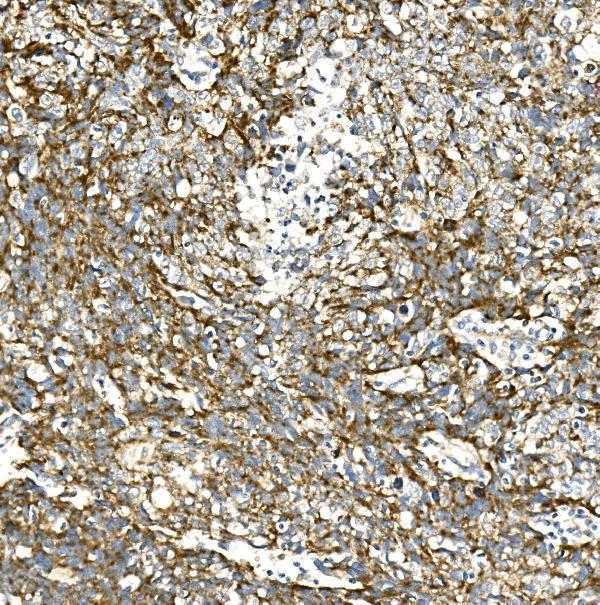
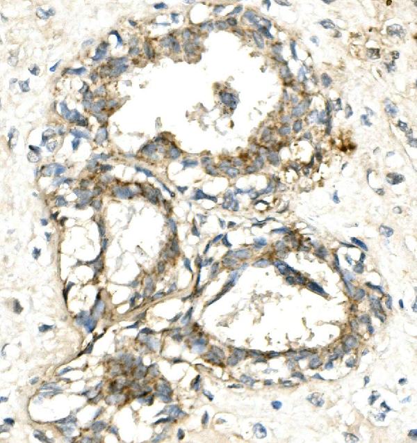
A03216-1 has IHC images from human lung and mammary cancer paraffin sections (A03216-1 IHC captions). Human, mouse and rat reactivity is listed; no IF figure is supplied (catalog reactivity; IF image alts).

Real IHC data IHC analysis of VPS4A using anti-VPS4A antibody (A03216-1). VPS4A was detected in paraffin-embedded section of human lung cancer tissue. Heat mediated antigen retrieval was performed in EDTA buffer (pH8.0, epitope retrieval solution). The tissue section was blocked with 10% goat serum. The tissue section was then incubated with 1μg/ml rabbit anti-VPS4A Antibody (A03216-1) overnight at 4°C. Biotinylated goat anti-rabbit IgG was used as secondary antibody and incubated for 30 minutes at 37°C. The tissue section was developed using Strepavidin-Biotin-Complex (SABC) (Catalog # SA1022) with DAB as the chromogen.
Anti-VPS4A Antibody ®
Cat # A03216-1

A03216-1 is listed for IHC with human, mouse and rat reactivity (catalog applications/reactivity). Its IHC images show human lung and mammary cancer paraffin sections; neither caption reports the fixative (A03216-1 IHC captions).

Which to pick: For tissue IHC, choose A03216-1: its rabbit antibody is shown on human paraffin sections using EDTA retrieval at pH 8.0, with the fixative unreported (catalog host; A03216-1 IHC captions). No SKU can be recommended for IF/ICC because A03216-1 has no listed IF/ICC application or IF figure (catalog applications; IF image alts). For mouse or rat IHC, A03216-1 has listed reactivity, though its IHC images show human tissue and clonality is unreported (catalog reactivity; A03216-1 IHC captions; catalog clone).

Each figure is that product's own IHC / IF validation image from its datasheet.

References

  1. UniProt Consortium. UniProt entry Q9UN37 (VPS4A_HUMAN, Vacuolar protein sorting-associated protein 4A).
  2. Human Protein Atlas. VPS4A tissue IHC expression (reliability: Approved).
  3. Human Protein Atlas. VPS4A subcellular location (ICC-IF): Localized to the midbody. Caution: Based on antibodies targeting proteins from multiple genes..
  4. Human Protein Atlas. VPS4A antibody validation summary (3 antibodies).
  5. Vps4A functions as a tumor suppressor by regulating the secretion and uptake of exosomal microRNAs in human hepatoma cells. Hepatology (Baltimore, Md.) 2015 — PMC4511093.
  6. Synthetic lethality between VPS4A and VPS4B triggers an inflammatory response in colorectal cancer. EMBO molecular medicine 2020 — PMC7005644.
  7. A Novel Modulator of Resistance for Oxaliplatin-Based Therapy for Colorectal Cancer: The ESCRT Family Member VPS4A. Cells 2025 — PMC12190599.
  8. PubMed PMID:11563910 — UniProt-cited evidence.
  9. PubMed PMID:12594041 — UniProt-cited evidence.
  10. PubMed PMID:10931946 — UniProt-cited evidence.