VPS4B / Vacuolar protein sorting-associated protein 4B · IHC design guide

Design Immunohistochemistry for VPS4B

Plan chromogenic VPS4B IHC on paraffin sections using A03403-1 at 0.5–1 μg/ml (datasheet A03403-1). Compare esophageal squamous cells, reported as high, with adipocytes, reported as undetected (HPA tissue IHC).

Evidence assembled Oct 2026 · For research use; verify linked source records and product datasheet before use
Immunohistochemistry protocol sheet for VPS4B (IHC for VPS4B): expected localisation Cytoplasmic tissue staining (HPA tissue IHC), antibody A03403-1, validated IHC image, and IHC protocol steps
Printable VPS4B IHC protocol sheet — expected localisation Cytoplasmic tissue staining (HPA tissue IHC), antibody A03403-1, controls and protocol steps. Open the full VPS4B IHC guide →

VPS4B Immunohistochemistry Experimental Design Guide

Expected localisation, validated protocols, controls and antibodies — the at-a-glance facts below, then the full design guide.

Must know before staining
Expected localisation Cytoplasmic tissue staining (HPA tissue IHC)
Staining pattern Esophageal squamous cells show cytoplasmic staining (HPA tissue IHC)
Antigen retrieval EDTA pH 8.0 HIER, heat-mediated (datasheet A03403-1)
Positive control ⓘ Bone marrow+4 more · see all
Negative control ⓘ Adipose tissue+4 more · see all
Important caveats
Reasons your staining may differ from the expected pattern.
Fixation Keep fixation consistent across paraffin sections (standard IHC practice; not target-specific)
Caveat Staining and RNA show medium consistency (HPA tissue IHC)
Regulation Ubiquitously expressed (UniProt)
Isoform / epitope No isoforms or cleaved chains annotated (UniProt)
Section 1

Recommended VPS4B IHC & IF Protocols

The catalog antibody’s IHC-P protocol (datasheet: A03403-1) is accompanied by published VPS4B IHC details for cortical tissue (PMC12695678) and colorectal samples and xenografts (PMC7005644).

Recommended immunohistochemistry (IHC-P) protocol parameters
SampleParaffin-embedded human lung cancer tissue; fixative not specified (datasheet A03403-1)
FixationImage fixative and duration unreported (datasheet A03403-1); verify before use.
Sectioning4–5 µm sections on charged slides (standard)
DeparaffinisationXylene, graded ethanol series to water (standard)
Antigen retrievalHeat retrieval: EDTA pH 8.0 (datasheet A03403-1); 20 min, 95–100 °C (standard)
Peroxidase block3% H2O2, 10 min, room temperature (standard)
Blocking10% goat serum (datasheet A03403-1)
Primary antibodyRabbit anti-VPS4B, 0.5-1μg/ml (datasheet A03403-1)
Primary incubationOvernight at 4 °C (datasheet A03403-1)
DetectionStreptavidin-biotin complex (SABC), DAB chromogen (datasheet A03403-1)
CounterstainHematoxylin, blue, dehydrate and mount (standard)
Expected resultVPS4B-positive staining in hematopoietic cells of bone marrow (HPA tissue IHC: High). HPA tissue profile: Cytoplasmic expression in several different tissues types, including esophagus. No signal in the no-primary control.
💡Decision noteStart with heat-mediated EDTA retrieval at pH 8.0 for the catalog antibody (datasheet: A03403-1). Neither included article excerpt specifies retrieval conditions (PMC12695678; PMC7005644).
Section 2

What Is the Expected VPS4B Staining Pattern?

VPS4B is a membrane-associated protein of the late endosomal compartment with no transmembrane segment (UniProt O75351 localization and topology). In paraffin-section IHC, expect mainly cytoplasmic staining in selected cells, including esophageal squamous epithelium and bone marrow hematopoietic cells (HPA tissue IHC). HPA rates its tissue staining Approved, with medium consistency between antibody staining and RNA expression (HPA tissue IHC).

What am I looking at on my slide?
Cytoplasmic staining in esophageal or cervical squamous epithelial cells, or bone marrow hematopoietic cells (HPA tissue IHC).These are listed as High-staining cell populations (HPA tissue IHC). Judge the signal within the identified cells and against neighboring tissue and a negative control (general IHC practice); HPA intensity labels do not prescribe a chromogen score for every section.
Strong, predominantly nuclear staining with little cytoplasmic signal.A nuclear-dominant pattern conflicts with HPA's cytoplasmic tissue profile and UniProt's late-endosome association (HPA tissue IHC; UniProt O75351 localization). Review morphology, counterstain and controls before assigning that signal to VPS4B (general IHC practice).
Strong staining in adipocytes or heart-muscle cardiomyocytes.HPA lists these specific cell populations as Not detected (HPA tissue IHC). Consider nonspecific antibody binding or endogenous detection activity, especially if the negative control also stains (general IHC practice); do not generalize a cell-level label to every cell in that organ.
Diffuse chromogen across cells, extracellular spaces or the whole section.A broad haze obscures the expected cell-associated cytoplasmic pattern (HPA tissue IHC). Inadequate washing, excessive detection reagent or background from the detection system can cause this appearance (general IHC practice); compare with a negative control.
No convincing signal in esophageal squamous epithelium or bone marrow hematopoietic cells.Both are listed as High in HPA tissue IHC (HPA tissue IHC). An absent signal calls for checks of tissue preservation, antigen retrieval, antibody preparation and detection controls (general IHC practice); HPA's Approved rating does not guarantee positivity in every specimen.
💡Expected VPS4B appearanceCall a section positive when identifiable squamous epithelial or hematopoietic cells show appreciable cytoplasmic chromogen (HPA tissue IHC: High); nuclear-only staining or equally strong signal in HPA Not detected cell populations is suspect (HPA tissue IHC; UniProt O75351 localization).
How each factor affects the staining
Compartment and topology (UniProt O75351 localization and topology).VPS4B associates with the late endosome membrane and has no transmembrane segment (UniProt O75351). HPA describes tissue IHC as cytoplasmic (HPA tissue IHC); paraffin IHC need not resolve individual endosomal vesicles (general IHC practice).
Tissue and cell identity (HPA tissue IHC).HPA calls esophageal, cervical, tonsillar and vaginal squamous epithelial cells High, while appendix glandular cells are Medium (HPA tissue IHC). Select and score the named cell population; whole-organ averages can hide the relevant pattern (general IHC practice).
Evidence strength (HPA tissue IHC; HPA antibody validation).HPA calls tissue staining Approved with medium RNA agreement, and lists HPA057649 and CAB046445 as IHC Approved (HPA tissue IHC; HPA antibody validation). Those labels support comparison with HPA images but do not establish an Enhanced IHC validation claim.
Does ICC-IF predict the IHC appearance? (HPA subcellular ICC-IF).HPA reports mainly vesicular localization in ICC-IF and marks that location Approved (HPA subcellular ICC-IF). Use it as compartment context; the supplied tissue IHC profile describes cytoplasmic staining, so assess the paraffin-section result at its available resolution (HPA tissue IHC; general IHC practice).
Protein forms (UniProt O75351 processing and isoforms).The supplied UniProt record lists one chain spanning residues 1–444, no signal peptide or propeptide, and no listed isoforms (UniProt O75351). These annotations give no basis to expect a secreted staining pattern or separate isoform-specific tissue pattern.
Why is my staining missing, weak or wrong?
SituationLikely causeNext action
Known High-staining cells show no chromogen (HPA tissue IHC).The assay may have failed, or the particular section may lack detectable antigen; the supplied sources do not identify a VPS4B-specific fixation effect (general IHC practice; source-scope limit).Check tissue identity and morphology, run an appropriate positive section, and verify retrieval, antibody application and detection reagents (general IHC practice).
A brown signal appears predominantly over nuclei.This differs from the cytoplasmic tissue profile and late-endosome association (HPA tissue IHC; UniProt O75351 localization); counterstain or nonspecific signal may complicate reading (general IHC practice).Inspect a negative control and matched morphology, then reassess staining location before scoring VPS4B positivity (general IHC practice).
Adipocytes or cardiomyocytes stain strongly.Those named cell populations are listed as Not detected (HPA tissue IHC); nonspecific binding or endogenous detection activity is possible (general IHC practice).Compare a negative control and a known High cell population; address background in the chosen detection system before interpreting the unexpected cells (general IHC practice).
The entire section has diffuse brown haze.General IHC causes include inadequate washing, excessive antibody or detection reagent, and incomplete suppression of endogenous detection activity (general IHC practice).Check the negative control, washing and detection controls; adjust reagent conditions according to the assay documentation, then confirm that cell-associated contrast returns (general IHC practice).
A weak signal appears in salivary gland, pancreatic exocrine or prostate glandular cells.HPA lists these cell populations as Low, so faint staining may be compatible with its profile (HPA tissue IHC); section and assay background still affect interpretation (general IHC practice).Score only identifiable cells against a negative control and a High-staining reference; avoid treating a Low HPA label as a guaranteed positive control (HPA tissue IHC; general IHC practice).
Paraffin IHC looks diffuse, while the expected ICC-IF signal is vesicular.HPA reports cytoplasmic tissue IHC and mainly vesicular ICC-IF (HPA tissue IHC; HPA subcellular ICC-IF); tissue chromogen may not resolve small vesicles (general IHC practice).Assess cytoplasmic cell-associated contrast and controls in the section; use HPA's ICC-IF localization as context, not as a required punctate IHC scoring rule (HPA tissue IHC; HPA subcellular ICC-IF).

Sample controls for VPS4B IHC & IF

🧪Run bone marrow first: hematopoietic cells should stain strongly (HPA: High in bone marrow hematopoietic cells). Run adipose tissue as a low-signal comparator (HPA: Not detected in adipocytes); where marrow adipocytes are present, they should remain at background level, though that internal expectation is extrapolated from the adipose-tissue row (HPA: Not detected in adipocytes).
Positive control tissue: Bone marrow (Hematopoietic cells, HPA High)
Negative control tissue: Adipose tissue (HPA Not detected)
ICC-IF cell lines (HPA subcellular resource): HPA ICC-IF images show VPS4B in HaCaT, RT-4, U2OS, KOLF2.1J, with annotated localisation: Vesicles (approved) (HPA subcellular).
Technical controls: Include a no-primary, secondary-only slide and a concentration-matched nonimmune rabbit IgG isotype control (caption: rabbit primary antibody; standard IHC control practice). A VPS4B knockout specimen provides a biological specificity control (standard IHC control practice); quench endogenous peroxidase in bone marrow, particularly in myeloid cells, and assess endogenous biotin if using the caption's biotin-based detection (standard IHC practice; caption: biotinylated secondary and streptavidin-biotin detection).
⚠️Feasibility: No target-specific fixation window or fixation effect is reported, and the selected A03403-1 paraffin-section caption leaves the fixative unreported (caption: fixative not stated). That caption uses heat retrieval in EDTA at pH 8.0, but does not establish that retrieval is required; paraffin IHC is more directly supported here than frozen sections, while HPA ICC-IF images support vesicular localization without showing that IF is easier (caption: EDTA retrieval; HPA: vesicles, approved). In bone marrow, endogenous peroxidase in myeloid cells can mimic chromogenic signal unless quenched (standard IHC practice).

HPA tissue IHC evidence for VPS4B

Comprehensive Human Protein Atlas IHC scoring per tissue (reliability: Approved — Medium consistency between antibody staining and RNA expression data.). Rows are taken directly from the HPA tissue chart — click any row's HPA link to view the source.

Positive expression · recommended positive controls

TissueCell typeLevelEvidenceSource
Bone marrow Hematopoietic cells High Protein (IHC) HPA →
Cervix Squamous epithelial cells High Protein (IHC) HPA →
Esophagus Squamous epithelial cells High Protein (IHC) HPA →
Nasopharynx Respiratory epithelial cells High Protein (IHC) HPA →
Spleen Cells in red pulp High Protein (IHC) HPA →

Undetected expression · recommended negative controls

TissueCell typeLevelEvidenceSource
Adipose tissue Adipocytes Not detected Protein (IHC) HPA →
Adrenal gland Glandular cells Not detected Protein (IHC) HPA →
Endometrium Cells in endometrial stroma Not detected Protein (IHC) HPA →
Epididymis Glandular cells Not detected Protein (IHC) HPA →
Gallbladder Glandular cells Not detected Protein (IHC) HPA →
Section 3

Advanced VPS4B IHC Tips

Troubleshoot VPS4B staining in paraffin sections using the catalog IHC conditions, expected vesicular localisation, tissue patterns, and appropriate controls.

Which retrieval condition should I try first for weak VPS4B staining?
Start with heat-mediated antigen retrieval in EDTA pH 8.0 for paraffin sections (datasheet A03403-1). The catalog tissue image used this condition before overnight incubation with 1 µg/mL antibody at 4°C, providing a practical reference for comparing staining (caption A03403-1). Keep section thickness, heating duration, cooling, and detection consistent while comparing retrieval runs, and include a known positive section on each run (standard IHC practice; HPA: high in esophageal squamous epithelial cells). If staining remains weak, test a different retrieval buffer or pH on adjacent sections, judging improvement by cellular pattern and background as well as intensity (standard IHC practice; HPA: cytoplasmic expression).
Could fixation explain weak or uneven VPS4B staining?
VPS4B-specific sensitivity to fixation is unknown from the supplied evidence, and the paraffin-section caption does not state a fixative (caption A03403-1). Record the fixative, fixation duration, tissue thickness, and processing history for each specimen before comparing staining across blocks (standard IHC practice). On serial sections, hold retrieval at EDTA pH 8.0 and antibody concentration at 1 µg/mL while checking whether weak staining tracks with a processing batch (datasheet A03403-1; caption A03403-1; standard IHC practice). Include tissue with an expected positive cell population and assess morphology alongside signal, since damaged or poorly preserved areas can mislead interpretation (HPA: high in esophageal squamous epithelial cells; standard IHC practice).
Is diffuse cytoplasmic VPS4B staining plausible when I expect vesicles?
Interpret chromogenic signal at both the cell and subcellular levels: tissue IHC shows cytoplasmic expression, while cell imaging places VPS4B mainly in vesicles (HPA: tissue IHC; HPA: approved vesicle localisation). VPS4B is associated with late endosome membranes and has no transmembrane segment, so intracellular puncta or granular cytoplasmic staining are biologically plausible (UniProt O75351: localisation and topology). Compare the pattern in well-preserved positive cells with adjacent negative controls at the same DAB development time (HPA: high in esophageal squamous epithelial cells; standard IHC practice). Uniform nuclear staining or signal confined to damaged section edges warrants a control review before being scored as VPS4B (UniProt O75351: localisation; standard IHC practice).
How should I investigate an unexpected staining pattern or possible epitope effect?
The supplied record lists 0 isoforms and one 1–444 chain, so an isoform-specific explanation lacks support here (UniProt O75351: isoforms and processing). VPS4B contains an MIT domain at residues 4–82 and reported phosphoserines at 93, 102, 108, and 410, but the antibody epitope is not supplied (UniProt O75351: domains and modified residues; caption A03403-1). Do not assign an observed change to a particular domain or phosphorylation site without epitope mapping and direct evidence (standard IHC interpretation). Compare an independent antibody with a documented, nonoverlapping epitope where available, while retaining matched retrieval and negative controls (standard IHC practice).
How can IF help resolve ambiguous VPS4B staining in tissue?
Use IF on matched sections to examine whether the IHC cytoplasmic pattern contains vesicular puncta, the main localisation reported by cell imaging (HPA: tissue IHC and approved vesicle localisation). Multiplex VPS4B with an epithelial cell marker when examining esophageal squamous epithelium, and include single-stain controls to assess bleed-through (HPA: high in esophageal squamous epithelial cells; standard IF practice). Choose a far-red fluorophore when shorter-wavelength tissue autofluorescence obscures signal, checking an unstained section first (standard IF practice). Because VPS4B is intracellular and membrane-associated without a transmembrane segment, titrate permeabilisation to expose the intracellular epitope while preserving puncta; its precise epitope is unspecified (UniProt O75351: topology and localisation; standard IF practice).
What should I check when DAB staining is widespread?
Check a no-primary section first, then review endogenous peroxidase blocking and DAB development time to identify detection-derived colour (standard chromogenic IHC practice). The catalog image used 10% goat serum blocking, 1 µg/mL primary overnight at 4°C, and a biotinylated secondary for 30 minutes at 37°C before SABC and DAB (caption A03403-1). If background persists, titrate primary concentration and shorten chromogen development on adjacent sections while retaining a positive control (standard IHC practice; HPA: high in esophageal squamous epithelial cells). With a biotin-based detection system, assess tissue-dependent endogenous biotin signal using an appropriate detection control (caption A03403-1; standard IHC practice).
How should I score VPS4B IHC across specimens? ⚠ ANSWER MARKED FOR VERIFICATION
Define the cell population and compartment before scoring, because VPS4B tissue staining is described as cytoplasmic and its cellular localisation is mainly vesicular (HPA: tissue IHC and approved vesicle localisation). For comparable regions, report the percentage of positive cells and an H-score using intensity categories 0–3, with a maximum score of 300 (standard IHC scoring practice). Normalise counts to the number of evaluable cells in that population, or report positive-cell density per mm² of viable tissue when cell counts are unsuitable (standard IHC quantification practice). Keep retrieval, imaging, thresholds, and DAB development consistent, and record excluded necrotic or damaged areas (standard IHC practice).
How do I distinguish true VPS4B staining from artefact?
Give greatest weight to reproducible cytoplasmic or granular signal in intact cells, consistent with tissue IHC and the approved vesicle localisation (HPA: tissue IHC and subcellular localisation). An esophageal squamous epithelial region can serve as a positive comparator, while absence in alveolar cells is consistent with the reported tissue panel; these patterns do not establish antibody specificity alone (HPA: high in esophageal squamous epithelial cells; HPA: not detected in alveolar cells; standard IHC interpretation). Inspect nuclear-only staining, section edges, and necrotic areas for artefact before scoring (UniProt O75351: late endosome localisation; standard IHC practice). Use no-primary and peroxidase controls to investigate DAB signal generated by the detection workflow (standard chromogenic IHC practice).
Boster reagents

Best VPS4B / Vacuolar protein sorting-associated protein 4B IHC Antibodies

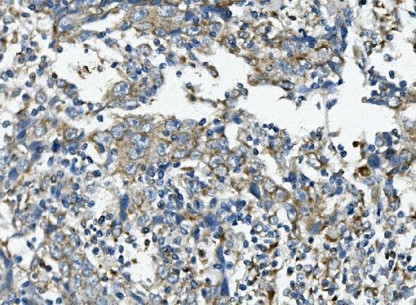
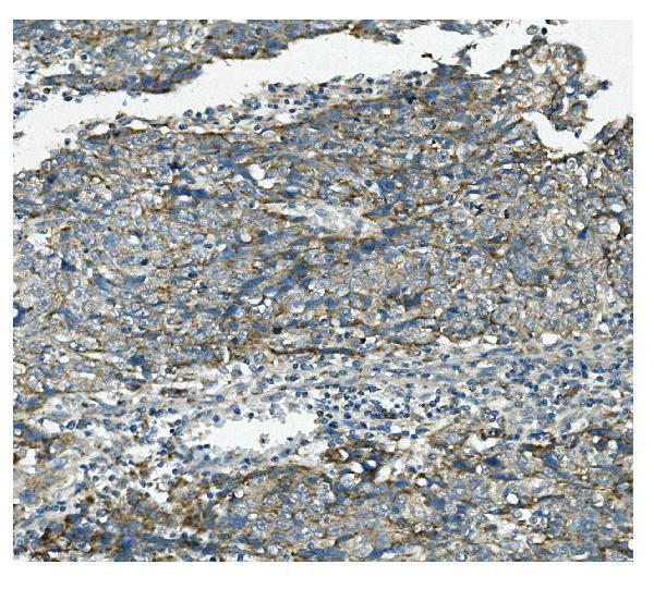
A03403-1 has real IHC data from a human lung cancer paraffin section (catalog IHC image caption); no IF image or IF/ICC application is reported (catalog applications and images).

Real IHC data IHC analysis of VPS4B/MIG1 using anti-VPS4B/MIG1 antibody (A03403-1). VPS4B/MIG1 was detected in paraffin-embedded section of human lung cancer tissue. Heat mediated antigen retrieval was performed in EDTA buffer (pH8.0, epitope retrieval solution). The tissue section was blocked with 10% goat serum. The tissue section was then incubated with 1μg/ml rabbit anti-VPS4B/MIG1 Antibody (A03403-1) overnight at 4°C. Biotinylated goat anti-rabbit IgG was used as secondary antibody and incubated for 30 minutes at 37°C. The tissue section was developed using Strepavidin-Biotin-Complex (SABC) (Catalog # SA1022) with DAB as the chromogen.
Anti-VPS4B/MIG1 Antibody ®
Cat # A03403-1

A03403-1 was demonstrated by IHC on a human lung cancer paraffin section (catalog IHC image caption). Its listed reactivity is human, mouse and rat, while the IHC dilution entry specifies human (catalog reactivity and dilution); no IF/ICC data are supplied (catalog applications and images).

Which to pick: Choose A03403-1 for human paraffin-section IHC: its own image caption reports EDTA retrieval at pH 8.0 and 1 μg/ml primary antibody overnight at 4 °C (catalog IHC image caption). The fixative is unreported (catalog IHC image caption). For mouse or rat tissue, reactivity is listed but tissue IHC evidence is not supplied; for IF/ICC, no validated option is listed (catalog reactivity, dilution, applications and images).

Each figure is that product's own IHC / IF validation image from its datasheet.

References

  1. UniProt Consortium. UniProt entry O75351 (VPS4B_HUMAN, Vacuolar protein sorting-associated protein 4B).
  2. Human Protein Atlas. VPS4B tissue IHC expression (reliability: Approved).
  3. Human Protein Atlas. VPS4B subcellular location (ICC-IF): Mainly localized to vesicles..
  4. Human Protein Atlas. VPS4B antibody validation summary (3 antibodies).
  5. Dysregulation of the endosomal sorting complex III is linked to neurodegeneration in progressive multiple sclerosis. Brain pathology (Zurich, Switzerland) 2026 — PMC12695678.
  6. Synthetic lethality between VPS4A and VPS4B triggers an inflammatory response in colorectal cancer. EMBO molecular medicine 2020 — PMC7005644.
  7. Vps4b heterozygous mice do not develop tooth defects that replicate human dentin dysplasia I. BMC genetics 2019 — PMC6330468.
  8. Cytoplasmic YAP1-mediated ESCRT-III assembly promotes autophagic cell death and is ubiquitinated by NEDD4L in breast cancer. Cancer communications (London, England) 2023 — PMC10174091.
  9. PubMed PMID:11563910 — UniProt-cited evidence.
  10. PubMed PMID:12594041 — UniProt-cited evidence.
  11. PubMed PMID:9653160 — UniProt-cited evidence.