This website uses cookies to ensure you get the best experience on our website.
- Table of Contents
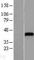
Facts about Proto-oncogene Wnt-1.
In certain developmental processes, is also a ligand for the coreceptor RYK, thus triggering Wnt signaling (By similarity). Plays an essential role in the development of the embryonic brain and central nervous system (CNS) (By similarity).
Human | |
---|---|
Gene Name: | WNT1 |
Uniprot: | P04628 |
Entrez: | 7471 |
Belongs to: |
---|
Wnt family |
Int-1; INT1Proto-oncogene Int-1 homolog; proto-oncogene Wnt-1; wingless-type MMTV integration site family, member 1 (oncogene INT1); wingless-type MMTV integration site family, member 1; Wnt1; Wnt-1
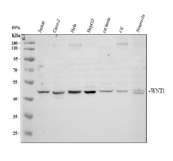
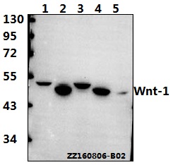
Mass (kDA):
40.982 kDA
Human | |
---|---|
Location: | 12q13.12 |
Sequence: | 12; NC_000012.12 (48978322..48982620) |
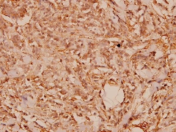
Secreted, extracellular space, extracellular matrix. Secreted.
In this article, we review Boster Bio's Anti-Wnt-1 Monoclonal Antibody and the effect of five Wnt ligands on the functioning of SMSCs in OA synovium. We also discuss the Boster Bio Anti-Desmin Antibody. This article is geared towards researchers and scientists from all over the world however it could be used in any area.
This antibody reacts with mouse ascites fluid, a sample that is derived from human, rabbit, or rats. The antibody is stored at 4degC or -20degC , and can be used for six months. It has Dvl-3 marker as well as B-catenin, which is used to test for cancerous cells and tumors in samples.
In addition to being a powerful immune therapy tool, this antibody could be beneficial in cancer treatment. Studies have proven that anti WNT1 antibodies can trigger apoptosis of a subset human sarcomas active in Wnt-1/bcatenin signaling. In a study of A-204 cells in which monoclonal antiwNT1 antibody caused significant apoptosis. Further, it induced cell death in SJSA-1 cells, another cancer cell line.
In the study of human immune cells, this antibody has been shown to be effective in treating a range of cancers, including melanoma , and leukemia. It has been proven that gd T cell are immune-related and have a WNT1 marker. These T cells are located in antigen-presenting cell lines by using CD11c microbeads.
This antibody is a boon for many reasons that include its ability to stop cancer metastasis from spreading and invading by targeting Wnt/bcatenin. Furthermore, it suppresses cell proliferation, and increases the resistance to anti-cancer medications. ARG is an inhibitor of phosphorylation of GSK3b and blocks Wnt/b-catenin from cancer cells.
This antibody could target CD4+ T cells to activate or differentiate in addition to targeting Wnt receptors. It also inhibits the release of Wnt-ligand which is crucial for CD4+ T cells differentiation. Other studies have demonstrated its ability to target mRNA-encoding NK cell lines.
Boster Bio Antismin Monoclonal Antibodies react to Human, Hamster, Mouse, and Rat proteins. They are available without BSA. A blocking peptide can be purchased separately at 1.0 mg/ml, and prices vary according to length. All Boster Bio's antibodies are tested on Immunofluorescence or WB before launch into the market.
The WNT1 gene plays an essential role in the protection of cardiac stem cells from H2O2-induced apoptosis. The Wnt pathway includes the proteins Wnt1/GSK3b and B-catenin. Wnt1 shields cardiac cells from Apoptosis inducing by H2O2.
We utilized immunofluorescence microscopy to determine whether the anti-Desmin antibody was specifically targeted at cardiac cells. We observed that most cardiac cells were positive for the c-kit that expressed desmin-negative proteins (GATA-4) and none of the cardiac muscle markers like cardiac troponin I were observed. Comparing the two groups of cells, we discovered that the Boster Bio Antismin Antibody utilizes WNT1 markers.
The expression of the OA synovium WNT1 marker Klotho decreased in mice following the anterior cruciate ligament was cut. The CTS intensity also affected the expression of MMP-9, MMP-2. These findings are in line with previous reports and suggest that Klotho may play a role in the development of OA. However, more research is required to confirm the role of Klotho in OA development.
This protein can be used to diagnose and treat OA. It is involved in cartilage loss and subchondral bone remodeling and synovitis. It regulates a complicated network of molecular pathways. This network is regulated by the WNTs protein which is a member of a family of 19 secreted glycoproteins that attach to frizzled (FZD) receptors. They also block the phosphorylation process of b-catenin that activates target genes.
WNT proteins migrate from synovium to cartilage. The Wnt proteins in the synovium are activated in OA, and b-catenin, one of the core components of the Wnt signaling pathway, is increased in OA. Mice suffering from severe OA had lower levels of WNT1 marker expression. However, this doesn't mean that OA synovial hyperexpression can affect OA pathology.
PRP is a blood derivative that is autologous, is also a promising option. PRP is an autologous blood component is proven to be effective in the treatment of OA. It decreases the production of cartilage degrading proteins and enhances the growth of chondrocytes. Further research is needed to determine whether PRP could be a helpful tool in this condition. Therefore, we are eager to discover the benefits it can bring to our patients. We are looking forward to reading more papers and hearing from you!
In the current study, we examined the effect of five Wnt ligands (Wnt5a and Wnt5b as well as Wnt10a, and Wnt11a) on SMSC functions. We found that the Wnt5b inhibitor inhibited the differentiation of the chondrogenic SMSCs and Wnt5a elevated the production of inflammatory cytokines. However, Wnt10a showed an effect of protecting cartilage. This study further suggests that the deregulation of the Wnt signaling pathway affects the functions of SMSCs and causes cartilage loss.
Wnt signaling has been implicated in osteoarthritis. SM04690 is an inhibitor of a small molecule of Wnt signaling. Activated SM04690 inhibits the Wnt signaling in the knee joint. This study suggests that SM04690 could be a treatment option for osteoarthritis.
The activation of GSK3b regulates a range of biological processes. Signaling through b-catenin and Wnt5A is involved in the inflammation response of macrophages, as well as human mononuclear cells. These proteins, along with CaMKII, are involved in the activation of anti-inflammatory protein C.
LRP5 protein is a crucial component of OA. It plays a significant role in the destruction and regeneration of cartilage. Its over-expression results in increased expression of catabolic factor and the production of cartilage degrading MMPs. Although LRP5's role is still not completely known studies of its function have revealed it to be essential for the homeostasis of cartilage. OA is caused by the constant activation of b-catenin pathways as well as an increase in SR protein levels. Moreover, the over-expression of FRZB leads to excessive WNT activation, which is associated with OA.
Recent research showed that Wnt signaling plays a crucial role in OA progression. FRZB also inhibited osteoclastogenesis (in mice) in research. It also suppressed abnormal osteoclast activity, and also reduced the expression of NFATc1 which is a marker of the Wnt pathway. Finally, the neutralizing FGF23 antibody ameliorated OA-related abnormalities in subchondral bone and reduced the expression of degradative/hypertrophic chondrogenic markers in high-molecular-weight joints in vivo. The study also revealed that Neura (NFATc1) and SOST inhibited osteoclastogenesis.
Developing secondary antibodies with WNT1 markings could assist in detecting cancer cells. The marker is expressed by tumor cells and is a potential target for immunotherapy against cancer. It plays many roles in the regulation of chemokine by cDCs. This study sheds new insights into the complex regulation of Wnt/bcatenin. It also opens avenues for improving the effectiveness of cancer immunotherapies.
Recent studies have revealed that Wnt1 expression is associated with tumor-infiltrating cells. Additionally, Wnt1 overexpression decreased the number of intratumoral T cells , and decreased their cytotoxicity. These findings demonstrate that this marker plays a crucial role in suppressing the immune response to lung tumors. But how does it work? The ability of these tumors suppress Wnt1-expressing cells within the T cell population is key to the immune response against cancer cells.
When NC-Ms express Wnt1, they transform into melanin-producing cells. This can hinder NC-Ms' becoming differentiated. NC-Ms with Wnt1 differ from other melanocytes. The Wnt1 signaling pathway is able to help researchers study the role of Wnt in NC-M development. Researchers can examine the mechanisms for the process of differentiation of NC/Ms into NC-Ms by using mice as models to develop secondary antibodies against WNT1 marker.
While Wnt1 is a specific marker for tumors, it could also play a role in the regulation of other cancers. The key role of Wnt/bcatenin activation in the development of cancer is crucial. Although most tumors don't have specific Wnt pathway mutations, they do show significant intracellular Wnt activation. Directly targeting Wnt receptors is more secure than blocking Wnt receptors.
PMID: 2998762 by van Ooyen A., et al. The nucleotide sequence of the human int-1 mammary oncogene; evolutionary conservation of coding and non-coding sequences.
PMID: 21244856 by Doubravska L., et al. Fatty acid modification of Wnt1 and Wnt3a at serine is prerequisite for lipidation at cysteine and is essential for Wnt signalling.
*More publications can be found for each product on its corresponding product page