This website uses cookies to ensure you get the best experience on our website.
- Table of Contents
1 Citations
Facts about Protein Wnt-5b.
May be a signaling molecule which affects the development of discrete regions of tissues. Is likely to signal over only few cell diameters.
Mouse | |
---|---|
Gene Name: | Wnt5b |
Uniprot: | P22726 |
Entrez: | 22419 |
Belongs to: |
---|
Wnt family |
MGC2648; protein Wnt-5b; wingless-type MMTV integration site family, member 5B; WNT-5B protein; Wnt5b; Wnt-5b
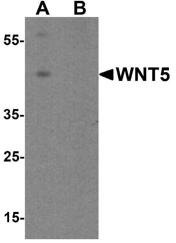
Mass (kDA):
40.343 kDA
Mouse | |
---|---|
Location: | 6 F1|6 56.86 cM |
Sequence: | 6; |
WNT5B is a nuclear translocation protein that plays several roles in the body. It activates the canonical pathway and inhibits b-catenin signaling. It is also expressed in adipocytes. Its functions are described in this article. It is applicable to scientists worldwide. Read on for more information. To get started with WNT5B, read this article.
The canonical pathway is a major regulator of cellular signaling. It activates the PI3K-AKT pathway through a series of signaling molecules. Among these molecules, RYK, ROR1 and p-Stat1 have extracellular WNT-binding domains. These proteins are deficient in intrinsic tyrosine kinase activity. When activated, these molecules phosphorylate b-catenin, resulting in nuclear translocation and transcriptional activation.
Activation of the canonical pathway has a multifaceted role in the development of tumors and is crucial in fighting various pathophysiological disorders. Activated tumors require a milieu of growth factors, secretory molecules, and crosstalk among cells. Activated Wnt signaling acts as a bridge between tumor cells and the microenvironment. Moreover, it regulates anti-tumor immune responses. The canonical Wnt ligands found in the tumor microenvironment are the source of the signaling cascade that leads to tumor development.
The melanoma-derived WNT5A promotes b-catenin signaling and induces immune evasion via Treg cells. Co-injection of porcupine inhibitor WNT-C59 or anti-CTLA4 mAb has demonstrated synergistic anti-melanoma effects in vivo. Pretreatment tumors of patients who failed to respond to PD-1 are often upregulated for WNT5A and ROR2. This suggests that the resistance to immune checkpoint blockers may involve non-canonical WNT signaling.
The canonical pathway promotes tumor growth by regulating the secretion of growth factors. Wnt signaling also promotes the migration of cancer cells to distant sites. It is important to understand how the canonical pathway is activated in vascular endohemia. Activation of the canonical pathway by the WNT5B marker can lead to the progression of cancer.
The WNT/b-catenin pathway regulates the expression of genes involved in cellular homeostasis. The WNT/b-catenin signaling pathway is activated in obstructive nephropathy, where it accumulates in fibrotic kidney tissues and is localized in the interstitium. Inhibition of b-catenin signaling by the WNT5B marker reduces the accumulation of b-catenin in the obstructive kidney tissue.
The Wnt pathway contributes to the development of breast cancer cells, where it is overexpressed. Mutations and overexpression of Wnt proteins have been found to cause the pathway to become aberrantly activated. Mutations of the WNT5B marker result in increased cytoplasmic and nuclear b-catenin levels, which correlate with a shorter disease-free survival. Moreover, overexpression of Wnt5B and c-myc genes in breast cancer cells is associated with reduced survival.
The WNT/b-catenin pathway is a high-priority molecular target for new drug development. While some inhibitors of the WNT/b-catenin pathway have entered preclinical and clinical trials, their monotherapy activities have been inadequate to warrant advancement to a registration trial. Moreover, beta-catenin signaling is widely used by multiple cell types, and thus, the WNT/b-catenin pathway inhibitors may have off-target effects.
The WNT/b-catenin pathway regulates the differentiation of CD4+ helper T cells. By inducing the expression of TCF-1, it promotes Th2 polarization in T cells and enables the activation of AT-rich sequence binding protein-1 (AT-rich sequence-B1), a key chromatin organizer involved in T cell development. Furthermore, sustained activation of b-catenin promotes Th27 polarization, increased RARC expression, and production of pro-inflammatory cytokines.
Inhibition of b-catenenin signaling by the WNT5b marker has been shown to suppress convergent extension movement in Xenopus gastrulation. This process is believed to be a result of the Wnt/b-catenin signaling pathway, which acts together with TGF-b. Similarly, the Wnt/b-catenin pathway also regulates cell adhesion through homophilic interactions between cadherin and WNT5a.
The Saa3 gene encodes a protein that is a member of the adipocyte-derived factor family. This group of proteins has been studied in both acute and chronic inflammatory diseases. Two members of the family, Saa1 and Saa2, are produced in the liver during acute inflammation, but Saa3 mRNA is highly expressed in extrahepatic tissues. These findings suggest that Saa3 is a key regulator of energy metabolism in adipocytes.
In a study of three different cell types, we found that FGF21 significantly decreased the expression of adipocyte-derived GTPase Rab5. This reflected a reduced FGF21 level in the adipocytes, which may have been a physiological response to excess intermediate metabolites. However, the FGF21 gene, also known as adiponectin, was not significantly different in the adipocytes from lean and obese controls.
To measure the expression of SOD3 in adipocytes, we treated adipocyte-derived MSCs with conditioned medium of RAW264.7 cells without LPS, or with adiponectin and TNF-a. After 24 hours of differentiation, we monitored the protein expression of SOD3 in the cell culture media using Western blotting. Using the same method for adiponectin, we calculated the expression levels of SOD3 and adiponectin in the cell culture media.
We found that Saa3 promoter activity was increased in obese adipose tissues, and this was correlated with macrophage infiltration. These results suggest that adipocytes may have an inflammatory role via the TLR4 signaling pathway. Further, we showed that naringenin inhibits the induction of adipocyte marker proteins. These findings suggest a role for this gene in adipocyte-related diseases.
The WNT5B gene encodes a transcription factor that is essential for the growth of lymphatic vessels. This gene is secreted by the tumor cells and enhances the formation of lymphatic tubes by regulating the migration of lymphatic endothelial cells. This finding may lead to the regeneration of lymphatic vessels and inhibit the growth of tumor cells. Further study of this gene is needed to determine its role in lymphangiogenesis.
WNT5B is a member of the WNT5 family of atypical proteins. It has overlapping functions in lymphatic development and is expressed in hematopoietic stem cells. It is also associated with the development of lymphoid lineage cells. Moreover, it has a role in immune response and tissue homeostasis. Hence, it is critical to fully understand its role in lymphangiogenesis and its downstream effects.
Moreover, WNT5B has been shown to contribute to lymphangiogenesis of secondary lymph nodes. Inflammation-induced LECs secrete chemokines like VEGF-C, a molecule involved in lymphatic flow. In addition, elevated interstitial fluid pressure can increase leukocyte flux into lymphatic vessels. These two mechanisms may play an important role in the regulation of lymphatic flow. In a mouse model of lymphedema, CCL21 is barely produced. Furthermore, Pflicke and Sixt found that CCR7 is critical for immigration of DCs. DCs with CCR7-deficient expression did not migrate to lymphatic vessels.
While the WNT5B marker is required for the development of lymphangiogenesis, its role is not fully understood. Recent studies have shown that Wnt5B induction promotes lymphangiogenesis in ADSC-endothelial co-culture system. Furthermore, in mice with defective lymphatic vessels, DC trafficking to the tissues increases, thereby promoting the growth of tumors.
If you need high affinity primary antibodies, Boster has the solution. These antibodies are highly cited and have been validated for use in various applications, including immunohistochemistry, western blotting, and ELISA. Boster has a vast array of secondary antibodies, allowing you to find the best match for your research needs. Additionally, Boster's antibodies are produced in their own facilities, ensuring high quality standards.
The Rabbit Anti-Goat IgG (H+L) Secondary Antibody is a Cy3 Conjugate that enables localization and quantification of target proteins in sliced samples. This antibody can also recognize multiple antigens in one specimen. Its versatile use enables researchers to test many samples simultaneously. And because it is compatible with multiple antigens, secondary antibodies can be used in many different studies.
PMID: 2279700 by Gavin B.J., et al. Expression of multiple novel Wnt-1/int-1-related genes during fetal and adult mouse development.
PMID: 10866835 by Tanaka K., et al. The evolutionarily conserved porcupine gene family is involved in the processing of the Wnt family.
*More publications can be found for each product on its corresponding product page