This website uses cookies to ensure you get the best experience on our website.


- Table of Contents
Plan YY1 chromogenic IHC in paraffin sections around its widespread nuclear pattern (HPA tissue IHC). Use high-staining follicle cells or urothelial cells as positive controls, and assess signal in nuclei (HPA tissue IHC).

Expected localisation, validated protocols, controls and antibodies — the at-a-glance facts below, then the full design guide.
| Expected localisation | Nuclear staining across tissues (HPA tissue IHC) | |
| Staining pattern | Widespread nuclear staining in tissue cells (HPA tissue IHC) | |
| Antigen retrieval | EDTA pH 8.0 HIER, heat-mediated (datasheet M00833-3) | |
| Positive control | Ovary+4 more · see all | |
| Negative control | None in HPA (detected in all 45 tissues); use no-primary + isotype controls |
| Fixation | Keep fixation consistent across sections. (standard IHC practice; not target-specific) | |
| Caveat | HPA staining may reflect proteins from more than one gene (HPA tissue IHC) | |
| Regulation | Low tissue specificity (HPA tissue RNA) | |
| Isoform / epitope | No isoforms annotated; one 1–414 chain (UniProt) |
The catalog antibody uses EDTA pH 8.0 heat retrieval (datasheet: M00833-3). The published chromogenic IHC protocols below provide YY1 staining conditions for gastric and esophageal tissue (PMC12754092; PMC3986816; PMC4196789).
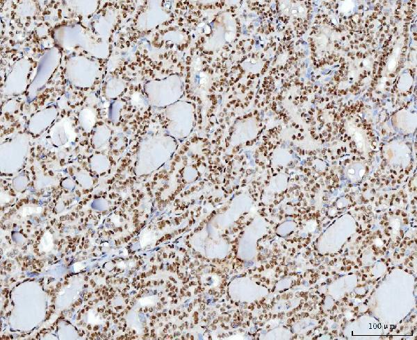
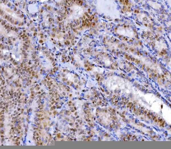
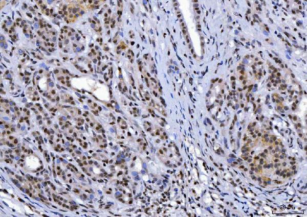
| Sample | Paraffin-embedded human thyroid cancer tissue; fixative not specified (datasheet M00833-3) |
| Fixation | Image fixative and duration unreported (datasheet M00833-3); verify before use. |
| Sectioning | 4–5 µm sections on charged slides (standard) |
| Deparaffinisation | Xylene, graded ethanol series to water (standard) |
| Antigen retrieval | Heat retrieval: EDTA pH 8.0 (datasheet M00833-3); 20 min, 95–100 °C (standard) |
| Peroxidase block | 3% H2O2, 10 min, room temperature (standard) |
| Blocking | 10% goat serum (datasheet M00833-3) |
| Primary antibody | Mouse monoclonal (clone 3F3E7) anti-YY1, 2 μg/ml (datasheet M00833-3) |
| Primary incubation | Overnight at 4 °C (datasheet M00833-3) |
| Detection | HRP-conjugated secondary, DAB chromogen (datasheet M00833-3) |
| Counterstain | Hematoxylin, blue, dehydrate and mount (standard) |
| Expected result | YY1-positive staining in follicle cells of ovary (HPA tissue IHC: High). HPA tissue profile: Ubiquitous nuclear expression. No signal in the no-primary control. |
YY1 is associated with the nuclear matrix (UniProt P25490) and shows ubiquitous nuclear staining in tissue IHC (HPA: Supported reliability). Expect stained nuclei across many cell types, with intensity varying by tissue: follicle cells, cytotrophoblasts, peritubular cells and urothelial cells are reported as High (HPA tissue IHC). YY1 has no transmembrane segment (UniProt P25490 topology).
| Discrete nuclear chromogen in the expected cells, with some nuclei darker than others. | This fits the ubiquitous nuclear profile; intensity need not be uniform across tissues or cell populations (HPA tissue IHC). Score the fraction of stained nuclei and nuclear intensity separately, using the same scoring approach across sections (standard IHC practice). |
| Strong cytoplasmic or membrane staining with little nuclear staining. | Treat this as an unexpected compartment pattern: YY1 is nuclear matrix associated and lacks a transmembrane segment (UniProt P25490). Review the negative control, detection background and antibody conditions before interpreting the extranuclear signal as YY1 (standard IHC practice). |
| An unexpected cell population stains more strongly than the mapped population. | Check cell identity and compartment before calling it positive: HPA reports ubiquitous nuclear expression, so an unlisted stained cell type alone does not establish an error (HPA tissue IHC). Staining confined to unexpected cells, especially outside nuclei, warrants checks for cross-reactivity or endogenous detection activity (standard IHC practice). |
| Diffuse colour covers nuclei, cytoplasm and surrounding tissue. | Diffuse staining is difficult to score as nuclear YY1 and may reflect detection background or inadequate blocking or washing (standard IHC practice). Compare with a section processed without primary antibody; HPA describes a nuclear tissue pattern (HPA tissue IHC). |
| No nuclear signal appears in a section expected to stain strongly. | An absent signal in a high-staining HPA population, such as placental cytotrophoblasts, suggests a run or specimen issue before a biological negative is assigned (HPA: High in cytotrophoblasts; standard IHC practice). Check a known-positive control processed with the same run. |
| Tissue and cell choice | HPA reports High staining in ovarian follicle cells, placental cytotrophoblasts, testicular peritubular cells and bladder urothelial cells, but Low staining in hepatocytes and several listed neural populations (HPA tissue IHC). A faint result in a Low population therefore has less diagnostic value than a failed High control. |
| Antibody evidence and specificity | Two listed antibodies have Supported IHC status, while the tissue profile carries Supported reliability (HPA antibodies; HPA tissue IHC). HPA also cautions that the assay targets protein from more than one gene; nuclear staining supports a YY1 compatible pattern but does not, by itself, establish exclusive molecular identity (HPA tissue IHC). |
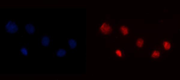
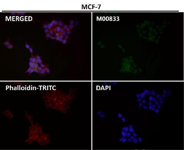
| IF/ICC Q&A: Where should fluorescence appear? | Mainly in the nucleoplasm, with additional signal reported at nucleoli fibrillar centers and nuclear bodies (HPA subcellular ICC-IF). That cell imaging detail helps assess compartment plausibility; it should not be required as a separately resolved pattern in chromogenic tissue IHC (HPA subcellular ICC-IF; standard IHC practice). |
| Situation | Likely cause | Next action |
|---|---|---|
| A High tissue control has no nuclear staining. | A staining-run problem is possible when an HPA High population is blank (HPA tissue IHC; standard IHC practice). | Review the run's positive control and reagent sequence, then assess retrieval and primary-antibody conditions as general IHC variables (standard IHC practice). No YY1-specific retrieval or fixation sensitivity is established by these sources. |
| Staining is weak in hepatocytes or listed neural populations. | Those populations are reported as Low by HPA (HPA tissue IHC). | Compare with a High population from the same run before changing conditions; interpret the weak signal in its cell and tissue context (HPA tissue IHC; standard IHC practice). |
| Cytoplasmic staining dominates the section. | That distribution conflicts with the reported nuclear localization (UniProt P25490; HPA tissue IHC). | Compare a no-primary control and a known-positive section, then review antibody dilution and wash conditions for nonspecific staining (standard IHC practice). |
| Diffuse chromogen obscures nuclear borders. | High background can obscure compartment scoring (standard IHC practice). | Inspect the no-primary control; review blocking, wash steps and detection development as general chromogenic IHC checks (standard IHC practice). |
| Colour persists in a no-primary control. | Primary-independent colour can arise from endogenous detection activity or the detection system (standard IHC practice). | Address the detection-system background using its appropriate control and blocking steps before scoring YY1 (standard IHC practice). |
| Only an unexpected cell population appears strongly positive. | HPA reports ubiquitous nuclear expression and cautions that staining may target protein from more than one gene; cell identity and specificity need review (HPA tissue IHC). | Confirm the stained cells on the counterstain, compare compartment and expected HPA populations, and review controls or an independently validated antibody if specificity remains uncertain (HPA tissue IHC; standard IHC practice). |


Comprehensive Human Protein Atlas IHC scoring per tissue (reliability: Supported — High consistency between antibody staining and RNA expression data. Caution, targets protein from more than one gene.). Rows are taken directly from the HPA tissue chart — click any row's HPA link to view the source.
| Tissue | Cell type | Level | Evidence | Source |
|---|---|---|---|---|
| Ovary | Follicle cells | High | Protein (IHC) | HPA → |
| Placenta | Cytotrophoblasts | High | Protein (IHC) | HPA → |
| Testis | Peritubular cells | High | Protein (IHC) | HPA → |
| Urinary bladder | Urothelial cells | High | Protein (IHC) | HPA → |
| Adrenal gland | Glandular cells | Medium | Protein (IHC) | HPA → |
| Tissue | Cell type | Level | Evidence | Source |
|---|---|---|---|---|
| None in HPA: YY1 is detected in all 45 scored tissues. Use a no-primary (secondary-only) and an isotype control instead. | ||||
Troubleshoot YY1 staining in paraffin sections by checking retrieval, nuclear localisation and controls before comparing chromogenic IHC results (datasheet M00833-3; UniProt P25490).
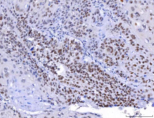
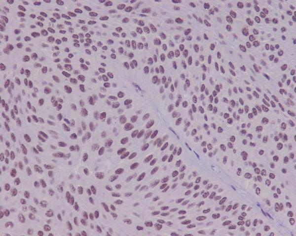
Anti-YY1 antibodies have IHC images from human paraffin sections and an IF image from A431 cells (catalog image captions); the catalog lists human, mouse and rat reactivity (catalog applications and reactivity).
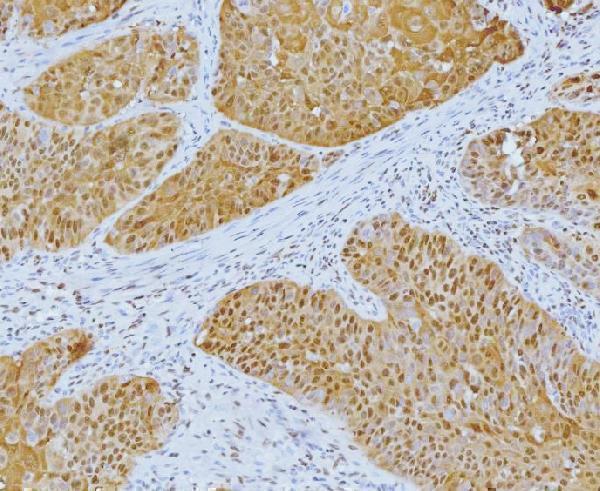
M00833-3 and M00833-2 show IHC in human thyroid cancer sections; M00833-1 shows human laryngeal squamous cell carcinoma, and M00833 shows human bladder (each SKU’s IHC image caption). PB9909 shows IF in A431 cells (PB9909 IF image caption).
Which to pick: For tissue IHC, choose a SKU with an image from the relevant paraffin section: M00833-3, M00833-2, M00833-1 or M00833 (each SKU’s IHC image caption); the captions do not report the fixative. For IF/ICC, PB9909 has both applications listed and an A431 IF image (PB9909 catalog applications and IF image caption). For work across species, M00833 is a rabbit monoclonal with IHC and IF listed and human, mouse and rat reactivity (M00833 catalog); its IHC image documents a human paraffin section only (M00833 IHC image caption).