ZAP70 / Tyrosine-protein kinase ZAP-70 · IHC design guide

Design Immunohistochemistry for ZAP70

Plan chromogenic ZAP70 IHC in paraffin sections using cytoplasmic staining in immune-cell subsets as the tissue reference (HPA tissue IHC). The guide covers fixation, staining controls and interpretation of activation-linked membrane recruitment (standard IHC practice; UniProt).

Evidence assembled Oct 2026 · For research use; verify linked source records and product datasheet before use
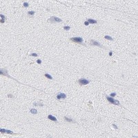
Immunohistochemistry protocol sheet for ZAP70 (IHC for ZAP70): expected localisation Cytoplasm in immune-cell subsets (HPA tissue IHC), antibody PB9968, validated IHC image, and IHC protocol steps
Printable ZAP70 IHC protocol sheet — expected localisation Cytoplasm in immune-cell subsets (HPA tissue IHC), antibody PB9968, controls and protocol steps. Open the full ZAP70 IHC guide →

ZAP70 Immunohistochemistry Experimental Design Guide

Expected localisation, validated protocols, controls and antibodies — the at-a-glance facts below, then the full design guide.

Must know before staining
Expected localisation Cytoplasm in immune-cell subsets (HPA tissue IHC)
Staining pattern Selective cytoplasmic staining in immune-cell subsets (HPA tissue IHC)
Antigen retrieval EDTA pH 8.0 HIER, heat-mediated (datasheet PB9968)
Positive control ⓘ Appendix+4 more · see all
Negative control ⓘ Adipose tissue+4 more · see all
Important caveats
Reasons your staining may differ from the expected pattern.
Fixation Matched tissue-IHC evidence does not establish the fixation claim. Validate the specimen-specific method before use. (selected-SKU IHC image PB9968)
Caveat Presumed off-target binding can confound staining (HPA tissue IHC)
Regulation TCR activation recruits ZAP70 to the membrane (UniProt)
Isoform / epitope 3 isoforms; epitope coverage unspecified (UniProt)
Section 1

Recommended ZAP70 IHC & IF Protocols

The catalog antibody protocol (datasheet: PB9968) is accompanied by published ZAP70 IHC protocols for endometrial tissue (PMC10131270) and laryngeal cancer sections (PMC9047300).

Recommended immunohistochemistry (IHC-P) protocol parameters
SampleParaffin-embedded human prostate cancer tissue; fixative not specified (datasheet PB9968)
FixationImage fixative and duration unreported (datasheet PB9968); verify before use.
Sectioning4–5 µm sections on charged slides (standard)
DeparaffinisationXylene, graded ethanol series to water (standard)
Antigen retrievalHeat retrieval: EDTA pH 8.0 (datasheet PB9968); 20 min, 95–100 °C (standard)
Peroxidase block3% H2O2, 10 min, room temperature (standard)
Blocking10% goat serum (datasheet PB9968)
Primary antibodyRabbit anti-ZAP70, 2-5μg/ml (datasheet PB9968)
Primary incubationOvernight at 4 °C (datasheet PB9968)
DetectionHRP-conjugated secondary, DAB chromogen (datasheet PB9968)
CounterstainHematoxylin, blue, dehydrate and mount (standard)
Expected resultZAP70-positive staining in lymphoid tissue of appendix (HPA tissue IHC: High). HPA tissue profile: Selective cytoplasmic expression in subsets of immune cells. No signal in the no-primary control.
💡Decision noteStart with heat-mediated EDTA retrieval at pH 8.0 (datasheet: PB9968); the laryngeal cancer protocol reports antigen retrieval without conditions (PMC9047300: methods).
Section 2

What Is the Expected ZAP70 Staining Pattern?

ZAP70 should appear mainly in the cytoplasm of subsets of immune cells in paraffin sections (HPA: Enhanced reliability; selective cytoplasmic expression). T and natural killer cells are expected sources; early thymocytes and pro/pre B cells can also express it (UniProt P43403: tissue specificity). ZAP70 has no transmembrane segment; stimulated T cells can recruit it to the plasma membrane (UniProt P43403: topology and subcellular location).

What am I looking at on my slide?
Discrete cytoplasmic staining in immune cells of appendix lymphoid tissue or lymph node non-germinal center areas.This fits the reported selective pattern. Both sites have High staining, but the result should be judged by cell and compartment, not a uniformly stained section (HPA: tissue IHC profile and levels).
Membrane-associated staining in a subset of immune cells.This can fit ZAP70 recruitment after T-cell receptor activation; cytoplasmic staining remains expected in quiescent T cells. An IHC pattern alone does not establish activation state (UniProt P43403: subcellular location and function).
Predominantly nuclear staining, without a convincing cytoplasmic immune-cell pattern.Treat this as a compartment mismatch and investigate artefact or nonspecific detection. The supplied localization is cytoplasmic, with activation-associated membrane recruitment; nuclear localization is not reported here (HPA: tissue IHC profile; UniProt P43403: subcellular location).
Strong staining in cells outside the expected immune-cell distribution.Check cell identity and assay controls before calling it ZAP70. HPA notes presumed off-target binding in its Enhanced tissue assessment; kidney proximal-tubule microvilli and testis Leydig-cell entries should not, by themselves, override the selective immune-cell profile (HPA: reliability description, profile and tissue IHC).
Widespread haze, or no signal in a known-positive immune-cell area.Haze obscures the selective cytoplasmic pattern; absent signal prevents interpretation of a negative test area. Confirm that appendix lymphoid tissue or lymph node non-germinal center cells stain in the same run (HPA: tissue IHC).
💡Expected ZAP70 appearanceCall a result positive when a subset of immune cells shows clear cytoplasmic staining in a High reference area; broad nuclear, epithelial or background staining warrants review (HPA: tissue IHC profile and levels; UniProt P43403: localization).
How each factor affects the staining
Cell distribution and reference tissueAppendix lymphoid tissue and lymph node or tonsil non-germinal center cells are High; bone-marrow hematopoietic cells and spleen red-pulp cells are Medium. Compare like cell populations when judging intensity (HPA: tissue IHC).
Subcellular location and activationQuiescent T-cell ZAP70 is cytoplasmic; stimulated T-cell receptor complexes recruit it to the membrane. Membrane signal may vary with cell state, so require the expected immune-cell context (UniProt P43403: subcellular location and function).
Antibody validation and unusual sitesHPA grades tissue staining Enhanced and lists HPA003134 and CAB002625 as IHC Enhanced, while acknowledging presumed off-target binding. Use the selective cytoplasmic immune-cell profile when reviewing unexpected staining (HPA: reliability description, antibodies and tissue IHC).
Protein form and epitope interpretationThe protein has no transmembrane segment or signal peptide; UniProt lists 3 isoforms and several modified residues. These facts alone do not establish which forms or phosphorylation states an unspecified antibody detects (UniProt P43403: topology, isoforms and modified residues).
IF/ICC evidence — can this IHC pattern be applied to IF/ICC?Use cytoplasmic and activation-associated membrane localization as biological context (UniProt P43403: subcellular location). HPA supplies no ICC-IF images or main-location assignment here, so this IHC result does not validate an IF/ICC staining pattern (HPA: subcellular record).
Why is my staining missing, weak or wrong?
SituationLikely causeNext action
No staining in appendix or lymph-node immune cells.The run lacks a positive reference signal; a negative result elsewhere is uninterpretable (HPA: High tissue IHC levels).Check that a reference section and its detection controls worked; review retrieval and antibody conditions as general IHC checks. ZAP70-specific fixation sensitivity is unreported in the supplied sources.
Only the test area is negative while the positive reference stains.The sampled area may contain few ZAP70-expressing cells; expression is selective within immune-cell subsets (HPA: tissue IHC profile; UniProt P43403: tissue specificity).Use the counterstain to identify the relevant cells, then score their cytoplasm against the stained reference area (general IHC practice; HPA: tissue IHC profile).
Diffuse color covers cells and extracellular space.Background can hide the selective cytoplasmic pattern; the image alone cannot identify its source (HPA: tissue IHC profile; general IHC practice).Review blocking, washes and detection controls, then compare signal within cells with the surrounding section (general IHC practice).
Strong nuclear signal dominates.The compartment conflicts with the reported cytoplasmic and activation-associated membrane locations (HPA: tissue IHC profile; UniProt P43403: subcellular location).Review counterstain and control sections; withhold a ZAP70-positive call until the expected immune-cell cytoplasmic pattern is demonstrated.
Tubular, Leydig-cell or other non-immune staining dominates.HPA lists some staining at those sites but also reports presumed off-target binding and a selective immune-cell profile; the staining alone cannot resolve specificity (HPA: tissue IHC and reliability description).Compare with immune-cell positive areas and detection controls before interpreting the unusual cell population (HPA: tissue IHC; general IHC practice).
Signal appears at the cell edge instead of throughout the cytoplasm.T-cell receptor stimulation can recruit ZAP70 to the plasma membrane; edge signal alone does not prove stimulation (UniProt P43403: subcellular location and function).Confirm immune-cell identity and assess cytoplasmic staining in nearby cells before scoring the edge pattern (UniProt P43403: tissue specificity; HPA: tissue IHC profile).

Sample controls for ZAP70 IHC & IF

🧪Run lymph node first: non-germinal center cells should stain (HPA: High in lymph node non-germinal center cells). Use adipose tissue as a negative comparator because adipocytes are not detected (HPA: Not detected in adipose tissue adipocytes); within the lymph node slide, assess morphologically nonlymphoid stromal cells for absence of specific staining (UniProt P43403: expression in T and natural killer cells).
Positive control tissue: Appendix (Lymphoid tissue, HPA High)
Negative control tissue: Adipose tissue (HPA Not detected)
ICC-IF cell lines (HPA subcellular resource): HPA carries no ICC-IF cell line for ZAP70; derive a cell-line control from the positive tissue's cell type (Lymphoid tissue) and confirm it by RNA or western blot first.
Technical controls: Include a no-primary secondary-only slide, a concentration-matched nonimmune rabbit IgG isotype control, and ZAP70-knockout material as a biological negative when available (caption: rabbit anti-ZAP70 PB9968; standard IHC practice). Quench endogenous peroxidase for chromogenic detection in lymph node and check background before scoring (caption: peroxidase secondary and DAB; standard IHC practice).
⚠️Feasibility: A target-specific fixation window or fixation effect is unreported, and the selected PB9968 paraffin-section caption does not state a fixative (PB9968 tissue-IHC caption). The caption reports heat retrieval in EDTA at pH 8.0, but it does not establish whether retrieval is required; the supplied evidence also does not establish that frozen sections or IF/ICC are easier (PB9968 tissue-IHC caption; HPA subcellular: no ICC-IF images listed). Dense lymph node cell populations can complicate assignment of chromogenic staining to individual cells, so score staining against morphology and the control slides (HPA: High in lymph node non-germinal center cells; standard IHC practice).

HPA tissue IHC evidence for ZAP70

Comprehensive Human Protein Atlas IHC scoring per tissue (reliability: Enhanced — High consistency between antibody staining and RNA expression data. Presumed off target binding observed and disregarded.). Rows are taken directly from the HPA tissue chart — click any row's HPA link to view the source.

Positive expression · recommended positive controls

TissueCell typeLevelEvidenceSource
Appendix Lymphoid tissue High Protein (IHC) HPA →
Kidney Proximal tubules (microvilli) High Protein (IHC) HPA →
Lymph node Non-germinal center cells High Protein (IHC) HPA →
Tonsil Non-germinal center cells High Protein (IHC) HPA →
Bone marrow Hematopoietic cells Medium Protein (IHC) HPA →

Undetected expression · recommended negative controls

TissueCell typeLevelEvidenceSource
Adipose tissue Adipocytes Not detected Protein (IHC) HPA →
Adrenal gland Glandular cells Not detected Protein (IHC) HPA →
Breast Adipocytes Not detected Protein (IHC) HPA →
Bronchus Respiratory epithelial cells Not detected Protein (IHC) HPA →
Caudate Glial cells Not detected Protein (IHC) HPA →
Section 3

Advanced ZAP70 IHC Tips

Troubleshoot ZAP70 staining in paraffin sections by checking retrieval, cell identity, cytoplasmic localisation and detection controls before interpreting signal.

What should I change when ZAP70 staining is weak in paraffin sections?
Start with heat mediated retrieval in EDTA pH 8.0 for paraffin sections (datasheet PB9968). The selected image used this retrieval before overnight incubation at 4°C with 2 μg/ml antibody; its fixative was not reported (datasheet PB9968). If staining remains weak, compare retrieval duration on matched sections while holding antibody concentration and detection conditions constant (standard IHC practice). Include a lymphoid control with identifiable immune cells, since ZAP70 is expressed in T and natural killer cells and HPA reports selective cytoplasmic staining in immune cell subsets (UniProt P43403; HPA tissue IHC).
Could fixation explain weak or uneven ZAP70 staining?
Target specific fixation sensitivity is unknown because the selected paraffin section caption does not report a fixative (datasheet PB9968). Record the fixative, fixation duration and processing history for each specimen, then compare sections processed together when investigating weak staining (standard IHC practice). Check morphology and retrieval consistency before increasing antibody concentration, because uneven processing can complicate comparisons across paraffin sections (standard IHC practice). Use a positive lymphoid control in the same staining run and interpret its cytoplasmic signal alongside the test section (HPA tissue IHC; standard IHC practice). Do not assign a fixation effect to ZAP70 without a direct comparison (datasheet PB9968).
Should ZAP70 appear at the membrane or in the cytoplasm?
Expect predominantly cytoplasmic staining in resting T cells: ZAP70 is cytoplasmic in quiescent T lymphocytes, and HPA describes selective cytoplasmic staining in immune cell subsets (UniProt P43403; HPA tissue IHC). Following T cell receptor activation, ZAP70 can be recruited to the plasma membrane through CD247/CD3Z (UniProt P43403). It has no transmembrane segment, so a crisp membrane outline alone needs careful assessment against cell identity and morphology (UniProt P43403 topology; standard IHC practice). Compare the suspected pattern with nearby immune cells and a same run lymphoid control before interpreting focal membrane staining as activation (HPA tissue IHC; standard IHC practice).
Could isoforms or phosphorylation change what this antibody detects?
ZAP70 has 3 recorded isoforms and contains two SH2 domains plus a kinase domain, but the supplied caption does not identify the antibody epitope (UniProt P43403; datasheet PB9968). Recorded modified residues include phosphotyrosines at positions 248, 292, 315 and 319 (UniProt P43403). Without an epitope map, do not infer isoform coverage or phosphorylation sensitivity from a positive paraffin section alone (UniProt P43403; datasheet PB9968). If staining differs across specimens, first compare retrieval, controls and the distribution of immune cells, then seek direct epitope validation before assigning a molecular explanation (standard IHC practice).
How should I adapt a ZAP70 panel for immunofluorescence?
Treat IF as a separate application: the selected PB9968 evidence describes chromogenic staining of a paraffin section and supplies no IF validation (datasheet PB9968). Multiplex ZAP70 with a marker that identifies the expected T cell population, and assess whether signals occupy the same cells rather than merely adjacent cells (UniProt P43403; standard IF practice). Choose a fluorophore channel after inspecting tissue autofluorescence and include single stain controls when setting exposure and assessing bleed through (standard IF practice). Because ZAP70 is cytoplasmic in quiescent T cells and has no transmembrane segment, use permeabilisation suitable for an intracellular epitope, then check cell morphology (UniProt P43403; standard IF practice).
How can I reduce diffuse or misleading DAB staining?
First inspect a no primary control and the distribution of staining across the section; diffuse colour or staining at damaged edges warrants a background check (standard IHC practice). The selected PB9968 image used 10% goat serum, a peroxidase conjugated secondary and DAB development after overnight primary incubation (datasheet PB9968). Optimise blocking and washing, and include a peroxidase block before DAB detection as part of the general chromogenic workflow (standard IHC practice). Titrate the primary around the reported 2 μg/ml condition while retaining a positive control, so reduced background is assessed alongside retained immune cell staining (datasheet PB9968; HPA tissue IHC; standard IHC practice).
What is a defensible way to quantify ZAP70 IHC? ⚠ ANSWER MARKED FOR VERIFICATION
Define the cell population and tissue region before scoring, since HPA reports selective cytoplasmic expression in subsets of immune cells (HPA tissue IHC; standard IHC practice). For a cell based endpoint, report the percentage of positive cells or an H score from intensity categories; for infiltrates, report positive cell density per mm² of evaluable tissue (standard IHC practice). Normalise cell based results to the number of eligible cells and density results to measured viable tissue area, excluding folds and necrosis by a prespecified rule (standard IHC practice). Keep retrieval, detection, threshold and scoring rules consistent across specimens, and report immune cell abundance separately when it varies (standard IHC practice).
How do I distinguish genuine ZAP70 signal from artefact?
Look for cytoplasmic staining in morphologically credible immune cells, consistent with HPA's tissue profile and ZAP70 expression in T and natural killer cells (HPA tissue IHC; UniProt P43403). A membrane associated pattern may occur with T cell receptor recruitment, but morphology and cell identity remain necessary checks (UniProt P43403; standard IHC practice). Treat staining confined to section edges or necrotic areas as suspect, and check a no primary control for endogenous enzyme or detection background (standard IHC practice). HPA also reports presumed off target binding in its tissue assessment, so isolated staining in an unexpected cell type needs independent validation before a biological conclusion (HPA tissue IHC).
Boster reagents

Best ZAP70 / Tyrosine-protein kinase ZAP-70 IHC Antibodies

The IHC-validated antibody PB9968 has a paraffin-section image from human prostate cancer tissue (catalog image caption); its listed reactivity also includes mouse and rat (catalog applications/reactivity).

Real IHC data IHC analysis of ZAP70 using anti-ZAP70 antibody (PB9968). ZAP70 was detected in a paraffin-embedded section of human prostate cancer tissue. Heat mediated antigen retrieval was performed in EDTA buffer (pH 8.0, epitope retrieval solution). The tissue section was blocked with 10% goat serum. The tissue section was then incubated with 2 μg/ml rabbit anti-ZAP70 Antibody (PB9968) overnight at 4°C. Peroxidase Conjugated Goat Anti-rabbit IgG was used as secondary antibody and incubated for 30 minutes at 37°C. The tissue section was developed using HRP Conjugated Rabbit IgG Super Vision Assay Kit (Catalog # SV0002) with DAB as the chromogen.
Anti-ZAP70 Antibody ®
Cat # PB9968

PB9968 is the sole rendered card, with an IHC image from a human prostate cancer paraffin section (PB9968 image caption). M00754-3 lists human IHC and IF/ICC applications, but has no IHC or IF image in the payload (catalog applications/image alts).

Which to pick: Choose PB9968 for tissue IHC when its documented paraffin-section workflow is useful; the image caption reports EDTA pH 8.0 retrieval and 2 μg/ml primary antibody, but does not report the fixative (PB9968 image caption). For IF/ICC, M00754-3 lists those applications and a 1:50 IF dilution, though no IF image is supplied (catalog applications/dilution/image alts). PB9968 lists human, mouse and rat reactivity, while M00754-3 lists human reactivity and rabbit monoclonal clone ACAF-26 (catalog reactivity/clone).

Each figure is that product's own IHC / IF validation image from its datasheet.

References

  1. UniProt Consortium. UniProt entry P43403 (ZAP70_HUMAN, Tyrosine-protein kinase ZAP-70).
  2. Human Protein Atlas. ZAP70 tissue IHC expression (reliability: Enhanced).
  3. Human Protein Atlas. ZAP70 subcellular location (ICC-IF): Highest expression in JURKAT: 134.1 nTPM.
  4. Human Protein Atlas. ZAP70 antibody validation summary (2 antibodies).
  5. Immunohistochemical detection of ZAP70 in chronic lymphocytic leukemia predicts immunoglobulin heavy chain gene mutation status and time to progression. Modern pathology : an official journal of the United States and Canadian Academy of Pathology, Inc 2010 — PMC2966512.
  6. Serial minimal residual disease (MRD) monitoring during first-line FCR treatment for CLL may direct individualized therapeutic strategies. Leukemia 2018 — PMC6192870.
  7. ZAP70 interaction with 13 mRNAs as a potential immunotherapeutic target for endometrial cancer. Oncology letters 2023 — PMC10131270.
  8. AQP9 and ZAP70 as immune-related prognostic biomarkers suppress proliferation, migration and invasion of laryngeal cancer cells. BMC cancer 2022 — PMC9047300.
  9. PubMed PMID:1423621 — UniProt-cited evidence.
  10. PubMed PMID:14985102 — UniProt-cited evidence.
  11. PubMed PMID:15815621 — UniProt-cited evidence.