ZBTB7A / Zinc finger and BTB domain-containing protein 7A · IHC design guide

Design Immunohistochemistry for ZBTB7A

Plan ZBTB7A IHC in paraffin sections around widespread nuclear staining, with colon glandular cells as a high-staining reference (HPA tissue IHC). The catalog antibody’s IHC-P example uses 1 μg/ml primary antibody and DAB detection (datasheet PB9910).

Evidence assembled Oct 2026 · For research use; verify linked source records and product datasheet before use
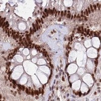
Immunohistochemistry protocol sheet for ZBTB7A (IHC for ZBTB7A): expected localisation Nuclear staining in tissue (HPA tissue IHC), antibody PB9910, validated IHC image, and IHC protocol steps
Printable ZBTB7A IHC protocol sheet — expected localisation Nuclear staining in tissue (HPA tissue IHC), antibody PB9910, controls and protocol steps. Open the full ZBTB7A IHC guide →

ZBTB7A Immunohistochemistry Experimental Design Guide

Expected localisation, validated protocols, controls and antibodies — the at-a-glance facts below, then the full design guide.

Must know before staining
Expected localisation Nuclear staining in tissue (HPA tissue IHC)
Staining pattern Widespread nuclear staining across cell types (HPA tissue IHC)
Antigen retrieval EDTA pH 8.0 HIER, heat-mediated (datasheet PB9910)
Positive control ⓘ Appendix+4 more · see all
Negative control ⓘ None in HPA (detected in all 45 tissues); use no-primary + isotype controls
Important caveats
Reasons your staining may differ from the expected pattern.
Fixation Keep fixation consistent across sections (standard IHC practice; not target-specific)
Caveat Intensity varies by cell type, from high to low (HPA tissue IHC)
Regulation Upregulated in some lymphomas/carcinomas (UniProt)
Isoform / epitope No annotated isoforms; one 1–584 chain (UniProt)
Section 1

Recommended ZBTB7A IHC & IF Protocols

The catalog antibody protocol uses EDTA pH 8.0 heat retrieval (datasheet PB9910). The published protocols below describe ZBTB7A staining in mouse liver and human colorectal cancer samples (PMC6973189; PMC13520279; PMC7650168).

Recommended immunohistochemistry (IHC-P) protocol parameters
SampleParaffin-embedded human intestinal cancer tissue; fixative not specified (datasheet PB9910)
FixationImage fixative and duration unreported (datasheet PB9910); verify before use.
Sectioning4–5 µm sections on charged slides (standard)
DeparaffinisationXylene, graded ethanol series to water (standard)
Antigen retrievalHeat retrieval: EDTA pH 8.0 (datasheet PB9910); 20 min, 95–100 °C (standard)
Peroxidase block3% H2O2, 10 min, room temperature (standard)
Blocking10% goat serum (datasheet PB9910)
Primary antibodyRabbit anti-ZBTB7A, 0.5-1μg/ml (datasheet PB9910)
Primary incubationOvernight at 4 °C (datasheet PB9910)
DetectionStreptavidin-biotin complex (SABC), DAB chromogen (datasheet PB9910)
CounterstainHematoxylin, blue, dehydrate and mount (standard)
Expected resultZBTB7A-positive staining in glandular cells of appendix (HPA tissue IHC: High). HPA tissue profile: Ubiquitous nuclear expression. No signal in the no-primary control.
💡Decision noteStart with EDTA pH 8.0 heat retrieval for the catalog antibody (datasheet PB9910). Citrate pH 6.0 is a published alternative for colorectal cancer sections (PMC7650168).
Section 2

What Is the Expected ZBTB7A Staining Pattern?

ZBTB7A should stain nuclei in paraffin sections: UniProt places it in the nucleus and reports no transmembrane segment (UniProt O95365). HPA describes ubiquitous nuclear expression, with high staining in several epithelial populations and cerebral cortical neurons (HPA tissue IHC). Tissue IHC reliability is Supported, with medium consistency between antibody staining and RNA expression; use that qualification when interpreting an unexpected result (HPA tissue IHC).

What am I looking at on my slide?
Distinct nuclear staining in appendix or colon glandular cells, with little staining outside nuclei.This matches the expected compartment and two high-staining cell populations (UniProt O95365; HPA: High in appendix and colon glandular cells). Compare nuclei within the relevant cell population; a tissue-wide impression can hide differences between cell types.
Predominantly cytoplasmic, membranous, or extracellular signal, with weak or absent nuclear staining.Treat this as a discordant pattern and investigate artefact or nonspecific staining before scoring it as ZBTB7A. UniProt places ZBTB7A in the nucleus and reports no transmembrane segment; HPA describes nuclear tissue staining (UniProt O95365; HPA tissue IHC).
Strong signal in an unexpected cell population, while the anticipated nuclei remain unstained.Check cell identity and controls before assigning target expression. Cross-reactivity or endogenous chromogenic activity are possible technical explanations (general IHC practice). HPA reports low staining in adipocytes and breast glandular cells, but low does not mean absent (HPA tissue IHC).
Diffuse brown haze spans nuclei, cytoplasm, and surrounding tissue without clear nuclear boundaries.This is difficult to score as target-specific staining because the reported pattern is nuclear (HPA tissue IHC; UniProt O95365). Inspect the no-primary control and detection background, then reassess blocking, washes, and chromogen development (general IHC practice).
No nuclear signal in a section expected to contain high-staining cells.Check whether the relevant cells are present before calling the result negative. Appendix and colon glandular cells, respiratory epithelium in bronchus, and cerebral cortical neurons are reported as high (HPA tissue IHC). A failed positive control points to the staining run (general IHC practice).
💡Expected ZBTB7A appearanceCall a positive result when identifiable nuclei stain in an HPA high-staining cell population; strong cytoplasmic or extracellular color without matching nuclear staining is suspect (HPA tissue IHC; UniProt O95365).
How each factor affects the staining
Tissue and cell populationHPA reports high staining in selected glandular, respiratory epithelial, squamous epithelial, and neuronal populations, alongside low staining in several others (HPA tissue IHC). Choose and score the specified cells within each section; low-staining populations are poor stand-alone negative controls.
Expected compartmentUniProt assigns ZBTB7A to the nucleus and reports no transmembrane segment; HPA calls tissue expression ubiquitously nuclear (UniProt O95365; HPA tissue IHC). This supports nuclear scoring, without requiring every nucleus to show identical intensity.
Antibody evidenceHPA lists HPA046387, CAB037214, and CAB037285 as Supported for IHC; the overall tissue profile has medium staining-to-RNA consistency (HPA antibodies; HPA tissue IHC). These ratings support comparison with the reference pattern but do not establish specificity for every section.
IF/ICC Q&A: where should signal appear?In nucleoplasm: HPA reports an approved nucleoplasmic ICC-IF location and lists HPA046387 as ICC Approved (HPA subcellular; HPA antibodies). That observation helps assess localization; this section does not specify an IF/ICC protocol.
Why is my staining missing, weak or wrong?
SituationLikely causeNext action
Expected nuclei are unstained in an appendix or colon section.The relevant glandular cells may be absent from the field, or the staining run may have failed; both tissues contain high-staining glandular cells in the HPA record (HPA tissue IHC).Locate intact glandular cells and check a concurrent positive control. If that control also fails, review section preparation, retrieval, primary incubation, and detection as general IHC workflow checks (general IHC practice).
Color is mainly cytoplasmic or outlines membranes.The distribution conflicts with nuclear localization and the absence of a transmembrane segment (UniProt O95365; HPA tissue IHC). Background or nonspecific binding is possible (general IHC practice).Do not score this pattern as a nuclear positive. Compare with a no-primary control, inspect nuclear counterstain, and repeat with an IHC-supported antibody if needed (HPA antibodies; general IHC practice).
Brown staining persists in cells that lack convincing nuclear signal.Cross-reactivity or endogenous chromogenic activity may contribute (general IHC practice); the observed pattern alone cannot distinguish them.Run no-primary and detection controls, review the appropriate endogenous-activity block, and check whether any nuclear staining agrees with HPA cell-type patterns (HPA tissue IHC; general IHC practice).
The whole section has diffuse background.A poorly resolved chromogenic reaction can obscure nuclei and prevent comparison with HPA's nuclear pattern (HPA tissue IHC; general IHC practice).Check the no-primary control, washes, blocking, and chromogen development; adjust the general IHC workflow before interpreting weak nuclear color (general IHC practice).
A low-staining population appears negative.Low staining is documented for adipocytes, breast glandular cells, and several other populations; HPA does not label these populations negative (HPA tissue IHC).Report the observed intensity in the named cell type. Include a high-staining population in the run before interpreting an absent signal as a technical failure (HPA tissue IHC; general IHC practice).
Two sections or antibodies give different nuclear intensities.HPA rates tissue IHC as Supported with medium consistency against RNA expression, and separately lists three IHC-supported antibodies (HPA tissue IHC; HPA antibodies). The supplied evidence does not establish a target-specific fixation effect.Compare the same cell type, section quality, and controls; document the antibody and staining conditions. Treat intensity differences cautiously when the nuclear pattern remains plausible (HPA tissue IHC; general IHC practice).

Sample controls for ZBTB7A IHC & IF

🧪Run colon first and score its glandular cells for nuclear staining (HPA: High in colon glandular cells; UniProt O95365: nucleus). HPA detects ZBTB7A in all 45 scored tissues, so use no-primary and isotype controls for the negative; on the positive slide, cytoplasm and extracellular areas should lack specific nuclear signal, but do not assume another cell type is ZBTB7A-negative (HPA: no negative tissue rows; HPA: nucleoplasm).
Positive control tissue: Appendix (Glandular cells, HPA High)
Negative control tissue: None in HPA: ZBTB7A is detected in all 45 scored tissues. Use a no-primary (secondary-only) and an isotype control instead.
ICC-IF cell lines (HPA subcellular resource): HPA ICC-IF images show ZBTB7A in A-431, U-251MG, U2OS, with annotated localisation: Nucleoplasm (approved) (HPA subcellular).
Technical controls: Include a no-primary, secondary-only section, a concentration-matched rabbit IgG isotype section, and a ZBTB7A knockout sample or validated peptide-block control where available (selected PB9910 caption: rabbit primary and goat anti-rabbit secondary; standard IHC specificity controls). Check endogenous peroxidase and biotin background in colon before interpreting DAB staining because the selected detection uses a streptavidin–biotin complex with DAB (selected PB9910 caption: SABC/DAB).
⚠️Feasibility: A target-specific fixation window or fixation effect is unreported, and the selected PB9910 paraffin-section caption does not state the fixative (selected PB9910 caption: fixative unreported). The reported IHC procedure uses heat retrieval in EDTA at pH 8.0; that example supports testing retrieval but does not establish that ZBTB7A requires it (selected PB9910 caption: EDTA heat retrieval). The supplied evidence does not establish that frozen sections or IF are easier; for colon, assess glandular nuclear staining against nonspecific DAB signal and the control sections (HPA: High in colon glandular cells; HPA: nucleoplasm; selected PB9910 caption: DAB detection).

HPA tissue IHC evidence for ZBTB7A

Comprehensive Human Protein Atlas IHC scoring per tissue (reliability: Supported — Medium consistency between antibody staining and RNA expression data.). Rows are taken directly from the HPA tissue chart — click any row's HPA link to view the source.

Positive expression · recommended positive controls

TissueCell typeLevelEvidenceSource
Appendix Glandular cells High Protein (IHC) HPA →
Bronchus Respiratory epithelial cells High Protein (IHC) HPA →
Cerebral cortex Neuronal cells High Protein (IHC) HPA →
Cervix Squamous epithelial cells High Protein (IHC) HPA →
Colon Glandular cells High Protein (IHC) HPA →

Undetected expression · recommended negative controls

TissueCell typeLevelEvidenceSource
None in HPA: ZBTB7A is detected in all 45 scored tissues. Use a no-primary (secondary-only) and an isotype control instead.
Section 3

Advanced ZBTB7A IHC Tips

Troubleshoot ZBTB7A staining by checking nuclear localisation, retrieval conditions and cell specific signal against the supplied IHC evidence.

What should I change when nuclear ZBTB7A staining is weak?
Start with heat mediated antigen retrieval in EDTA at pH 8.0 (datasheet PB9910). The PB9910 paraffin section image used that retrieval, followed by 1 μg/ml primary antibody overnight at 4°C; these conditions provide a documented starting point, although the caption does not report the fixative (PB9910 caption). If nuclei remain faint, compare shorter and longer heating within the same buffer while keeping section handling and detection constant (standard IHC practice). If staining is still weak, test another retrieval buffer as a fallback and judge improvement by nuclear contrast and tissue preservation (UniProt O95365; standard IHC practice).
Could fixation explain patchy or absent ZBTB7A staining?
Target specific sensitivity to fixation is unknown: the PB9910 tissue caption says paraffin embedded but gives no fixative or fixation duration (PB9910 caption). Compare sections with documented processing histories, and keep retrieval in EDTA at pH 8.0 and primary incubation at 1 μg/ml overnight at 4°C when assessing a fixation difference (datasheet PB9910; PB9910 caption; standard IHC practice). Record fixation conditions and compare preserved nuclear morphology alongside staining, since damaged or detached tissue makes the result hard to interpret (standard IHC practice). Do not infer fixation tolerance from widespread tissue staining or from ZBTB7A’s nuclear localisation (HPA tissue IHC; UniProt O95365).
How should I investigate strong cytoplasmic staining with weak nuclei?
Treat nuclei as the expected compartment: ZBTB7A is annotated as nuclear, and the tissue IHC profile reports ubiquitous nuclear expression (UniProt O95365; HPA tissue IHC). Compare the DAB signal with the nuclear counterstain at the same magnification, including an omission control to assess detection background (standard IHC practice). If cytoplasmic colour dominates, revisit EDTA retrieval at pH 8.0, primary antibody concentration and washing before assigning that signal to ZBTB7A (datasheet PB9910; standard IHC practice). Nuclear recruitment to DNA double strand break sites is documented, but a routine chromogenic section cannot establish damage specific recruitment from a staining pattern alone (UniProt O95365; standard IHC practice).
Can an isoform or modified epitope explain inconsistent staining?
The supplied record lists 0 isoforms and one 1–584 chain, so an annotated isoform switch does not explain the pattern (UniProt O95365). ZBTB7A has a BTB domain at residues 34–101 and reported phosphoserines including 337 and 341, but the PB9910 caption does not identify its binding epitope (UniProt O95365; PB9910 caption). Compare retrieval and primary antibody titration on adjacent sections before attributing variable nuclear staining to epitope masking (standard IHC practice). If an epitope sequence becomes available, assess whether it overlaps a documented modification; without that mapping, modification dependent recognition remains untested (UniProt O95365; standard IHC practice).
How can IF help resolve ambiguous chromogenic nuclear staining?
Use IF as a separate validation experiment: the HPA subcellular profile places ZBTB7A in the nucleoplasm and lists ICC/IF images from A-431, U-251MG and U2OS cells (HPA subcellular). Multiplex with a marker for the cell population being examined and a nuclear counterstain, then assess nuclear overlap cell by cell (standard IF practice). Choose fluorophores in channels with low specimen autofluorescence and include single channel controls before interpreting apparent overlap (standard IF practice). Because ZBTB7A is nuclear and has no transmembrane segment, optimise permeabilisation for antibody access to nuclei; the paraffin IHC caption supplies no IF fixation or permeabilisation conditions (UniProt O95365; PB9910 caption; standard IF practice).
What causes diffuse brown colour despite a nuclear target?
First compare an omission control with the stained section to identify colour arising from the detection workflow (standard IHC practice). The documented PB9910 workflow uses a biotinylated secondary, streptavidin biotin complex and DAB, so assess endogenous biotin and peroxidase background when diffuse brown colour appears (PB9910 caption; standard IHC practice). Check whether the 10% goat serum block, 1 μg/ml primary incubation and 30 minute secondary incubation were followed as reported before changing antibody concentration (PB9910 caption). Judge improvements by clearer nuclear contrast rather than loss of all colour, because broad nuclear tissue staining is expected in the HPA profile (HPA tissue IHC; standard IHC practice).
How should I score heterogeneous ZBTB7A staining across sections? ⚠ ANSWER MARKED FOR VERIFICATION
Score nuclear staining within a defined cell population, since the expected tissue pattern is nuclear and expression varies among listed cell types (UniProt O95365; HPA tissue IHC). An H-score can combine the percentage of nuclei at each intensity, while percentage positive nuclei or positive nuclei per mm² can answer simpler questions (standard IHC practice). Set the positivity threshold and intensity scale using the same reference sections, exposure and counterstain across the comparison (standard IHC practice). Normalise counts to the number of evaluable nuclei in that cell population, or normalise density to its measured area, and exclude damaged regions using a prespecified rule (standard IHC practice).
When is an apparent positive section convincing for ZBTB7A?
A convincing result places signal in preserved nuclei within the cells being scored, consistent with ZBTB7A’s nuclear annotation and the HPA tissue profile (UniProt O95365; HPA tissue IHC). Compare cell identity carefully: HPA reports high staining in colon glandular cells but low staining in liver cholangiocytes, and its tissue IHC reliability is Supported with medium RNA concordance (HPA tissue IHC). Discount colour restricted to cut edges, necrotic areas or structures stained in an omission control; these patterns warrant investigation as processing or detection artefacts (standard IHC practice). If brown colour persists without primary antibody, check endogenous peroxidase and biotin contributions before calling the section positive (PB9910 caption; standard IHC practice).
Boster reagents

Best ZBTB7A / Zinc finger and BTB domain-containing protein 7A IHC Antibodies

Human-reactive anti-ZBTB7A antibodies are listed for IHC and IF; PB9910 has pictured IHC in paraffin-section human intestinal cancer tissue and IF in A431 cells (catalog: applications/reactivity; PB9910 IHC/IF captions).

Real IHC data IHC analysis of ZBTB7A using anti-ZBTB7A antibody (PB9910). ZBTB7A was detected in a paraffin-embedded section of human intestinal cancer tissue. Heat mediated antigen retrieval was performed in EDTA buffer (pH 8.0, epitope retrieval solution). The tissue section was blocked with 10% goat serum. The tissue section was then incubated with 1 μg/ml rabbit anti-ZBTB7A Antibody (PB9910) overnight at 4°C. Biotinylated goat anti-rabbit IgG was used as secondary antibody and incubated for 30 minutes at 37°C. The tissue section was developed using Strepavidin-Biotin-Complex (SABC) (Catalog # SA1022) with DAB as the chromogen.
Anti-ZBTB7A Antibody ®
Cat # PB9910

Only PB9910 renders a card; its IHC figure shows staining in a paraffin section of human intestinal cancer tissue (PB9910 IHC caption). Its separate IF figure shows A431 cells, and the catalog lists human reactivity and IHC, IF, and ICC applications (PB9910 IF caption; catalog: PB9910 applications/reactivity).

Which to pick: For tissue IHC, choose PB9910: its own figure documents paraffin sections, EDTA retrieval at pH 8.0, and 1 μg/ml primary antibody; the fixative is unreported (PB9910 IHC caption). For IF/ICC, PB9910 has a pictured A431 cell experiment at 5 μg/ml; M03081 is a rabbit monoclonal listed for IHC and ICC/IF but has no supplied IHC or IF image (PB9910 IF caption; catalog: M03081 clone/applications/image alts). Neither SKU has documented cross-species reactivity: both list human only (catalog: PB9910 and M03081 reactivity).

Each figure is that product's own IHC / IF validation image from its datasheet.

References

  1. UniProt Consortium. UniProt entry O95365 (ZBT7A_HUMAN, Zinc finger and BTB domain-containing protein 7A).
  2. Human Protein Atlas. ZBTB7A tissue IHC expression (reliability: Supported).
  3. Human Protein Atlas. ZBTB7A subcellular location (ICC-IF): Localized to the nucleoplasm..
  4. Human Protein Atlas. ZBTB7A antibody validation summary (3 antibodies).
  5. ZBTB7A promotes malignant phenotypes in ovarian cancer through transcriptional activation of CRLF1. Oncology reports 2026 — PMC13051280.
  6. Obesity-Induced Upregulation of ZBTB7A Promotes Lipid Accumulation through SREBP1. BioMed research international 2020 — PMC6973189.
  7. Programmable ZBTB7A RNA N(6)-methyladenosine site-specific demethylation suppresses colorectal cancer liver metastasis. Molecular cancer 2026 — PMC13520279.
  8. ZBTB7A functioned as an oncogene in colorectal cancer. BMC gastroenterology 2020 — PMC7650168.
  9. PubMed PMID:9973611 — UniProt-cited evidence.
  10. PubMed PMID:15489334 — UniProt-cited evidence.
  11. PubMed PMID:9927193 — UniProt-cited evidence.