This website uses cookies to ensure you get the best experience on our website.
- Table of Contents
Ready-to-use Renilla luciferase reporter cell lines for quantitative pathway activity and small-molecule screening.
Begin Inquiry
Quantitatively measure signaling pathway activity with validated Renilla reporter cell lines.
Get real-time data with Boster Bio's ready-to-use validated reporter cell lines with Renilla luciferase.


1 images

1 images

1 images

1 images

1 images

1 images

1 images

1 images
1 images

1 images
Displaying Items 1-10 of
Still can't find what you're looking for?
We can make Custom Reporter Cell Lines
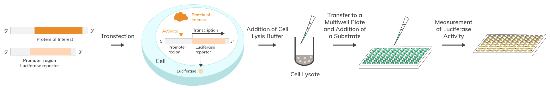
Reporter cell lines are common stable cell lines that have been labelled with a reporter gene, such as Renilla luciferase, one of several widely used types of reporter genes. The Renilla luciferase reporter cell line is a stably transfected cell line which expresses the Renilla luciferase reporter gene under the transcriptional control of a selected promoter. The reporter provides a quantitative readout of promoter or response-element activity via bioluminescence. Renilla luciferase reporter cell lines measure transcriptional activity with bioluminescence, in contrast to chromogenic systems such as the reporter gene lacZ.
Reporter cell lines are stably transfected common cell lines that allow for highly sensitive quantification of engrafted target cells. Researchers utilize reporter gene assays to study signaling pathways, gene regulation, and structure of regulatory elements. These reporter genes monitor receptor-mediated cellular processes at the transcriptional level, illustrating core reporter gene function in living cells.
Renilla luciferase is an enzyme from the shallow-water soft coral Renilla reniformis, commonly known as the sea pansy. Renilla's molecular biology and chemistry is well-studied. The monomeric, 36 kDa protein catalyzes a chemical reaction between Renilla luciferase and its substrate coelenterazine responsible for its bioluminescence. The resulting photon emission is a result of the oxidation of coelenterazine to produce light. Renilla luciferase is a powerful monitoring system for biological processes, as no additional post-translational processing is required for enzymatic activity, making it a classic reporter gene example in many assays, alongside other formats such as the reporter gene GFP.
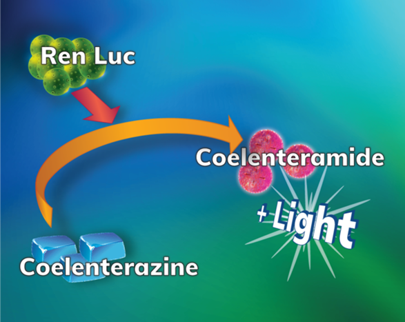
A Renilla luciferase reporter gene assay, utilizes the luciferase enzyme and the substrate coelenterazine to study gene regulation at the transcriptional level.
The Renilla luciferase reporter gene assay monitors your promoter of choice induction activity by screening of the promoter's signaling pathway for activators and inhibitors. The assay allows exploration of a protein's ability to activate or inhibit the expression of a target gene. Using Renilla luciferase as a reporter protein results in a chemical reaction between the enzyme and a coelenterazine substrate, resulting in the emission of photons - bioluminescence. This bioluminescence is quantified as a direct measure of its enzymatic activity.
Gene expression and regulation analysis
Signal pathway mapping
Promoter and regulatory element structure analysis
Protein folding and protein-protein interactions, or environmental sensing with an AHR reporter cell line
SNP (single nucleotide polymorphism) analysis
Antiviral research and therapy, or immune checkpoint studies using a PD-1 reporter cell line
Cytotoxicity assay or cytokine signaling studies with a TSLP reporter cell line
Drug discovery
This protocol may vary per application and experimental objective, but should be similar for either a single reporter or dual reporter assay (dual reporter assays typically use Renilla luciferase as an internal control to normalize experimental variations arising from sample handling and transfection efficiency)
| ISRE | STAT4 | TLR/NF-KB | MRE |
| SBE | CRE | SRF-RE | SRE |
| TLR9/NF-KB | TLR8/NF-KB | TLR7/NF-KB | TLR3/NF-KB |
| TLR2/NF-KB | NF-KB | TLR3/ISRE | TLR3/INFB |
| TCF/LEF | STAT3 | NFAT | GATA3 |
| TNF-β | TNF-α | AP-1 |
| ISRE | INOS | STAT1 | NFAT |
| TNF-β | TNF-α | MIP-2 | IL-8 |
| NF-KB |
| ATF6 | P53 | TLR-4/IL-8 | HRE |
| STAT1 |
| MDA/NF-KB | RIG-I/NF-KB |
| STAT4 | STAT5 |
| FOXP3 | NF-KB |
| Nrf2 |
| IL-6 |
Be Aware (& practice good aseptic technique)! Although possible with other cell types, HeLa cells are so notoriously prolific that countless studies over the years have been challenged after it was found that HeLa cells could float on dust particles in the air and contaminate other cell cultures. Good authentication and validation practices will additionally help detect any cross-contamination with other cell lines.
HeLa Cell Lines
HeLa cell lines are the first immortalized and most widely used human cell line. Obtained from a cervical tumor in 1951 during Henrietta Lacks' treatment of cancer at Johns Hopkins, the highly prolific HeLa cell line continues to make a wide-range of diverse contributions towards many different fields of research and medical discovery. For example, HeLa cell lines contributed significantly towards the development of the COVID-19, polio, tetanus, and many other vaccines. HeLa cells are also used for research of cancer and underlying disease mechanisms, AIDS, viral prevalence to cervical cancers (like HPV), the effects of radiation exposure, to test human sensitivity to certain drugs and chemicals, and many more significant discoveries.
The distinct difference between HeLa cells and normal human cells that directly contribute to HeLa's prolific success:
Doubling time ≈ 34 hours
HEK293 & HEK293T Cell Lines
Human embryotic kidney (HEK293) cell lines are an extensively used immortalized cell line grown in tissue culture. The original HEK cell line was derived by transformation with sheared Adenovirus 5 DNA. After repeated attempts to cultivate, a rapidly growing, single transformed clone was isolated and dubbed as HEK293. Transfection with adenovirus genes results in a highly prolific model system.
HEK293T cells are a daughter cell line derived from the original HEK293 cells and transfected with a SV40 plasmid vector. The SV40 transfection allows for an even higher production of recombinant proteins.
Doubling time ≈ 33 hours
BaF3 Cell Lines
The Ba/F3 cell line derives from the murine C3H species as a hematopoietic, immortalized pro-B cell line, dependent on IL3 for growth. Ba/F3 cells provide a model system as powerful indicators of kinase activity, such as potency, mutations and downstream signaling, or inhibition of kinase activity.
Protein kinases are popular targets for drug discovery as they have the ability to behave as dominant oncogenes across a wide variety of cancers.
Doubling time ≈ 8-10 hours
Q1: What is a reporter cell line?
Q2: How are your reporter cell lines validated?
Q3: How should cell lines be handled upon arrival?
It is recommended to thaw cells immediately upon receipt. Cells are shipped on dry ice (not as cold as liquid nitrogen) therefore thawing has begun in shipping. Immediately storing at -80 °C or on liquid nitrogen will cause cell damage thus decreasing cell viability. The thaw cycle is a crucial beginning step for successful propagation. Do not refreeze reporter cells once thawed.
A typical reporter cell line will require you to thaw frozen cells immediately upon receipt from frozen or liquid nitrogen in water bath at 37 °C.
Be sure to check the culture conditions included with your cell lines, or under the product listing on our website.
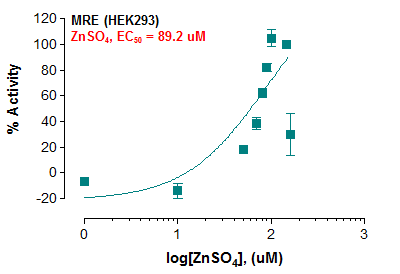
Q4: Can Boster Bio’s reporter cell lines be used for quantitative measurements?
Q5: Can Boster Bio’s reporter cell lines be used in other assays, such as Western Blotting or ELISA?
Q6: How is the luciferase activity measured?
Q7: How do I determine the amount of background activity?
Ready to accelerate your research?
Explore our validated reporter cell lines or request a custom solution tailored to your pathway of interest.