Click image to see more details
Product Info Summary
| SKU: | BA1105 |
|---|---|
| Size: | 0.5ml |
| Reactive Species: | Rabbit |
| Host: | Goat |
| Application: | Flow Cytometry, IF, WB |
Customers Who Bought This Also Bought


Product info
Product Overview
| Product Name | Goat Anti-Rabbit IgG (H+L) Secondary Antibody, FITC Conjugate |
|---|---|
| Synonyms | FITC-conjugated Goat Anti-Rabbit IgG; Goat Anti-Rabbit IgG-FITC Secondary Antibody; Fluorescein-labeled Goat Anti-Rabbit IgG Secondary Antibody |
| Description | Goat Anti-Rabbit IgG (H+L) Secondary Antibody, FITC Conjugate, for detection, localization and quantification of target proteins in a sample via indirect immunofluorescence in IHC-P, IHC-F, ICC, or FCM. |
| Reagent Type | Fluorophore-conjugated secondary antibody |
| Label | FITC |
| Host | Goat |
| Target Species | Rabbit |
| Antibody Class | IgG |
| Clonality | Polyclonal |
| Immunogen | Whole molecule rabbit IgG |
| Purification | Immunoaffinity chromatography, solid phase adsorbed with human serum proteins |
| Specificity | Rabbit IgG specific; no cross-reactivity with human/mouse/bovine IgG |
| Form Supplied | Liquid, concentrated buffered stock solution |
| Formulation | 0.5 mg FITC-conjugated secondary antibody
0.01 M PBS (PH 7.4) 5 mg/mL BSA 50% glycerol |
| Pack Size | 0.5 ml |
| Concentration | 1 mg/ml |
| Application | IF, Flow Cytometry *Our Boster Guarantee covers the use of this product in the above marked tested applications. |
| Storage | At -20˚C for one year from date of receipt. Avoid repeated freezing and thawing. Protect from light. |
| Shipping | Ships in room temperature. Can also ship with gel ice or dry ice but not necessary. |
| Precautions | FOR RESEARCH USE ONLY. NOT FOR DIAGNOSTIC OR CLINICAL USE |
Assay Information
| Sample Type | Human primary-antibody-probed Single cell suspension, Formalin-fixed paraffin-embedded (FFPE) tissue sections, Thawed frozen samples (IHC-F) |
|---|---|
| Assay Type | Immunoanalytical |
| Assay Purpose | Protein detection/quantification |
| Technique | Immunofluorescence |
| Equipment Needed | Excitation light source; Filter set and detector: fluorescence microscope (can be combined with confocal microscope), fluorescence plate-reader, flow cytometer, or cell sorter |
| Additional Materials Required | Primary antibody against target antigen raised in human; Diluent Buffer (PBS or TBS); Application-specific reagents and appliances; |
Main Advantages
| Specificity | High signal-to-noise ratio |
|---|---|
| High Signal Amplification | Multiple secondary antibodies can bind to a single primary antibody; Multiple FITC molecules bind to a single secondary antibody |
| Fast | Fewer processing steps - no need to add a substrate; Less optimization required compared to enzymatic detection; Generates strong signals in a relatively short time span; Fluorescence can be observed directly |
| Quantifieable | Allows quantification of detected signal |
| Easy to Use | Supplied in a workable liquid format |
| Multiplex Compatibility | Colocalization studies possible, even in close proximity: use primary antibodies from different host species for simultaneous detection by fluorophore-conjugated secondary antibodies; use multiple differently colored fluorophores in the same experiment for target differentiation |
| Dynamic range | Good linearity within detection limits |
Background
Most commonly, secondary antibodies are generated by immunizing the host animal with a pooled population of immunoglobulins from the target species. The host antiserum is then purified through immunoaffinity chromatography to remove all host serum proteins, except the specific antibody of interest. Purified secondary antibodies are further solid phase adsorbed with other species serum proteins to minimize cross-reactivity in tissue or cell preparations, and are then modified with antibody fragmentation, label conjugation, etc., to generate highly specific reagents. Secondary antibodies can be conjugated to a large number of labels, including enzymes, biotin, and fluorescent dyes/proteins. Here, the antibody provides the specificity to locate the protein of interest, and the label generates a detectable signal. The label of choice depends upon the experimental application.
Immunofluorescence is a technique used for light microscopy with a fluorescence microscope which utilizes fluorescent dyes as reporters. It is being employed in a variety of applications such as cellular imaging and flow cytometry and is commonly used to visualize the distribution of target molecules through a sample, to detect protein location and activation, to identify protein complex formation and conformational changes, and to monitor biological processes in vivo. Fluorescent dyes (also known as fluorochromes, fluorophores, or simply fluors) are molecules that can absorb light of a specific energy and wavelength, thereby undergoing excitation, and then re-emit it at a lower energy and longer wavelength upon returning to the ground state.
Fluorescent reporters widely used in biological research are of two types: organic compounds with a low molecular weight (0.2-1 kDa) typically containing numerous aromatic groups or plane or cyclic moieties with π bonds (e.g.FITC, TRITC, Alexa Fluor Dyes, DyLight Fluors), and biological fluorophores (e.g.green fluorescent protein (GFP), R-Phycoerythrin). FITC (Fluorescein isothiocyanate) is derivative of fluorescein where a hydrogen atom on the bottom ring of the structure is replaced by isothiocyanate functional group (-N=C=S), making it more reactive to amine and sulfhydryl groups on proteins. The excitation and emission wavelengths of FITC are 495 nm and 525 nm respectively. Like most fluorochromes, it is prone to photobleaching, i.e. losing fluorescing properties due to molecule structure degradation. Loss of activity caused by photobleaching can be controlled by reducing the intensity or time-span of light exposure, by increasing the concentration of the fluorophore, or by employing more robust fluorophores that are less prone to bleaching (e.g., Alexa Fluors, Seta Fluors, or DyLight Fluors). Analogs of FITC with greater photostability and higher fluorescence intensity tailored in various biological applications are Alexa 488 and DyLight 488.
Product Images
Validation Images & Assay Conditions
Click image to see more details
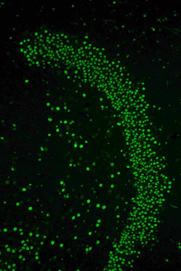
PKC Iota was detected in paraffin-embedded sections of rat brain tissues using rabbit anti-PKC Iota Antigen Affinity purified polyclonal antibody (Catalog # PB9321) at 1 μg/mL. FITC Conjugated Goat Anti-rabbit IgG (H+L) secondary antibody (Catalog # BA1105) was used to detect the primary antibody at 20μg/mL.
Specific Publications For BA1105
Loading publications
Customer Reviews
Have you used Goat Anti-Rabbit IgG (H+L) Secondary Antibody, FITC Conjugated?
Submit a review and receive an Amazon gift card.
- $30 for a review with an image
0 Reviews For Goat Anti-Rabbit IgG (H+L) Secondary Antibody, FITC Conjugated
Customer Q&As
Have a question?
Find answers in Q&As, reviews.
Can't find your answer?
Submit your question
1 Customer Q&As for Goat Anti-Rabbit IgG (H+L) Secondary Antibody, FITC Conjugated
Question
For BA1105 Goat Anti-Rabbit IgG (H+L) Secondary Antibody, FITC Conjugate, the customer wanted to know if it is highly cross adsorbed. Do you have any information?
Verified Customer
Verified customer
Asked: 2020-04-29
Answer
BA1105 has been highly cross-adsorbed against human IgG only.
Boster Scientific Support
Answered: 2020-04-29
