This website uses cookies to ensure you get the best experience on our website.
- Table of Contents
IHC technical resources
For all of your research needs, lab developed tests (LDT) and companion diagnostic tests (CDx), BosterBio offers reliable IHC service and is ready to discuss your project.
Fixation prevents autolysis and necrosis of excised tissues, preserving their morphology and antigenicity as well as increasing their resistance to processing. The choice of fixative is critical as different fixatives are more optimal for some antigens than others. Proper tissue fixation also minimizes epitope masking, preserves protein-protein interaction, and supports consistent immunohistochemistry staining.
Keywords: IHC, optimization, fixative, pfa, perfusion, immersion, fixation method
Learn More About IHC FixativesOnce your tissue sample is fixed using the appropriate method and fixative, it is ready for sectioning and mounting. In order to preserve tissue morphology during sectioning, the tissue must be embedded in paraffin. This guide gives you insight on antigen retrieval, fixation and embedding. Optimized embedding helps reduce background staining and improves specific staining during antibody staining workflows.
Keywords: IHC, optimization, ICH-P, IHC-F, FFPE, frozen section, paraffin embedding, paraffin embedded vs frozen
Learn More About IHC EmbeddingThere are two common methods of epitope retrieval: heat-induced epitope retrieval (HIER), and proteolytic-induced epitope retrieval (PIER). Selecting the right method is critical to get the best results for your application. Effective antigen retrieval reduces non-specific background signals and improves antibody specificity.
Keywords: IHC, optimization, HIER vs PIER, heat-induced epitope retrieval, proteolytic-enduce epitope retrieval, antigen retrieval
Learn More About IHC Antigen RetrievalChoosing the right primary antibody is a critical step in achieving accurate and reproducible immunohistochemistry (IHC) results. Because primary antibodies determine target recognition, their performance directly impacts staining specificity, signal intensity, and overall data reliability across the full IHC workflow — from tissue preparation and antigen retrieval to visualization and interpretation.
Researchers may select monoclonal antibodies when high antibody specificity and consistent epitope recognition are required, especially for diagnostic or quantitative applications. Alternatively, polyclonal antibodies may be preferred for detecting low-abundance targets due to their enhanced sensitivity and broader epitope recognition. Ensuring proper antibody validation, including cross-referencing with Western blotting or known positive controls, further strengthens confidence in target detection.
Optimizing antibody concentration, incubation time, and incubation temperature is essential for balancing strong signal intensity with minimal background interference. Overconcentrated antibodies or excessive incubation can lead to non-specific binding, whereas insufficient antibody levels may result in weak or inconsistent staining. Fine-tuning these parameters helps achieve consistent Primary Antibody Staining, improves specific staining, and supports reliable interpretation of biologically relevant signals.
In addition, proper antibody selection can help mitigate common technical challenges such as epitope masking, cross-reactivity, and interference from tissue autofluorescence, ensuring more accurate visualization of antigen expression across different tissue types and experimental conditions.
The secondary antibody plays a vital role in signal amplification and detection, directly influencing staining clarity, contrast, and reproducibility. Selecting a compatible secondary antibody that matches the host species and isotype of the primary antibody is essential to maintain detection accuracy and prevent false-positive results.
Optimizing secondary antibody dilution and incubation time helps reduce background staining while preserving strong target signal. Overly concentrated secondary antibodies can amplify non-specific background signals, whereas insufficient concentration may weaken detection sensitivity. Proper blocking steps and washing protocols further minimize detection artifacts and improve overall staining precision.
In detection workflows, enzyme- or fluorophore-conjugated secondary antibodies contribute to signal development, supporting downstream chromogenic or fluorescent visualization. Careful optimization ensures reliable IHC staining, minimizes technical noise, and enhances reproducibility across experimental runs.
When integrated effectively with upstream steps such as tissue fixation, antigen retrieval, and primary antibody optimization, secondary antibody refinement strengthens the entire immunohistochemistry pipeline, improving result consistency, interpretability, and experimental confidence.

Download troubleshooting handbooks for IHC, Western blot and ELISA for FREE.
Troubleshooting Guides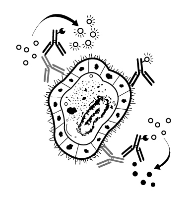
Protocols, optimization tips, troubleshooting guides , and more for IHC.
Technical ResourcesThe principle behind Immunohistochemistry (IHC) entails detection of antigen or happens in cells of a tissue section by exploiting the principle of antibodies binding specifically to antigens in biological tissues. Learn more about this in this guide.
Learn the concept of IHC PrincipleBosterBio has a detailed stepwise IHC protocol with a clearly illustrated IHC workflow with recommended reagents. Learn how to effectively implement a successful immunostaining for tissue sections and cell climbing slices.
Learn our IHC ProtocolThis guide provides a thorough list of Immunohistochemistry troubleshooting tips, including weak staining, high background, nonspecific staining among others. Learn how to take control of your IHC process.
Check our IHC troubleshooting tipsLearn the best IHC and ICC sample preparation techniques. Get a detailed procedure of preparing different types of preserved tissues which is key to getting high quality staining during Immunohistochemistry (IHC)..
Check out our IHC Sample Preparation