| SKU | M02401 |
|---|---|
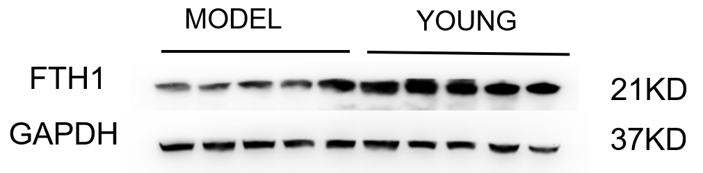
| Application | Western Blot |
| Sample | Mouse hippocampus tissue |
| Sample Processing Description | The tissue was minced and sonicated, then lysed on ice for 1 hour using RIPA buffer. After centrifugation to collect the supernatant and protein quantification by BCA, samples were mixed with loading buffer at the appropriate ratio and denatured by boiling in a water bath. Fifteen microliters of each protein sample were loaded per lane onto SDS-PAGE gel. |
| Primary Antibody | Anti-Ferritin FTH1 Rabbit Monoclonal Antibody |
| Primary Incubation | overnight at 4 ℃ |
| Secondary Antibody | HRP-conjugated Anti-Rabbit IgG Secondary Antibody |
| Secondary Incubation | 1 hour in room temperature |
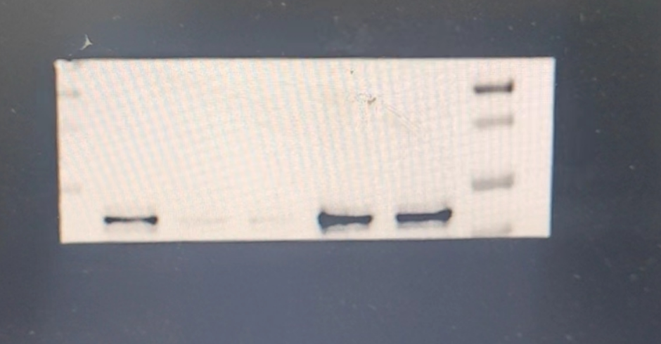
| Detection | Substrate: Ultra-sensitive ECL luminescent reagent (Cat# AR1191), Imaging system:Tanon |
| Results Summary | The FTH1 antibody was used to detect the expression of the target protein in human uterine tissue. The WB bands were single and clear, and compared with other domestic and international brands, this antibody offers excellent cost-performance. |