| SKU | PA1239 |
|---|---|
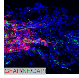
| Application | Immunofluorenscence |
| Sample | Rat Brain |
| Sample Processing Description | Fixed in 4% paraformaldehyde for 48 hours, followed by paraffin embedding and sectioning. |
| Primary Antibody | Anti-GFAP antibody |
| Primary Incubation | 1:500, overnight at 4 ℃ |
| Blocking Agent | Goat serum |
| Secondary Antibody | DyLight 550-conjugated goat anti-rabbit antibody. |
| Secondary Incubation | Incubate at room temperature for 1 hour |
| Detection | Laser confocal microscopy |
| Results Summary | During the experiment, this primary antibody can be conveniently used with the complimentary antibody diluent provided. The results obtained showed good quality and reproducibility, with no false-positive signals. Overall, this primary antibody offers excellent cost performance and provides key data and strong support for publication. |