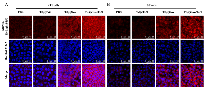
| SKU | A00066-1 |
|---|---|
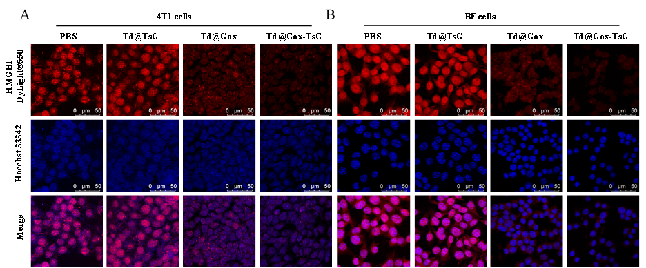
| Application | Immunofluorenscence |
| Sample | Mouse 4T1 cell climbing slides |
| Sample Processing Description | Mouse 4T1 cells fixed with 4% paraformaldehyde for 15 minutes |
| Primary Antibody | Anti-HMGB1 Antibody Picoband® |
| Primary Incubation | 1:500, overnight at 4 ℃ |
| Blocking Agent | Goat serum |
| Secondary Antibody | DyLight 550-conjugated goat anti-rabbit antibody. |
| Secondary Incubation | Incubate at room temperature for 1 hour |
| Detection | Laser confocal microscopy |
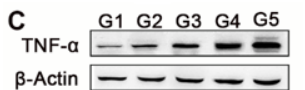
| Results Summary | The ordering and delivery cycle of the antibodies is very short — I was able to receive the products within a week, which saved a lot of time. The pre-sales and after-sales services are convenient and efficient. Since my background is in chemistry, I consulted Boster’s technical team about many details of biological experiments. The technical specialists were patient and thorough in their analysis and explanations, which helped me avoid many detours during the actual experimental procedures. Most importantly, these antibodies offer a high cost-performance ratio, with good binding performance, excellent imaging quality, and a high experimental success rate, providing a solid foundation for conducting related experiments. |