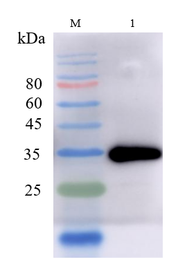
| SKU | M03918 |
|---|---|
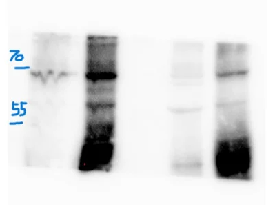
| Application | Western Blot |
| Sample | Mouse brown adipose tissue |
| Sample Processing Description | Tissue and cell proteins were extracted using RIPA buffer. After BCA quantification, 5× loading buffer was added, and the samples were boiled for 5 minutes for denaturation. |
| Primary Incubation | The membrane was incubated with the PLIN1 primary antibodies (1:1000) overnight at 4 °C. |
| Secondary Antibody | Goat Anti-Rabbit IgG Antibody (1:5000) |
| Secondary Incubation | Incubate at room temperature for 1 hour |
| Other Reagents used | Non-fat milk |
| Detection | Signal was developed using ECL substrate |
| Results Summary | This antibody shows excellent consistency and reproducibility across batches, ensuring reliable and stable experimental results. Its performance remains consistent across different experiments, providing strong technical support for our research. |