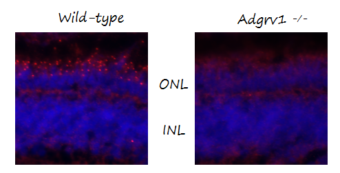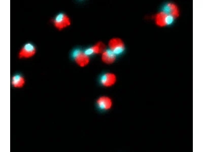
| SKU | DZ4103 |
|---|---|
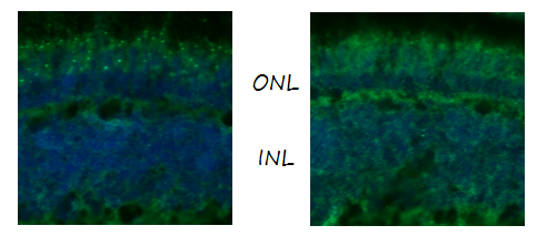
| Application | Immunofluorescence |
| Sample | wildtype and KO mutant zebrafish retina |
| Primary Incubation | 1:100 |
| Blocking Agent | 10% normal goat serum and 2% bovine serum albumin in PBS |
| Secondary Antibody | Alexa Fluor 488 goat anti-rabbit and was used in a 1:800 dilution. |