Customer Testimonials

Peer Reviews from Real Labs

Browse peer-submitted results linked to specific products—ratings, images, and practical notes included. Contribute your own result to strengthen the evidence base and claim rewards after verification.

Video Interviews

See real workflows and decision points—what researchers tried, what worked, and why. Share your story in a short interview and claim rewards!

Get Interview Rewards

Filter by category, reactivity, and host to find the most relevant peer results.

The antibody worked well even though we used fresh frozen brain sections cut at 20 micron thickness thaw-mounted onto microscope slide that were then post-fixed with 4% paraformaldehyde. The antibody worked equally well when used on fixed brains sectioned

Excellent, submitted by on

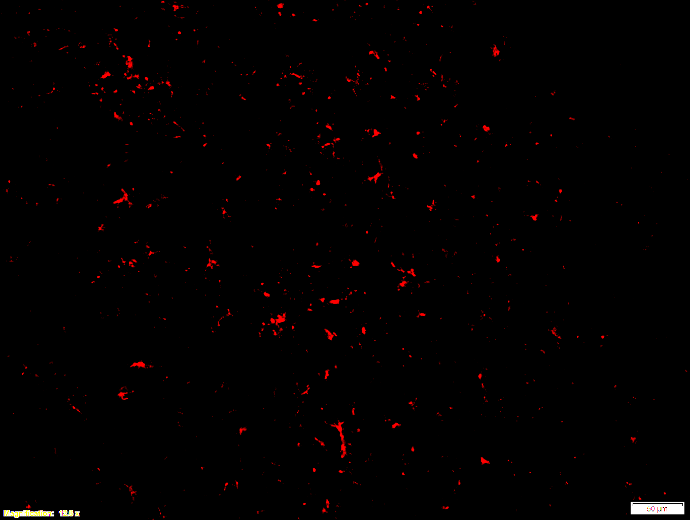
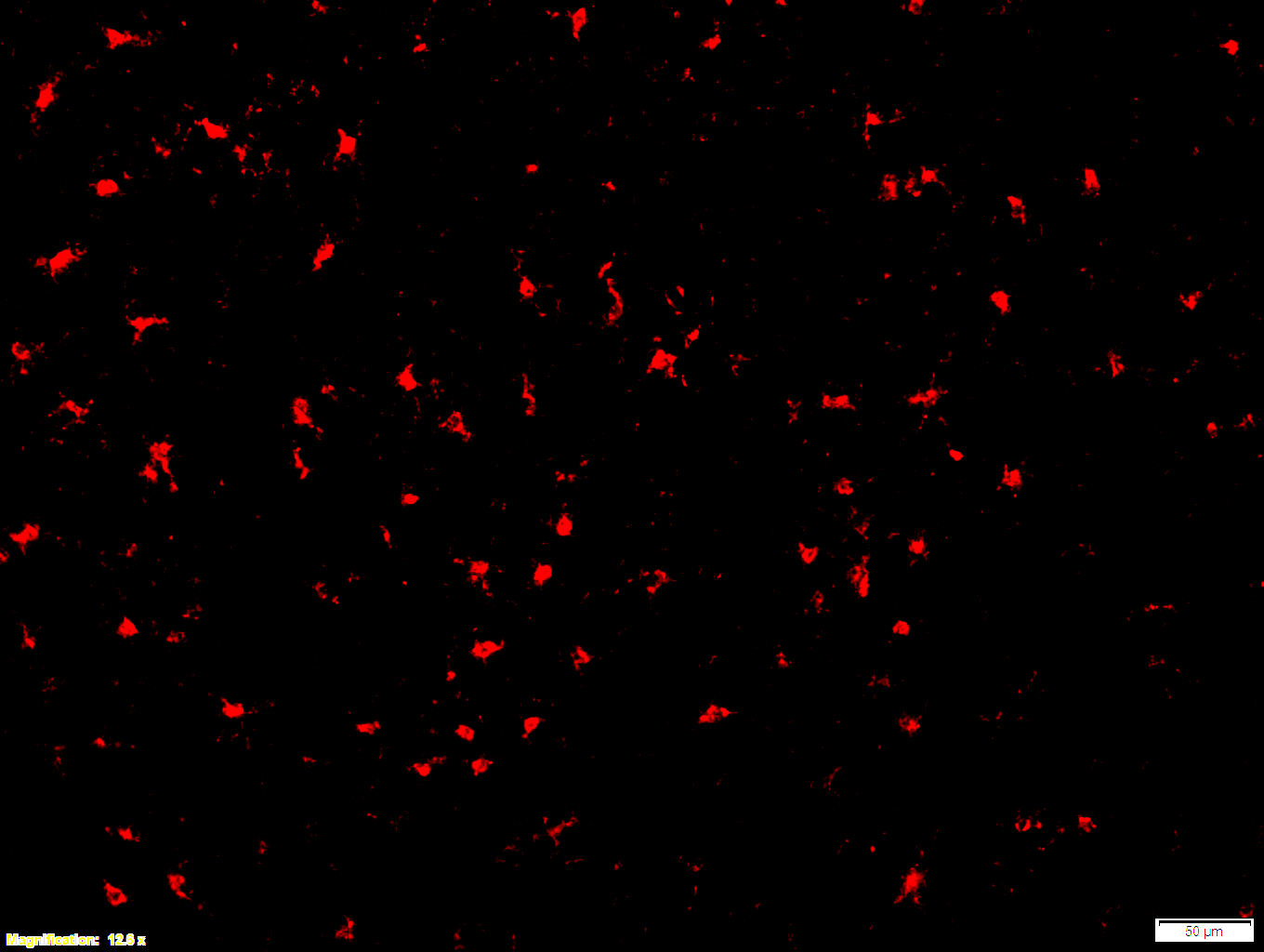
Boster bio Iba1 1:100 PFA (3-4) - cortex 12x - with scale bar

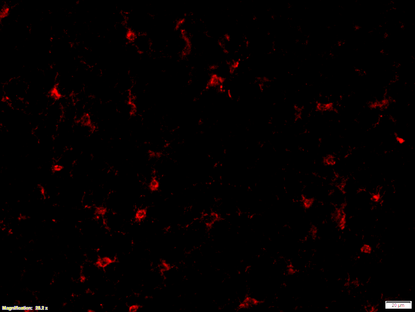
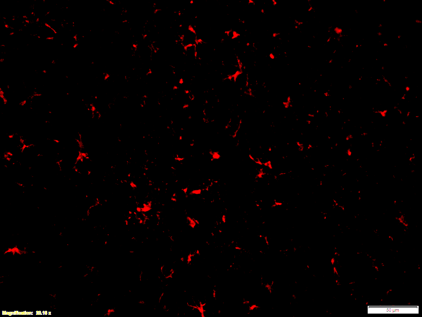
Boster bio Iba1 1:100 PFA (3-4) - cortex 25x - with scale bar

SKU A01394
Application Immunofluorescence
Sample Mouse brain
Primary Antibody Dilution 1:100

Images that were made from fresh frozen cryostat sections, were thaw-mounted onto microscope slides. Mounted sections were later post-fixed with 300 µL of 4% paraformaldehyde (PFA) in PBS at room temperature for 10 minutes. After fixation and prior to incubation with antibodies, slides were washed 3 times with 300 µL of 1X PBS with 0.01% sodium azide for 3 minutes per rinse, followed by permeabilization with 300 µL of 0.3% Triton X-100 in 1X PBS with 0.01% sodium azide for 30 minutes at room temperature, then blocking with 700 µL of 4% donkey serum diluted in 1X PBS with 0.01% sodium azide + Triton X-100 (blocking buffer) for 30 minutes at room temperature. Slides were incubated with 325 µL of Iba1 (1:100). Concentrations of 1:100 were achieved by diluting 10 µL of antibody in 1,000 µL of blocking buffer; concentrations of 1:250 were achieved by diluting 4 µL of antibody in 1,000 µL of blocking buffer. Sections were incubated overnight at 4 ˚C. On the next day, slides were washed 3 times with 300 µL of 1X PBS with 0.01% sodium azide for 3 minutes per rinse, then incubated with the secondary antibodies at room temperature, in the dark, for 1 hour. Washing step was repeated then slides were left in the dark to dry. Mounting media was added to cover slip the sections. Slides were kept in the dark at 4 ˚C prior to imaging.

The antibody worked well even though we used fresh frozen brain sections cut at 20 micron thickness thaw-mounted onto microscope slide that were then post-fixed with 4% paraformaldehyde. The antibody worked equally well when used on fixed brains sectioned

Excellent, submitted by on

Boster bio GFAP 1:100 PFA (7-2) - cortex 12x - with scale bar


Boster bio GFAP 1:100 PFA (7-2) - cortex 20x - with scale bar


Boster bio GFAP 1:250 12x (free-floating)

SKU PB9082
Application Immunofluorescence
Sample Mouse brain
Primary Antibody Dilution 1:100

Images that were made from fresh frozen cryostat sections, were thaw-mounted onto microscope slides. Mounted sections were later post-fixed with 300 µL of 4% paraformaldehyde (PFA) in PBS at room temperature for 10 minutes. After fixation and prior to incubation with antibodies, slides were washed 3 times with 300 µL of 1X PBS with 0.01% sodium azide for 3 minutes per rinse, followed by permeabilization with 300 µL of 0.3% Triton X-100 in 1X PBS with 0.01% sodium azide for 30 minutes at room temperature, then blocking with 700 µL of 4% donkey serum diluted in 1X PBS with 0.01% sodium azide + Triton X-100 (blocking buffer) for 30 minutes at room temperature. Slides were incubated with 325 µL of GFAP (1:100). Concentrations of 1:100 were achieved by diluting 10 µL of antibody in 1,000 µL of blocking buffer; concentrations of 1:250 were achieved by diluting 4 µL of antibody in 1,000 µL of blocking buffer. Sections were incubated overnight at 4 ˚C. On the next day, slides were washed 3 times with 300 µL of 1X PBS with 0.01% sodium azide for 3 minutes per rinse, then incubated with the secondary antibodies at room temperature, in the dark, for 1 hour. Washing step was repeated then slides were left in the dark to dry. Mounting media was added to cover slip the sections. Slides were kept in the dark at 4 ˚C prior to imaging.

Immunohistochemistry was also performed with free-floating sections, which were exposed to the same primary antibodies diluted (1:250) in blocking buffer overnight and, subsequently, exposed to secondary antibodies diluted in blocking buffer for 1 hour, following the same procedure as the mounted sections. After incubation, sections were washed 3 times with 300 µL of 1X PBS with 0.01% sodium azide for 3 minutes per rinse and transferred with a painting brush to a container filled with 1X PBS with 0.01% sodium azide, from which they were mounted onto slides. Slides were left to dry in the dark, after which mounting media was added to cover slip the sections. As with slide-mounted sections, these slides were kept in the dark at 4 ˚C prior to imaging.

"Very happy with this product"

Excellent, submitted by on

Source: Customer Feedback Submission

"Very happy with this product"

--Ailin Lepletier de Oliveira

SKU A00421-3
Application IHC
Sample Mouse spleen and lung

"Very happy with this product. We were using an IL-17A from Abcam but found very high endogenous non-specific staining. This product was a much better stain in comparison. We used this antibody (1:200) on the Leica BOND Rxm autostainer, blocked with Sniper/1% BSA and it worked beautifully."

"Works well for immunofluorescence"

Excellent, submitted by on

Source: Customer Feedback Submission

"Works well for immunofluorescence"

--Jason Tennessen

SKU DZ41223
Application IF
Sample Drosophila larval fat body

"We’ve verified the specificity of this custom antibody that Boster generated for us. The antibody "Anti-Fruit Fly Gpdh1 Antibody (DZ41223)” works well for immunofluorescence. A 1:100 dilution of this antibody stains fat body tissues of wild-type larvae but not Gpdh1 mutant. I’m happy to advertise this antibody to the fly community."

Immunofluorescence for Anti-CG6353 Antibody

--Amin Ghabrial  Pathology and Cell Biology  Columbia University  Principal Investigator

Excellent, submitted by on

Source: Biocompare.com

"My lab is characterizing a mutant we named disjointed. The disjointed gene is also known as CG6353 or Archease. We have used this antibody for IF, and it appears to work well. It gives us a staining pattern for the endogenous protein that matches the pattern we see with an HA-tagged fusion protein detected by anti-HA staining."

SKU DZ33921-1
Application Immunofluorescence
Sample Drosophila larvae
Primary Incubation   1:100 at 4 degrees overnight
Blocking Agent 4% horse serum
Secondary Incubation   1:1000 anti-Rb-A488
Tertiary Incubation   N/A
Detection IF
Results Summary Antibody staining reveals subcellular localization of the protein. In salivary gland and trachea, localization was primarily nuclear.

Anti-Smoothened/SMO Antibody for Flow Cytometry

--Yair Fisher, GliXogen Therapeutics, CSO

Excellent, submitted by on

Source: Customer Feedback Submission

SKU A00451-1
Application Flow Cytometry
Sample C3H10T1/2 mouse cell line

"Overlay histogram showing C3H10T1/2 cells stained with A00451-1 (red line). The cells were blocked with 5% normal goat serum, then incubated with rabbit anti-Smoothened/SMO Antibody (A00451-1, 1:50, 2 μg/0.5x10^6 cells) for 30 min at RT. Alexa Fluor® 647 AffiniPure F(ab')₂ Fragment Goat Anti-Rabbit IgG (H+L) (1:250) was used as a secondary antibody for 30 minutes at RT. Isotype control antibody (blue line) used under the same conditions. Unlabelled sample (yellow line) was also used as a control."

Western blot for Polyclonal Anti-Drosophila melanogaster (Fruit fly) emp Antibody

--Yuzuru Imai, Department of Research for Parkinson's Disease Juntendo University Graduate School of Medicine

Excellent, submitted by on
SKU DZ41152
Application Western blot
Sample Drosophila melanogaster (head and thorax)
Detection Immobilon Forte Western HRP substrate

"There are non-specific bands and immunostaining may not be possible, but Western blotting is sufficient with this antibody."

Good Zebrafish Mef2 Antibody from Boster Bio

--Jingli Cao, Assistant Professor, Weill Cornell Medical College

Excellent, submitted by on
SKU DZ01398-1
Application Immunofluorescence
Sample Zebrafish heart
Detection Confocal Microscopy

"Mef2a was detected in a cryosection of zebrafish heart using the rabbit anti-Mef2a antibody (cat# DZ01398-1, 1:200, 4 degrees overnight). AlexaFluor546 Conjugated Goat Anti-Rabbit IgG was used as a secondary antibody at 1:200 dilution and incubated for 1.5 h at room temperature."

Immunohistochemistry for Anti-CD31/PECAM1 Antibody

Excellent, submitted by on
SKU M01513-4
Application Immunohistochemistry (Paraffin-embedded)
Blocking step 5% BSA as a blocking agent for 30 min at 37°C
Sample Human Lung
Fixative Fixed with 4% paraformaldehyde
Primary Ab Incubation 37°C for 30 minutes
Primary Ab Incubation diluent 5% BSA in TBS
Primary Ab Concentration 1ug/ml
Secondary Antibody SABC kit from Boster Bio, (SA1022)
Secondary Ab Dilution The kit was ready to use, no dilution needed
Secondary Ab Incubation at 37°C for 30 min

Good GATA3 Antibody from BosterBio

--Xiangrong Geng Internal medicine University of Michigan Postdoctoral Researcher

Excellent, submitted by on

Source: Biocompare.com

SKU M00593
Application Western Blot
Sample HEK293T
Detection ECL

"I used this antibody for western blotting and immunoprecipitation. It works well in both of them. Very good antibody."