Customer Testimonials

Peer Reviews from Real Labs

Browse peer-submitted results linked to specific products—ratings, images, and practical notes included. Contribute your own result to strengthen the evidence base and claim rewards after verification.

Video Interviews

See real workflows and decision points—what researchers tried, what worked, and why. Share your story in a short interview and claim rewards!

Get Interview Rewards

Filter by category, reactivity, and host to find the most relevant peer results.

Antibody Optimized and validated

Excellent, submitted by on
SKU DZ41314
Application Western Blot
Sample Drosophila Ovaries
Sample Processing Description Dissection, Homogenization, Sample boiling in 1X Lamelli, SDS-PAGE, Standard Western Blotting
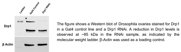
Primary Antibody Anti-Fruit Fly Drp1 Antibody
Primary Incubation 1:1000 in 5% BSA and 0.1% PBST Either 2 hours at RT or Overnight at 4 Degrees
Secondary Antibody 1:10000 Anti-rabbit/mouse IgG horseradish peroxidase-conjugated
Secondary Incubation 1 hour at room temperature
Other Reagents used ECL Clarity Substrate and enzyme for Chemiluminescence
Detection Biorad ChemiDoc
Results Summary In Drosophila ovaries, loss of Drp1 function was confirmed. Western blot analysis validated Drp1 knockdown, showing reduced Drp1 protein levels (~85 kDa) in ovaries expressing Drp1 miRNA, with β-Actin serving as a loading control.

Antibody Optimized and validated

Excellent, submitted by on
SKU DZ41314
Application Immunofluorescence
Sample Drosophila Fat body
Sample Processing Description Dissection, Fixation in 4%PFA, Permeabilization in PBS-TX, Blocking, Primary, Washes, Secondary, Washes, Nuclei Staining, Slide Preparation
Primary Antibody Anti-Fruit Fly Drp1 Antibody
Primary Incubation 1:100 in 2%BSA and 0.1% PBS-TX for Fat body
Secondary Antibody 1:1000 Anti-Rabbit/Mouse Alexa Fluor 488/Cy3/Cy5
Secondary Incubation 1 hour at room temperature
Detection Zeiss LSM 900 Confocal Microscope with Airyscan 2
Results Summary In Drosophila fatbody, Drp1 was seen as punctate structures, as expected from the literature on mitochondria.

A strong nuclear signal for Cyclin E was observed in proliferating cells by IF . Results were reproducible and matched expected patterns.

Excellent, submitted by on
SKU DZ41312
Application Immunofluorescence
Sample Human HaCaT cell
Sample Processing Description Cells were fixed in 4% paraformaldehyde for 15 minutes at room temperature, permeabilized with 0.1% Triton X-100 for 10 minutes and blocked in 1% BSA for 1 hour before incubation with the primary antibody.
Primary Antibody Human CCNE1 Antibody
Primary Incubation 1:100-1:200, overnight at 4 ℃
Secondary Antibody Goat anti-rabbit IgG conjugated to Alexa Fluor 488
Secondary Incubation 1:1000, 1-2 hours in room temperature
Other Reagents used PBS, 0.1% Triton X-100, 1% BSA, DAPI for nuclear counterstaining
Detection Fluorescence microscopy using Alexa Fluor 488 (excitation 488 nm, emission 519 nm) Using Confocal Microscope Zeiss LSM 900
Results Summary The CyclinE-FL antibody (DZ41312) worked in IF application using human cell lines. In IF, the nuclear localization of Cyclin E matched the known expression pattern with good specificity. A fluorescent secondary antibody (Alexa Fluor 488) was used for detection and gave clear signal. Overall, a reliable antibody for full length Cyclin E detection in human cell systems.

A strong nuclear signal for Cyclin E was observed in proliferating cells by IF. Results were reproducible and matched the expected patterns.

Excellent, submitted by on
SKU DZ41311
Application Immunofluorescence
Sample Human HaCaT cell
Sample Processing Description Cells were fixed in 4% paraformaldehyde for 15 minutes at room temperature, permeabilized with 0.1% Triton X-100 for 10 minutes and blocked in 1% BSA for 1 hour before incubation with the primary antibody.
Primary Antibody Human CCNE1 Antibody
Primary Incubation 1:100-1:200, overnight at 4 ℃
Secondary Antibody Goat anti-rabbit IgG conjugated to Alexa Fluor 488
Secondary Incubation 1:1000, 1-2 hours in room temperature
Detection Fluorescence microscopy using Alexa Fluor 488 (excitation 488 nm, emission 519 nm) Using Confocal Microscope Zeiss LSM 900
Results Summary The CyclinE-D15 antibody (DZ41311) worked well in IF application using human cell lines. In IF, the nuclear localization of Cyclin E matched the known expression pattern. The antibody showed good specificity with a minimal background. A fluorescent secondary antibody (Alexa Fluor 488) was used for detection and gave strong, clear signal. Overall, a reliable antibody for Cyclin E detection in human cell systems.

This antibody is highly specific and efficient, suitable for detecting ATP4B protein in rat colon by Western blot, with only minimal nonspecific bands.

Excellent, submitted by on
SKU A08719-2
Application Western Blot
Sample rat colon tissue
Sample Processing Description RIPA lysis buffer with protease inhibitor PMSF (100:1) was used to lyse the sample for 10 minutes, followed by centrifugation at 12,000 rpm for 15 minutes. The supernatant was mixed with 5× loading buffer, denatured at 100°C for 10 minutes, and then loaded onto SDS-PAGE.
Other Reagents Blocking buffer
Primary Antibody ATP4B Antibody Picoband®
Primary Incubation 1:1000, overnight at 4 ℃
Secondary Antibody HRP Conjugated AffiniPure Goat Anti-Rabbit IgG (H+L)
Secondary Incubation 1:5000, 1 hour in room temperature
Detection Substrate: ECL, Imaging system:ChemiDoc MP
Results Summary The figure shows a schematic representation of Western blot results for the target protein ATP4B and the loading control Actin in rat colon across the normal group, model group, low-, medium-, and high-dose traditional Chinese medicine groups, and the western medicine group. Expression was increased in the model group, and among the traditional Chinese medicine groups, the high-dose group showed the best efficacy. The target bands are clear and distinct, and the experimental results are satisfactory.

Our lab will continue to use this antibody, and we will recommend it to other researchers at the university working on similar studies.

Excellent, submitted by on
SKU A31732-2
Application Immunofluorescence
Sample Normal rat liver and alcoholic rat liver
Sample Processing Description The samples consist of myocardium from normal SD rats and myocardium from SD rats subjected to an alcoholic liver disease model. The alcoholic liver disease model was established by gavaging 50% ethanol twice daily, while allowing the rats to freely consume alcoholic beverages, for a total of 14 weeks.
Other Reagents Goat Serum, DAPI, Anti-fade mounting medium
Primary Antibody Anti-PTRF/CAVIN1 Antibody Picoband®
Primary Incubation 1:200, overnight at 4 ℃
Secondary Antibody DyLight 488 Goat Anti-Rabbit IgG (H+L)
Secondary Incubation 1:500, 45 minutes in room temperature
Detection Imaging system:Leica DM2500
Results Summary PTRF/CAVIN1 is a multifunctional protein that shuttles between the plasma membrane and the nucleus, assisting in the recruitment of repair proteins when the plasma membrane is damaged. The results of this IF experiment indicate that alcohol gavage induces myocardial injury and leads to increased expression of PTRF.

The SLC40A1 antibody was used to detect the expression of the target protein in human uterine tissue. Although two bands were observed, the differences in expression levels were still clearly discernible.

Excellent, submitted by on
SKU A01953-2
Application Western Blot
Sample human uterine tissue
Sample Processing Description The tissue was minced and further disrupted by sonication, then lysed on ice for 1 hour using RIPA buffer. After centrifugation, the supernatant was collected and quantified using the BCA method. The appropriate amount of loading buffer was added, and the samples were boiled in a water bath to denature the proteins. Finally, 15 μL of each protein sample was loaded into each lane of the SDS-PAGE gel.
Other Reagents 5% Non-fat milk
Primary Antibody Anti-SLC40A1 Antibody Picoband®
Primary Incubation overnight at 4 ℃
Secondary Antibody HRP-conjugated Anti-Rabbit IgG Secondary Antibody
Secondary Incubation 1 hour in room temperature
Detection Substrate: Ultra-sensitive ECL luminescent reagent (Cat# AR1191), Imaging system:Tanon
Results Summary The SLC40A1 antibody was used to detect the expression of the target protein in human uterine tissue. Although two bands were detected, the differences in expression levels were still clearly observable and did not affect the analysis of the experimental results.

The SLC7A11 antibody was used to detect the expression of the target protein in human uterine tissue. Although two bands were observed in the experiment, this did not affect the interpretation of the results.

Excellent, submitted by on
SKU A03036-2
Application Western Blot
Sample human uterine tissue
Sample Processing Description The tissue was minced and further disrupted by sonication, then lysed on ice for 1 hour using RIPA buffer. After centrifugation, the supernatant was collected and quantified using the BCA method. The appropriate amount of loading buffer was added, and the samples were boiled in a water bath to denature the proteins. Finally, 15 μL of each protein sample was loaded into each lane of the SDS-PAGE gel.
Other Reagents 5% Non-fat milk
Primary Antibody Anti-xCT/SLC7A11 Antibody Picoband®
Primary Incubation 1:1000, overnight at 4 ℃
Secondary Antibody HRP-conjugated Anti-Rabbit IgG Secondary Antibody
Secondary Incubation 1 hour in room temperature
Detection Substrate: Ultra-sensitive ECL luminescent reagent (Cat# AR1191), Imaging system:Tanon
Results Summary The SLC7A11 antibody was used to detect the expression of the target protein in human uterine tissue. Although two bands were observed in the experiment, this did not affect the interpretation of the results.

Western blot analysis was performed using the GP9 antibody to detect GP9 protein expression in the mouse hippocampus. This antibody is highly efficient and specific, making it suitable for quantitative WB detection of GP9 in mouse tissues.

Excellent, submitted by on
SKU M00024-1
Application Western Blot
Sample Mouse hippocampus tissue
Sample Processing Description The mouse hippocampus was lysed with RIPA buffer containing a protease inhibitor cocktail. After protein quantification, samples were mixed with 5× protein loading buffer and heated for 10 minutes to denature. Load 5 μL of protein per lane and apply to SDS-PAGE.
Primary Antibody Anti-AKT1 Monoclonal Antibody
Primary Incubation overnight at 4 ℃
Secondary Antibody HRP-conjugated Anti-Rabbit IgG Secondary Antibody
Secondary Incubation 1 hour in room temperature
Detection Substrate: Ultra-sensitive ECL luminescent reagent (Cat# AR1191), Imaging system:Tanon
Results Summary CD71 antibody was used to detect protein expression in mouse uterine tissue. The bands were clear and free of non-specific signals. After recovery, the antibody could be reused and still showed good performance.

A strong nuclear signal for Cyclin E was observed in proliferating cells by IF. Results were reproducible and matched the expected patterns.

Excellent, submitted by on
SKU DZ41310
Application Immunofluorescence
Sample Human HaCaT cell
Sample Processing Description Cells were fixed in 4% paraformaldehyde for 15 minutes at room temperature, permeabilized with 0.1% Triton X-100 for 10 minutes and blocked in 1% BSA for 1 hour before incubation with the primary antibody.
Primary Antibody Human CCNE1 Antibody
Primary Incubation 1:100-1:200, overnight at 4 ℃
Secondary Antibody Goat anti-rabbit IgG conjugated to Alexa Fluor 488
Secondary Incubation 1:1000, 1-2 hours in room temperature
Other Reagents used PBS, 0.1% Triton X-100, 1% BSA, DAPI for nuclear counterstaining
Detection Fluorescence microscopy using AlexaFluor488 (excitation 488 nm, emission 519 nm) using Zeiss LSM 900 Confocal Microscope with Airyscan 2
Results Summary The CyclinE-N15 antibody (DZ41310) worked fine in IF application using human cell lines. In IF, the nuclear localization of Cyclin E matched the known expression pattern. A fluorescent secondary antibody (Alexa Fluor 488) was used for detection and gave clear signal. Overall, a reliable antibody for D15 transcript Cyclin E detection in human cell systems.