| SKU | M00057-2 |
|---|---|
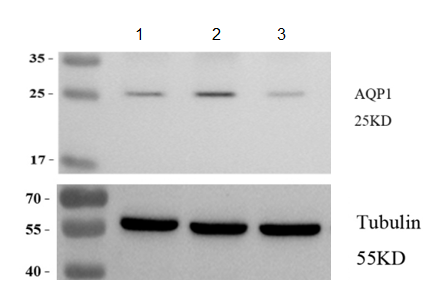
| Application | Western Blot |
| Sample | mouse brain tissue |
| Sample Processing Description | Mouse brain tissues were lysed in RIPA buffer containing a protease inhibitor cocktail at 4°C for 2 hours, centrifuged to collect the supernatant, and protein concentration was determined. After adjusting the concentration, samples were mixed with 5× protein loading buffer, denatured by heating at 95–100°C for 10 minutes, and 15 μL of protein was loaded per lane for SDS-PAGE. |
| Other Reagents | 5% non-fat milk |
| Primary Antibody | GAPDH Antibody Picoband® |
| Primary Incubation | 1:1000, overnight at 4 ℃ |
| Secondary Antibody | goat anti rabbit secondary antibodies |
| Secondary Incubation | 1:5000, 1 h in RT |
| Detection | Substrate: ECL substrate, Image system:ChemiDoc MP |
| Results Summary | This antibody is highly sensitive, produces clear WB bands, is reusable, offers excellent cost-effectiveness, and demonstrates a clear advantage over similar international products, making it highly recommended for use. |