| SKU | M00003 |
|---|---|
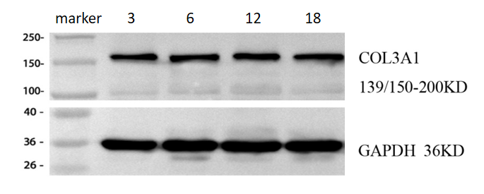
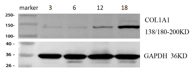
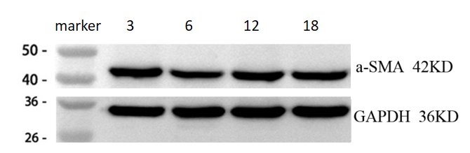
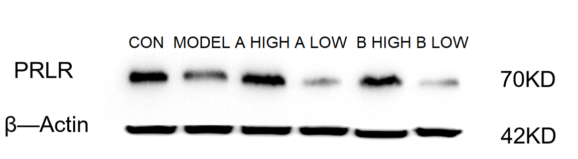
| Application | Western Blot |
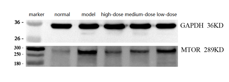
| Sample | Rat skeletal muscle tissue |
| Sample Processing Description | Total protein was extracted from rat skeletal muscle tissue: ① normal, ② injury model, ③ high-dose drug treatment, ④ medium-dose drug treatment, ⑤ low-dose drug treatment. |
| Other Reagents | RIPA lysis buffer, Protease inhibitor, Running buffer, Transfer buffer, Blocking buffer |
| Primary Antibody | mTOR/Tor Rabbit Monoclonal Antibody |
| Primary Incubation | 1:2000, overnight at 4 ℃ |
| Secondary Antibody | HRP Conjugated AffiniPure Goat Anti-Rabbit IgG (H+L) (BA1054) |
| Secondary Incubation | 1:10000, 1 h in RT |
| Detection | Substrate: ECL substrate, Image system:ChemiDoc MP |
| Results Summary | The results show clear MTOR bands, with higher levels in the injury model compared to normal, and a dose-dependent decrease in the treatment groups, as expected. |