| SKU | PB9561 |
|---|---|
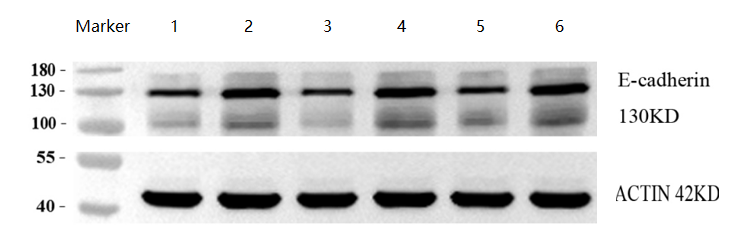
| Application | Western Blot |
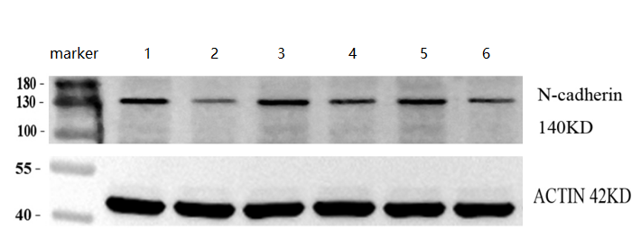
| Sample | human cervical cancer and adjacent tissues |
| Sample Processing Description | Three samples each of human cervical cancer and adjacent tissues were collected, and total protein was extracted. |
| Other Reagents | RIPA lysis buffer, Protease inhibitor, Running buffer, Transfer buffer, Blocking buffer |
| Primary Antibody | E Cadherin 1/CDH1 Antibody Picoband® |
| Primary Incubation | 1:1000, overnight at 4 ℃ |
| Secondary Antibody | HRP Conjugated AffiniPure Goat Anti-Rabbit IgG (H+L) (BA1054) |
| Secondary Incubation | 1:10000, 1 h in RT |
| Detection | Substrate: ECL substrate, Image system: ChemiDoc MP |
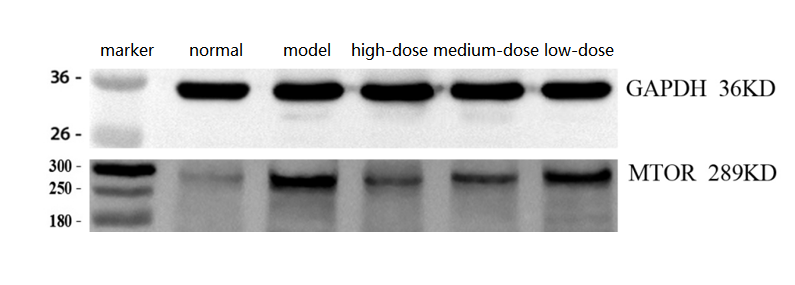
| Results Summary | Based on this study, the expression of E-cadherin in human cervical cancer and adjacent tissues was compared, and results from three cases showed that E-cadherin levels were significantly higher in adjacent tissues than in cancer tissues. |